Anti-inflammatory Activities of an Ethanol Extract of Sargassum macrocarpum in Lipopolysaccharide (LPS)-stimulated RAW 264.7 Macrophages
Ji Min Cheon1, Hyang Suk Kim2, Eun Ok Choi2, Da Hye Kwon2, Yung Hyun Choi2,3,5, Byung Woo Kim4,5 and Hye Jin Hwang1,5*
1Department of Food and Nutrition, Dong-eui University, Busan 614-714, Korea
2Anti-againg Research Center, Dong-eui University, Busan 614-714, Korea
3Department of Oriental Medicine, Dong-eui University, Busan 614-052, Korea
4Division of Applied Bioengineering, Busan 614-714, Korea
5Blue Bio Industry RIC., Dong-eui University, Busan 614-714, Korea
Received September 5, 2017 /Revised October 10, 2017 /Accepted October 18, 2017
Sargassum macrocarpum is a widely distributed marine brown algae found in the North Pacific. The objective of this study was to evaluate the anti-inflammatory activity of an ethanol extract of S. macro- carpum (EESM). First, we investigated the anti-inflammatory activities of EESM in lipopolysaccharide (LPS)-stimulated RAW 264.7 macrophages. EESM treatment suppressed nitric oxide (NO) and prosta- glandin E2 (PGE2) production and inhibited the expressions of the inducible nitric oxide synthase (iNOS) and cyclooxygenase-2 (COX-2) at the mRNA and protein levels. In addition, the expression of pro-inflammatory cytokines, such as tumor necrosis factor-α (TNF-α) and interleukin-1 beta (IL-1β), was decreased in a dose dependent manner. Investigation of the signaling pathways of nuclear factor kappa B (NF-ĸB), phosphoinositide-3-kinase (PI3K)/Akt, and mitogen-activated protein kinases (MAPKs) revealed suppression of NF-ĸB translocation from the cytosol to nucleus by EESM treatment. The phosphorylation of the Akt and ERK proteins was also inhibited by EESM treatment. EESM treatment also stimulated the expression of the heme oxygenase-1 (HO-1) enzyme and its upstream transcription factor, nuclear factor-E2-related factor 2 (Nrf2). These results suggest that EESM has anti-inflammatory activity and could have potential uses in the field of nutraceuticals.
Key words : COX-2, iNOS, pro-inflammatory cytokines, RAW 264.7 macrophages, Sargassum macrocarpum
*Corresponding author
*Tel : +82-51-890-1594, Fax : +82-51-890-2646
*E-mail : [email protected]
This is an Open-Access article distributed under the terms of the Creative Commons Attribution Non-Commercial License (http://creativecommons.org/licenses/by-nc/3.0) which permits unrestricted non-commercial use, distribution, and reproduction in any medium, provided the original work is properly cited.
Journal of Life Science 2017 Vol. 27. No. 12. 1437~1444 DOI : https://doi.org/10.5352/JLS.2017.27.12.1437
서 론
염증(inflammation)은 조직이나 생채에 물리적 작용 또는 화학 물질, 유해 자극 등 외부 물질이 체내로 유입되었을 때 일어나는 생체 방어 반응이지만 비정상적인 염증 반응이 일어 나면 과도한 염증 매개 물질이 생성되고, 그 결과 만성 염증성 질환이 나타난다[2, 14]. 대식세포는 조직과 장기에 존재하는 면역세포로서 손상된 부위를 회복시키기 위해 활성화된다[1].
Nitric oxide (NO), prostaglandin E2 (PGE2)와 tumor necrosis factor-α (TNF-α) 및 interleukin-1β (IL-1β)와 같은 cytokines 등의 염증 매개 물질의 분비를 촉진한다. 염증 반응에 의해 생성되는 NO는 매우 반응성이 큰 물질로 inducible NO syn- thase (iNOS)로부터 생산되며 그 자체로 조직 손상과 염증을
유발시킬 뿐만 아니라 염증 매개체의 생합성을 촉진하여 염증 을 악화시키는 것으로 알려져 있다[1]. PGE2는 효소인 cyclo- oxygenase (COX)에 의해 arachidonic acid로부터 만들어지며 정상적인 생체기능을 하는 COX-1과 PGE2를 형성시키는 COX-2가 있다[9]. 또한 전염증성 cytokine은 세포에서 분비되 는 면역 유발 인자로 tumor necrosis factor-alpha (TNF-α), in- terleukin (IL), interferons (IFN) 등이 있으며 염증반응을 일으 키는데 중요한 역할을 한다[6].
이러한 염증 매개 물질들의 형성은 nuclear factor κB (NF-κ B), phosphoinosited-3-kinase (PI3K)/AKt 및 mitogen-acti- vated protein kinases (MAPKs) 등의 다양한 세포 내 신호 전달계의 활성을 통해 생산됨으로서 염증 반응을 자극한다 [15-17]. 그람 음성균으로 세포막에 존재하는 내독소인 lip- opolysaccharide (LPS)를 마우스 대식세포주의 일종인 RAW 264.7 macrophage에 처리하면 항체 생산, 패혈증, 국소 염증과 같은 다양한 증상을 일으킨다[25].
최근 화학적으로 합성된 항염증 제제를 장기간 복용했을 때 위, 신장, 심장 및 혈관의 기능 저해와 같은 심각한 부작용들이 밝혀지면서 상대적으로 부작용이 적은 천연자원으로부터 항 염증 제제를 개발하기 위한 연구가 진행되고 있다[4, 13, 20].
해조류는 오래 전부터 우리나라를 비롯한 극동지역에서 중 요한 식품자원으로 이용되어왔으며 그 중 갈조류는 fucoidan, phycocolloids, phlorotannins 등의 생리활성물질을 가지며 항 산화, 항응고, 항암 등의 다양한 효과를 보인다고 알려져 있다 [12]. 갈조류에 속하는 큰열매모자반(Sargassum macrocarpum) 은 국내에는 남해안, 제주도연안에 분포되어 있으며, 현재까 지 항균활성[29]과 신경세포보호[26]의 기능이 알려진 바 있으 나 항염증 효과에 대한 연구는 부족한 실정이다. 이에 본 연구 에서는 큰열매모자반 에탄올 추출물이 LPS로 염증이 유도된 RAW 264.7 대식세포에서 미치는 항염증 효과에 대하여 알아 보고자 하였다.
재료 및 방법
큰열매모자반 에탄올 추출물의 준비
본 실험에서 사용된 큰열매모자반 70% 에탄올 추출물(Eth- anol Extract of Sargassum macrocarpum,EESM)은 2007년 제주 도 북촌 다려도에서 채집한 것으로 제주테크노파크 생물종다 양성연구소(Jeju Biodiversity Research Institue)에서 구입하 여 -20℃에서 저장하며 사용하였다.
세포배양
염증 반응에 미치는 EESM의 영향을 조사하기 위해 사용된 murine macrophage cell line인 RAW 264.7 세포는 American Type culture collection (Manassas, VA, USA)에서 구입하여 Dulbecco’s Modified Eagle’s Medium (DMEM, WELGENE, Daegu, Republic of Korea)에 10% Fetal bovine serum (FBS, WELGENE)을 첨가하여 37℃, 5% CO2의 환경에서 48시간 배 양하였다.
세포 독성 측정
시료의 세포 독성과 실험에 사용될 농도를 결정하기 위해 MTT(3-(4,5-dimethylthazol-2-yl)-2,5-diphenyl-tetrazolium bromide, Sigma-Aldrich Co., St. Louis, MO, USA) assay를 실시하였다. 세포 배양용 6 well plate에 RAW 264.7 세포를 1×10⁵cell/ml이 되도록 분주 한 후 EESM을 농도별로 24시간 처리하였다. 또한 같은 조건하에 LPS 처리에 따른 세포생존율 을 측정하기 위해 EESM을 농도별로 처리하고, 1시간 후 LPS (100 ng/ml)를 처리하여 24시간 배양하였다. 24시간 후 배지 를 제거하고 배지와 희석된 0.5 mg/ml 농도의 MTT 시약을 well당 2 ml씩 분주하여 37℃, 5% CO2조건하에서 2시간 배양 하였다. 모든 시약을 깨끗이 제거한 후 dimethyl sulfoxide (DMSO, Sigma-Aldrich Co.)를 2 ml 분주하여 well에 생성된 formazan을 용해한 후 96 well plate에 200 μl씩 옮겨 담아 enzyme-linked immunosorbent assay (ELISA) reader (Molecular Devices, Sunnyvale, CA, USA)로 540 nm에서 흡
광도를 측정하였다. 또한 RAW 264.7 세포의 형태적 변형을 위상차 현미경(Carl Zeiss, Oberkochen, Germany)을 사용해 관찰하였다.
Nitric Oxide 생성량 측정
NO의 농도 측정은 배양액 내의 nitrite 농도를 Griess Reagent (Sigma-Aldrich Co.)를 이용하여 측정하였다. 6 well plate에 RAW 264.7 세포를 confluent 상태까지 배양하여 EESM 및 LPS를 처리한 후 24시간 후 세포 배양액을 수거하였 다. 배양액과 동량의 Griess Reagent를 넣어 반응시키고 ELISA reader로 540 nm에서 흡광도를 측정하였다. NO의 함량은 NaNO2 standard curve를 이용하여 시료의 농도를 계산하고 값을 구하여 나타내었다.
Prostaglandin E2와 cytokine 생성량 측정
LPS로 유도된 RAW 264.7 세포에서 EESM의 농도별 처리 에 의한 PGE2및 TNF-α, IL-1β의 전염증성 cytokine의 생성량 을 Prostaglandin E2 EIAkit (Cayman, AnnArbor, Michigan, USA)와 Quantikine ELISA kit (R&D systems, Minneapolis, USA)를 사용하여 측정하였다. 동일한 조건에서 배양된 RAW 264.7 세포의 배양액을 이용하여 PGE2과 cytokine의 생성량을 제시된 방법에 따라 처리한 다음 ELISA reader를 이용하여 각각 420 nm, 450 nm 파장에서 흡광도를 측정하고 계산하여 나타내었다.
핵, 세포질 분리
염증 매개 물질의 전사인 NF-κB의 조절을 알아보기 위하여 핵과 세포질에서의 발현 양상을 NE-PER Nuclear and Cyto- splasmic Extraction Reagents kit (Thermo Scientific, IL, USA) 를 이용하여 비교하였다. RAW 264.7 세포에 EESM 및 LPS를 처리하여 배양한 후 세포를 모아 상층액을 제거한 후 1☓
phosphate buffer saline (PBS)로 세척으로 상층액을 완전히 제거한 후 제조사의 제시된 과정을 거쳐 세포질과 핵을 분리 하였다.
면역세포화학
NF-κB의 핵과 세포질 발현 양상을 시각적으로 확인하기 위해 면역세포화학을 실시하였다. 세포 배양용 4 well cell cul- ture slide (SPL, Gyeonggi-do, Republic of Korea)에 RAW 264.7 세포를 분주하여 배양 후 100 μg/ml 농도로 EESM을 처리하고 LPS를 처리하여 1시간 동안 배양하였다. 100% 메탄 올로 고정과정을 거치고 1× PBS로 세척 후 5% BSA를 500 μl 분주하여 실온에서 1시간 반응 후 제거 하였다. 이 후 NF-κ B/p65 (Santa Cruz, CA, USA)로 1시간 염색하고, fluorescein isothiocyanate (FITC) conjugated donkey anti-rabbit immun- oglobulin G (IgG, Jackson Immuno Research Laboratories,
Inc., West Grove, PA, USA)로 1시간 더 반응시켰다. 수세 후 4',6-diamidino-2-phenylindole (DAPI, Sigma-Aldrich Co.) 시 약을 소량 분주하여 20분간 암소에서 핵을 염색한 후 형광현 미경(Carl Zeiss)을 통해 NF-κB p65의 발현을 관찰하였다.
Reverse transcriptase-polymerase chain reaction (RT-PCR)에 의한 mRNA의 분석
EESM이 mRNA 수준에서 염증과 관련된 전사 인자들의 발현에 미치는 영향을 알아보기 위해 RT-PCR을 실시하였다.
농도별로 EESM을 처리하고 1시간 후 LPS를 처리하여 배지를 제거한 후 trizol (Invitrogen Co., Carlsbad, CA, USA) 1 ml을 첨가하여 4℃에서 1시간 반응 시킨 후 chloroform을 200 μl씩 첨가하여 4℃에서 5분 반응시켰다. 140,000 rpm, 4℃에서 30분 원심분리시켜 상층액을 모아 동량의 isopropanol을 가하여 1 시간 방치 후 같은 조건으로 원심분리하여 상층액을 제거하고 total RNA만을 분리시켰다. Diethylpyrocarbonate (DEPC) water (AMRESCO, Solon, OH, USA)를 이용하여 녹이고 정량 후, 각각의 primer, DEPC water 그리고 ONE-STEP RT-PCR PreMix Kit (Intron, Republic of Korea)를 넣고 Mastercycler gradient (Eppendorf, Hamburg, Germany)를 이용하여 역전 사 및 증폭하였다. 각 PCR 산물들을 1.5% agarose gel을 이용 하여 전기영동하였고, ethidum bromide (EtBr, Sigma-Aldrich Co.)로 염색시켜 UV 하에서 확인하였으며 housedeeping 유 전자인 glyceraldegyde-3-phosphate dehydrogenase (GAPDH) 를 internal control로 사용하였다.
Western blotting에 의한 단백질 발현 분석
단백질 수준에서 염증과 관련된 전사 인자들의 발현 관찰을 위하여 준비된 세포들을 25 mM Tris-Cl (pH 7.5), 250 mM NaCl, 5 mM EDTA, 1% Nonidet P-40, 1 mM phenylmethane- sulfonylfluoride (PMSF), 5 mM dithiothreitol (DTT)과 pro- tease inhibitor가 함유된 lysis buffer를 사용하여 4℃에서 1 시간 동안 반응시킨 후, 14,000 rpm으로 30 분간 원심분리하여 상층액에 있는 총 단백질을 분리하였다. 상층액의 단백질 농 도는 Bio-Rad 단백질 정량 시약(Bio-Rad, Hercules, CA, USA) 을 이용하여 측정하였다. 동량의 단백질들을 2☓ Laemmli sample buffer (Bio-Rad)를 섞어서 sample을 만들었다. Wes- tern blotting을 위해 동량의 sample을 sodium dodecyl sul- phate (SDS)-polyacrylamide gel을 이용하여 전기영동으로 분 리하고, PVDF membrane (Bio-Rad)으로 전이시켜, 5% Skim milk를 처리하여 비특이적인 단백질들에 대한 blocking을 실 시하여 1차 항체를 처리하여 4℃에서 over night 시킨 다음 PBST로 세척한 후 1차 항체에 맞는 2차 항체를 사용하여 4℃
에서 1시간 정도 반응시켰다. 반응 후 Enhanced Chemilumi- noescence (ECL) solution (Amersham Corp. Arlington Heights, IL, USA) 용액을 적용시켜 X-ray film에 감광시켜
특정 단백질의 발현 양을 분석하였다. 본 연구에서 사용한 1차 항체는 Santa Cruz Biotechnology Inc. (Santa Cruz, CA, USA) 및 Cell Signaling (Beverly, MA, USA)에서 구입하였고, 2차 항체는 Santa Cruz Biotechnology Inc.에서 구입하였다.
결과 및 고찰
세포 독성 측정
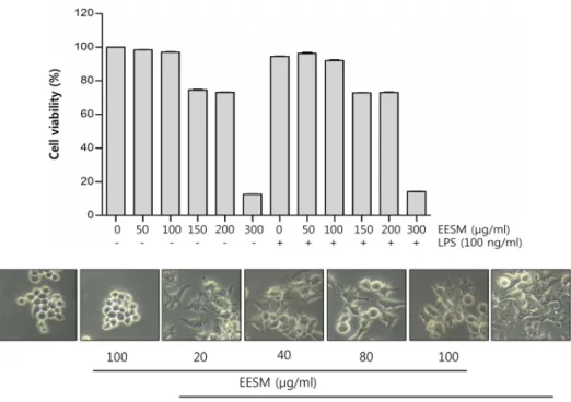
RAW 264.7 세포에서 EESM의 세포독성을 알아보기 위해 농도별의 EESM처리와 LPS (100 ng/ml)단독 및 LPS 1시간 후 처리 한 조건에서 MTT assay를 실시하였다. Fig. 1의 결과 에서 알 수 있듯이, 50~300 μg/ml 농도로 EESM을 처리하였을 때, 100 μg/ml 농도까지 97%의 세포 생존율을 보였고, 150 μg/ml 에서부터는 75% 이하의 세포 생존율을 보였다. LPS와 EESM을 같이 처리하였을 때도 마찬가지로 100 μg/ml 농도에 서 92% 이상의 세포 생존율을 보였고, 150 μg/ml 에서부터는 73% 이하의 세포 생존율을 보였다. 따라서 90% 이상으로 큰 세포 독성을 보이지 않는 100 μg/ml 농도까지 실험조건으로 설정하여 실험을 진행하였다. 또한 Fig. 1에 나타낸 바와 같이 농도별로 EESM과 LPS를 처리하였을 때 전형적인 대식세포 분화를 의미하는 형태학적 변화[5]를 관찰할 수 있었고 이를 현미경 사진으로 나타내었다.
EESM의 NO 생성저해활성 효과
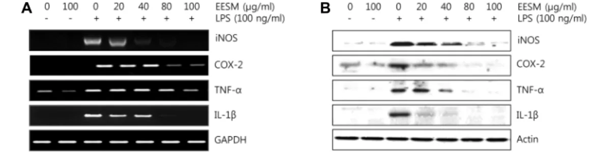
NO는 매우 반응성이 큰 생체 생성분자로써, 효소인 nitric oxide synthase (NOS)에 의해 L-arginine이 L-citrulline으로 합성될 때 생성된다[11]. Inducible NOS (iNOS)는 평소에 발 현되지 않다가 전염증성 cytokine이나 LPS 등에 의해 자극을 받으면 대식세포에서 과량 발현되면서 많은 양의 NO를 생성 하게 되고, 이는 염증을 유발하여 조직의 손상을 가져오는 것 으로 알려져 있다[28, 30]. 따라서 EESM 처리에 의한 NO 생성 정도를 측정하기 위하여 RAW 264.7 세포를 LPS로 자극시킨 후, NO와 iNOS 발현 양상을 확인하였다. 그 결과 Fig. 2A에서 볼 수 있듯이 LPS를 단독으로 처리하였을 때 NO가 현저하게 증가하였고, EESM을 처리하였을 때 농도 의존적으로 감소하 였다. 또한 RT-PCR과 Western blotting을 통해 mRNA와 단백 질 측면에서 iNOS 발현양상 비교했을 때, 유의하게 억제되는 것으로 나타났다(Fig. 3).
EESM의 PGE2와 염증성 cytokines의 생성저해활성 효과 Prostanoid는 생체 항상성 유지에 필요하지만, 과량의 pros- tanoid는 NO와 비슷하게 염증반응을 가속화시켜 류마티스 관 절염, 다발성 경화증과 같은 염증성 질병을 야기시키고[21, 23], 과도한 COX-2의 발현은 PGE2분비를 증가시킨다. 또한, 세포에서 분비되는 전염증성 cytokine은 염증 반응을 유발하 는 면역 조절 물질로 ‘종양괴사인자’인 TNF-α와 정상적인 상
Fig. 1. Effects of EESM on cell viability and morphology in RAW 264.7 macrophages. (A) After RAW 264.7 cells were seeded, the cells were treated with the indicated concentrations of EESM alone or pretreated with EESM for 1 hr before 100 ng/ml of LPS treatment. After 24 hr, the cell viability was assessed using an MTT reduction assay. Data were expressed as percentage of control. Each value indicates the mean ± S.D. and is representative of results obtained from three independent experiments.
(B) RAW 264.7 macrophages were treated with 20~100 μg/ml of EESM for 24 hr or with 100 μg/ml of LPS for 24 hr.
The morphological changes of RAW 264.7 macrophages surfaces were observed by taking photographs using an inverted microscope (original magnification, ×200).
A B
C D
Fig. 2. Effects of EESM on NO, PGE2 and pre-inflammatory cytokines production LPS-stimulated RAW264.7 macrophages. Cells were pre-treated with EESM for 1 hr and then incubation with LPS (100 ng/ml) for 24 hr. (A) NO production was measured using the Griess reagent in culture media. (B, C, D) The amount of PGE2, IL-1β, TNF-α production was measured by ELISA kit. Each value indicates the mean ± S.D. and is representative of results obtained from three independent experiments.
A B
Fig. 3. Effects of EESM iNOS, COX-2, TNF-α and IL-1β expression in LPS-stimulated RAW 264.7 macrophages. Cells were pre-treated with EESM for 1 hr and then incubation with LPS (100 ng/ml) for 24 hr. (A) Relative iNOS, COX-2, TNF-α and IL-1β mRNA expression were measured by RT-PCR. GAPDH was used as an internal control. (B) Relative iNOS, COX-2, TNF-α and IL-1β protein expression were measured by western blot. Actin was used as an internal control. Each value indicates the mean ± S.D. and is representative of results obtained from three independent experiments.
A
B
Fig. 4. Induction of nucleus translocation of NF-ĸB by EESM treatment in RAW 264.7 macrophages. Cells were pre-treated with EESM for 1 hr and then incubation with LPS (100 ng/ml) for 24 hr. (A) Lamin B and Actin were used as internal controls for the nucleus and cytosolic fractions, respectively. Cells were pre-treated with EESM for 1 hour and then incubation with LPS (100 ng/ml) for 1 hr. (B) Localization of NF-ĸB p65 was visualized with fluorescence microscopy after immunofluorescence staining with NF-ĸB p65 antibody (green). And cells were stained with DAPI for visualization of the nuclei (blue).
Representative of results obtained from three independent experiments.
태에서 낮은 농도로 생성되어 인체의 항상성유지에 작용하지 만, 염증성 cytokine으로 과도하게 분비되면 다양한 질병을 일으키는 IL-1β 등이 존재한다[8, 18, 27]. 이러한 원리에 근거 하여 LPS로 유도된 RAW 264.7 세포에서 PGE2와 TNF-α, IL-1 β의 염증성 cytokine의 생성량을 측정하였다. 그 결과 LPS를 단독으로 처리하였을 때 현저하게 증가한 PGE2발현이 EESM 을 처리하였을 때 농도 의존적으로 감소되었고, COX-2 발현 양상 역시 mRNA와 단백질 측면에서 비교했을 때 유의하게 억제되는 것으로 나타났으며, TNF-α, IL-1β 역시 같은 양상을 보였다(Fig. 2, Fig. 3)
EESM이 세포 내 신호전달계에 미치는 효과
앞선 염증성 매개 물질들이 어떤 기전을 통해 염증효과가
조절되는지 확인하기 위해 NF-ĸB와 PI3K/Akt경로 그리고 MAPKs 신호 경로를 확인하였다. NF-ĸB는 세포 내에서 반응 하는 전사 인자로서 염증성 효소와 부착분자의 발현을 조절한 다[10]. Fig. 4A에서 알 수 있듯이, 세포질 내 IκB-α의 발현이 LPS에 의해 감소하였고 EESM을 처리했을 때 증가됨을 확인 하였다. 또한 LPS에 의해 세포질에서 핵 내로 NF-κB p65가 이동함을 Western blotting을 통해 확인하였고, EESM에 의해 핵 내에서 NF-κB p65 발현이 감소됨을 관찰하였다. 이를 면역 형광염색법을 통해 NF-κB p65 세포 내 위치 변화를 조사한 결과 역시 EESM 처리에 의해 NF-κB p65 단백질이 핵 내로 이동되었음을 관찰할 수 있었다(Fig. 4B). PI3K는 세포막 구성 성분인 phosphoinostitide의 지질 성분을 인산화시키는 lipid kinase로, 2차 전달자를 생성하여 Akt 등 여러 하위 신호 전달
Fig. 5. Effects of EESM treatment on PI3K/Akt and MAPKs sig- naling pathways in RAW 264.7 macrophages. Cells were pre-treated with EESM for 1 hr and then incubation with LPS (100 ng/ml) for 24 hr. Total cellular proteins were resolved on SDS-polyacrylamide gels, followed by Western blotting using the specific antibodies, as in- dicated. Representative of results obtained from three in- dependent experiments.
A
B
Fig. 6. Effects of EESM on HO-1, Nrf2 and Keap1 protein ex- pressions in RAW 264.7 macrophages. (A) Cells were treated with 100 μg/ml EESM for 0.5, 1, 3, 6, 12 and 24 hr. (B) Cells were treated with EESM for 24 hr and then harvest cell lysates. Actin was used as an internal control. Each value indicates the mean ± S.D. and is rep- resentative of results obtained from three independent experiments.
체계를 활성화시킨다[7]. 아울러 ERK, JNK 및 p38을 포함한 MAPK 신호 경로 역시 염증성 시스템의 중요한 신호 전달 반응을 매개하고, 세포 활성 물질 생성과 다양한 생물학적 기 능조절의 주요인자로 작용한다[19]. Fig. 5에서 볼 수 있듯이 Akt 단백질의 인산화는 LPS 자극에 의해 현저히 증가되었지 만 EESM에 의해 억제되는 것으로 나타났다. MAPKs 경우 JNK, p38은 큰 차이를 보이지 않았으며 ERK의 인산화가 LPS 자극에 의해 증가하고 EESM에 의해 인산화가 억제되는 것으 로 나타났다. 이상의 결과에서 EESM이 RAW 264.7 세포에서 NF-κB 신호계를 포함한 PI3K/Akt 및 MAPK/ERK 신호경로 를 동시에 관여하고 있음을 알 수 있었다.
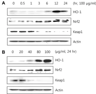
EESM이 HO-1, Nrf2 발현에 미치는 효과
Heme oxygenase-1 (HO-1)는 스트레스, 허혈 및 염증 반응 에서 유도되어지는 단백질로 지질과산화, 단백질산화, RNA 와 DNA 손상 등에 대해 방어작용을 한다[22]. HO-1 발현은 일차적인 전사단계에서 조절되며 전사인자인 nuclear fac- tor-E2-related factor 2 (Nrf2)에 의해 이루어진다[24]. 정상적 인 상태에서 Nrf2는 Keap1에 의해 세포질에서 비활성화 상태 로 존재하지만, 산화적 자극을 받으면 Keap1이 해리되어 핵 내로 이동하여 antioxidant response element (ARE)에 결합해 산화적 스트레스에 대한 생체방어기전의 핵심적인 역할을 한 다[3]. 이러한 원리에 따라 EESM의 HO-1과 Nrf2 신호전달경
로를 조사하였다. 그 결과 Fig. 6과 같이 100 μg/ml 농도의 EESM을 시간별로 처리했을 때, 12시간에서 HO-1의 단백질 발현이 가장 크게 나타났고, 20~100 μg/ml의 농도별로 EESM 을 처리했을 때, 농도의존적으로 HO-1 단백질 발현이 증가됨 을 확인하였다. 또한 상위 전사 인자인 Nrf2 단백질 발현은 3시간에서 점차 증가하여 12시간에서 가장 큰 발현을 보였고, 농도의존적으로 증가하였다. Keap1은 시간과 농도별로 Nrf2 와 반대의 양상을 나타내었다. 이와 같은 결과로 Nrf2가 Keap1에 의해 해리되어 핵 내로 이동하여 HO-1 발현을 조절 한다는 것을 알 수 있었다.
본 연구 결과 갈조류인 큰열매모자반은 LPS로 자극된 RAW 264.7 세포에서 NF-κB의 활성과 PI3K/Akt 및 MAPK/ERK 신호 경로의 활성화를 통하여 HO-1과 Nrf2의 단백질 발현과 함께 다양한 염증 관련 인자의 감소를 통해 염증을 억제시키 는 것으로 나타나, 항염증 건강 기능 식품 소재로서의 활용이 가능할 것이라고 사료된다.
감사의 글
본 연구는 산업통상자원부․부산광역시 지원 동의대학교 블루바이오 소재 개발 및 실용화 지원 센터(RIC 08-06-07)의 지원에 의해 연구되었으며 이에 감사드립니다.
References
1. Albina, J. E. and Reichner, J. S. 1995. Nitric oxide in in- flammation and immunity. New Horiz. 3, 461-464.
2. Arthur, J. S. and Ley, S. C. 2013. Mitogen-activated protein kinases in innate immunity. Nat. Rev. Immunol. 13, 679-692.
3. Balogun, E., Hoque, M., Gong, P., Killeen, E., Green, C. J.
Foresti, R., Alam, J. and Motterlini, R. 2003. Curcumin acti- vates the heme oxygenase-1 gene via regulation of Nrf2 and the antioxidant responsive element. Biochem. J. 371, 887-895.
4. Cerella, C., Sobolewski, C., Dicato, M. and Diederich, M.
2010. Targeting COX-2 expression by natural compounds:
a promising alternative strategy to synthetic COX-2 in- hibitors for cancer chemoprevention and therapy. Biochem.
Pharmachol. 15, 1801-1815.
5. Choi, H. S., Kim, S. R., Hong, S. H., Ku, J. M., Kim, M.
K., Seo, H. S., Cho, S. G., Shin, S., Shin, Y. C. and Ko, S.
G. 2013. Water extract of deer bones activates macrophages and alleviates neutropenia. Evid. Based Complement. Alternat.
Med. 2013, 617302.
6. Chung, H. T., Pae, H. O., Choi, B. M., Billiar, T. R. and Kim, Y. M.2001. Nitric oxide as a bioregulator of apoptosis.
Biochem. Biophys. Res. Commun. 282, 1075-1079.
7. Condliffe, A. M., Cadwallader, K. A., Walker, T. R., Rintoul, R. C., Cowburn, A. S. and Chilvers, E. R. 2000. Phosphoino- sitide 3-kinase: a critical signalling event in pulmonary cells.
Respir. Res. 1, 24-29.
8. Feghali, C. A. and Wright, T. M. 1997. Cytokines in acute and chronic inflammation, Front. Biosci. 2, 12-26.
9. Forstermann, U. and Kleinert, H. 1995. Nitric oxide syn- thase: expression and expressional control of the three iso- forms. Naunyn Schmiedebergs Arch. Pharmacol. 352, 351-364.
10. Fujihara, M., Muroi, M., Tanamoto, K., Suzuki, T., Azuma, H. and Ikeda, H. 2003. Molecular mechanisms of macro- phage activation and deactivation by lipopolysaccharide:
roles of the receptor complex. Pharmacol. Ther. 100, 171-194.
11. Guha, M. and Mackman, N. 2001. LPS induction of gene expression in human monocytes. Cell. Signal. 13, 85-94.
12. Halliwell B. and Gutteridge J. M. C. 1999. Antioxidant defenses. In Free radicals in Biology and Medicine. 3rd end.
Oxford Science Publications. 105-159.
13. Hink, U. and Münzel, T. 2006. COX-2, another important player in the nitric oxide-endothelin cross-talk: good news for COX-2 inhibitors?. Circ. Res. 98, 1344-1346.
14. Hoffmann, J. A., Kofatos, F. C., Janeway, C. A. and Ezeko- witz, R. A., 1999. Phylogenetic perspectives in innate im- munity. Science 284, 1313-1318.
15. Iwalewa, E. O., Mcgaw, L. J., Naidoo, V. and Eloff, J. N. 2007.
Inflammation: the foundation of diseases and disorders. A review of phytomedicines of south African origin used to treat pain and inflammatory conditions. African J. Biotechnol.
6, 2868-2885.
16. Kwon, D. H., Kang, H. J., Choi, Y. H., Chun, K. T., Lee, J.
H., Kang K. H., Hyun, S. K., Kim, B. W. and Hwang, H.
J. 2016. Immunomodulatory activity of water extract of Ulmus macrocarpa in macrophages. J. Life Sci. 26, 50-58.
17. Laroux, F. S. 2004. Mechanisms of inflammation: the good, the ban and the ugly. Front. Biosci. 9, 3156-3162.
18. Locksley, R. M., Killeen, N. and Lenardo, M. J. 2001. The TNF and TNF receptor superfamilies: integrating mamma- lian biology. Cell 104, 487-501.
19. Medzhitov, R. and Janeway, C. A. Jr. 1998. An ancient sys- tem of host defense. Curr. Opin. Immunol. 10, 12-15.
20. Miyasaka, N. and Hirata, Y. 1997. Nitric oxide and in- flammatory arthritides. Life Sci. 61, 2073-2081.
21. Needleman, P. and Isakson, P. C. 1997. The discovery and function of COX-2. J. Rheumatol. 24, 6-8.
22. Otterbein, L. E. and Choi, A. M. 2000. Heme oxygenase: col- ors of defense against cellular stress. Am. J. Physiol. Lung Cell Mol. Physiol. 279, 1029-1037.
23. Ren, K. and Torres, R. 2009. Role of interleukin-1β during pain and inflammation. Brain Res. Rev. 60, 57-64.
24. Shan, Y., Lambrecht, R. W., Donohue, S. E. and Bonkovsky, H. L. 2006. Role of Bach1 and Nrf2 in up-regulation of the heme oxygenase-1 gene by cobalt protoporphyrin. FASEB J. 20, 2651-2653.
25. Takeda, K., Kaisho, T. and Akira, S. 2003. Toll-like receptors.
Ann. Rev. Immunol. 21, 335-376.
26. Tsanga, C. K., Inaa, A., Gotob, T. and Kameia, Y. 2005.
Sargachromenol, a novel nerve growth factor-potentiating substance isolated from Sargassum macrocarpum, promotes neurite outgrowth and survival via distinct signaling path- ways in PC12D cells. Neuroscience 132, 633-643.
27. Turini, M. E. and DuBois, R. N. 2002. Cyclooxygenase-2:
a therapeutic target. Ann. Rev. Med. 53, 35-57.
28. Yoon, Y. I., Hwang, J. S., Ahn, M. Y., Lee, Y. B., Han, M.
S., Goo, T. W. and Yun, E. Y. 2015. Inhibition of in- flammation by Popillia flavosellata ethanol extract in LPS- in- duced RAW 264.7 macrophages. J. Life Sci. 25, 993-999.
29. Yuto, K., Miyuki, S., Ken-ichiro, H., Ryuta, T. and Hiroshi, N. 2009. The novel anti-Propionibacterium acnes compound, Sargafuran, found in the marine brown alga Sargassum macrocarpum. J. Antibiot. 62, 259-263.
30. Wang, Y., Vodovotz, Y., Kim, P. K., Zamora, R. and Billiar, T. R. 2002. Mechanisms of hepatoprotection by nitric oxide.
Ann. N. Y. Acad. Sci. 962, 415-422.
초록:Lipopolysaccaride로 유도된 Raw 264.7 세포에서 큰열매모자반 에탄올 추출물의 항염증 활성
천지민1․김향숙2․최은옥2․권다혜2․최영현3,5․김병우4,5․황혜진1,5*
(1동의대학교 식품영양학과, 2동의대학교 항노화연구소, 3동의대학교 한의학과, 4동의대학교 바이오응용공학부,
5블루바이오 소재개발 및 실용화지원센터)
본 연구에서는 갈조류인 큰열매모자반 에탄올 추출물(Ethanol extract of Sargassum Macrocarpum, EESM)의 항 염증 활성을 조사하였다. Lipopolysaccharide (LPS)로 염증이 유도된 RAW 264.7 대식세포에서 EESM에 의한 항 염증 효과를 조사한 결과 EESM은 nitric oxide (NO)와 prostaglandin E2 (PGE2)의 분비를 억제하는 결과를 보였 고, 이는 inducible NO synthase (iNOS)와 cyclooxygenase-2 (COX-2)의 발현 역시 억제하였다. 또한 전염증성 사 이토카인인 tumor necrosis factor-alpha (TNF-α)와 interleukin 1 beta (IL-1β)의 발현도 억제하였다. 이러한 염증 반응이 어떤 신호경로를 통해 일어나는지 알아보기 위해 nuclear factor kappa B (NF-ĸB), phosphoinositide-3- kinase (PI3K)/Akt 그리고 mitogen-activated protein kinases (MAPKs) 신호전달 경로를 조사한 결과 EESM에 의 해 NF-ĸB가 세포질에서 핵으로 이동과 Akt 단백질의 인산화가 억제되었고 MAPK에 속하는 kinase 중 ERK의 단백질 인산화가 억제되었다. 따라서 EESM이 NF-ĸB 신호계를 포함한 PI3K/Akt 및 MAPK/ERK 신호경로를 동 시에 관여하고 있음을 알 수 있었다. 또한 heme oxygenase-1 (HO-1)과 그 전사 인자인 nuclear factor-E2-related factor 2 (Nrf2)의 단백질 발현에 미치는 영향을 분석한 결과 EESM에 의해 HO-1 및 Nrf2의 발현이 증가됨을 보였 다. 본 연구 결과 EESM은 높은 항염증 활성을 갖는 것으로 확인되였으며 향후 잠재적인 기능성 소재로서 유용하 게 활용될 수 있을 것으로 사료된다.