Anti-inflammatory and Anti-oxidative Effects of Alpiniae Oxyphyllae Fructus Hot Aqueous Extract in Lipopolysaccharide (LPS)-stimulated Macrophages
Na Young Jo
1, Pyeong Jae Lee
2and Jeong Du Roh
1,*1
Dept. of Acupuncture & Moxibustion Medicine, Je-Cheon Hospital of Traditional Korean Medicine, Semyung University
2
School of Oriental Medicine and Bio Convergence Sciences, Semyung University
[Abstract]
Objectives : Alpiniae oxyphyllae Fructus (AOF) is an herbal medicine, which has been used for the treatment of fatigue, chills, and poor physical conditions. The objective of this study was to investigate the anti-inflammatory and anti-oxidative effects of AOF hot aqueous extract.
Methods : The cytotoxicity of AOF extract was evaluated using the MTT assay. Nitric oxide (NO) production was measured by the Griess reaction. Prostaglandin E
2(PGE
2) production was measured by a commercial competitive enzyme immunoassay. Cytokine production (IL- 1tion co6, and TNF- F- was measured by ELISA.
The anti-oxidative effect of AOF extracts was measured by the DPPH method. Polyphenol and flavonoid contents were measured by Folin-Ciocalteu’s phenol reagent and aluminum chloride, respectively.
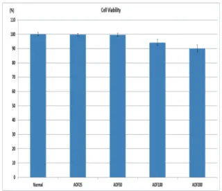
Results : AOF hot aqueous extract did not show toxicity at doses of 25, 50, 100, and 200 µg/mL.
AOF extract significantly inhibited NO production at doses of 100 and 200 µg/mL.PGE
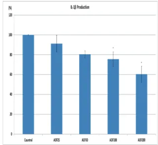
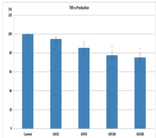
2pro- duction was inhibited by AOF extract treatment at doses of 100 and 200 µg/mL. AOF extracts reduced IL- 6 production in a dose-dependent manner. IL- 1ent maTNF- F- 1ent mannerd IL- 6 production in uction at doses of 100 and µg/mL. The DPPH free radical scavenging capability was above 50% at 200 µg/mL.
Conclusion : This study suggests that AOF hot aqueous extract may exert anti-inflammatory and anti-oxidative effects in a dose-dependent manner. Further studies are required for validating the safety and efficacy of AOF.
✱ Corresponding author : Dept. of Acupuncture & Moxibustion Medicine, Je-Cheon Hospital of Traditional Korean Medicine, Semyung University Semyungro66, Sinwoul-dong, Jecheon city, Chungbuk, 27136, Republic of Korea
Tel : +82-43-649-1816 E-mail : [email protected] Key words :
Alpiniae oxyphyllae Fructus;
Anti-inflammation;
Antioxidant;
Hot Aqueous Extract;
Korean Medicine
Received : 2017. 04. 20.
Revised : 2017. 05. 02.
Accepted : 2017. 05. 08.
On-line : 2017. 05. 20.
This is an Open-Access article distributed under the terms of the Creative Commons Attribution Non-Commercial License (http://creativecommons.org/licenses/by- nc/3.0) which permits unrestricted non-commercial use, distribution, and reproduction in any medium, provided the original work is properly cited.