환자 자세가 간의 방사선 치료 시 선량에 미치는 영향
경희의료원 방사선종양학과・연세암센터 방사선종양학과・대구보건대학교 방사선과
목 적 :간암 치료 시 호흡으로 인한 움직임을 최소화하기 위해 환자 체위 변화에서 종양의 움직임과 용적 변화를 분석하고자 하였다.
대상 및 방법 :간 세포암종(Hepatocellular Carcinoma) 환자 14명의 환자를 대상으로 시행하였다. 바로 누운 자세(Supine position)와 엎드린 자 세(Prone position)에서 2가지 방법으로 환자 고정기구를 제작하고 영상을 획득하여 간 종양의 움직임과 용적 그리고 선량을 분석하였다.
결 과 :바로 누운 자세(Supine position)와 엎드린 자세(Prone position) 에서 표적의 왼쪽-오른쪽(LR, Left-right) 움직임은 평균 2.76±1.25 mm, 2.21±0.93 mm 이고, 앞-뒤(AP, Anterior-posterior)와 상하(SI. Superior-inferior) 방향의 움직임은 각각 4.02±1.63 mm, 11.56±3.08 mm, 3.36
±1.17 mm, 7.45±1.96 mm 이었다. 이를 이용한 엎드린 자세(Prone position)에서 치료 용적(Treatment volume)은 감소하였고, 이에 따라 정상 간 용적은 증가 하였다.
결 론 : 호흡에 의한 간의 움직임을 최소화함으로써 치료 용적(Treatment volume)의 경계여유를 감소시킬 수 있었다. 즉 환자 자세 변화 특히 엎 드린(Prone) 자세는 간의 움직임을 감소 시켜주고 정상 간의 용적을 증가 시킬 수 있을 것으로 사료된다.
핵심용어 :바로 누운 자세(Supine position), 엎드린 자세(Prone position), 종양 용적(Tumor volume)
정원석・김주호・김영재・신령미・오정훈・정건아・조준영・김기철・최태규
서 론
호흡으로 인한 종양의 움직임은 인접한 정상조직(Organ) 의 방사선 흡수량을 증가시켜 방사선 독성으로 인한 부작용 을 증가시킨다. 육안적 종양체적 (GTV, Gross tumor volume) 주위의 여유면적(Margin)의 감소는 인접한 정상조 직의 방사선 독성을 제한하기 위해 필요하지만 해부학적 위 치 오차를 증가시켜 정상조직의 과선량을 유발할 수 있다.
현재 임상에서는 임상적 표적체적 (CTV, Clinical target volume)을 포함하여 위치 잡이 오차 (SM, Set-up margin) 와 내부종양체적(ITV, Internal target volume)을 고려하여 정상조직에 적은 방사선이 흡수되도록 치료계획을 설정하 고 있다. 이는 각기 다른 종양의 기하학적 형태와 장기의 움 직임만을 고려하여 설정된 것으로 개인적인 환자의 위치 변 화에 따른 내부종양체적의 움직임은 달라질 수 있는 제한점 을 지니고 있다.1) 특히 방사선이 조사되는 동안 인접한 정 상조직의 손상을 최소로 하고, 국소제어를 위해 치료 계획 용적(PTV, Planning target volume)을 작게 설정하여야 하
본 논문은 2014년 3월 21일 접수하여 2014년 4월 30일 채택되었음.
책임저자 : 정원석, 경희의료원 방사선종양학과 서울시 동대문구 경희대로 23, 130 - 872 Tel : 02) 958-8667, 010-3517-3798
며 반면 치료계획용적은 임상적 체적용적과 내부 여백 (Internal margin)으로 둘러쌓인 내부표적용적 (ITV, Internal Target Margin)을 포함한다. 이 내부표적용적은 치 료사이(Inter-fraction) 그리고 치료 중(Intra-fraction) 장기 움직임 변화에 의존하기 때문에 내부표적용적이 치료계획 용적을 설정하는데 있어 중요하다.2,3)
치료 중 종양은 원발된 장기의 특징, 종양의 크기, 비만도, 그리고 방사선 치료를 시행하면서 종양의 크기가 변화된 경 우 등에 의해서 종양 체적이 변할 수 있으며 물리적인 변화 요인으로는 연동운동, 심장의 움직임, 호흡에 의해 움직임의 변화가 생길 수 있다. 특히 흉복부에 위치한 종양은 호흡에 의해 위치 및 체적의 변화가 크다고 보고된 바 있다.4,5)
간암의 방사선 치료는 간조직의 정상조직 견딤 선량이 높 지 않아 질병의 표적설정이 어렵기 때문에 방사선치료에 제 한적인 특성이 있다. 최근 입체조형방사선치료(3DRT, 3- Dimensional Radiation Therapy)의 도입으로 정상조직에 입 사되는 방사선량이 감소되는 치료법이 활발히 시행하고 있 지만 종양 조직에 부여되는 처방선량은 호흡에 의해 인접한 정상조직이 표적용적에 침범되어 정상조직의 보호가 쉽지 않다. 이를 극복하기 위하여 치료계획용적(PTV, Planning Target Volume)의 변화를 도입하였는데 이는 환자 호흡, 연 동운동 등에 의한 기관이나 장기의 움직임을 충분히 고려하
여 설정한 체적으로 정의되며 유동적인 종양의 범위까지 포 함하여 설정한 치료용적의 범위이다. 치료계획용적은 종양 조직에 적합한 선량을 부여하는데 도움을 주지만 인접한 정 상조직의 방사선 흡수량을 증가시킬 수 있다. 즉, 치료계획 용적의 최소화는 정상조직의 방사선 흡수량을 적게 할 수 있 게 된다. 현재 임상에서는 환자의 호흡주기를 추적하거나 환 자의 체위를 고정하여 치료계획용적의 여백을 줄일 수 있는 방법을 시행중에 있다.6,7,8,9,10,11)
호흡으로 인한 움직임이 있는 복부 장기는 4DCT(Four dimensional computed tomography)를 이용하여 측정할 수 있다.12,13) 즉 환자의 호흡주기를 4차원 컴퓨터 단층촬영 술(4DCT, Four dimensional computed tomography)을 이 용하여 추적할 수 있다.12,13) 세기변조방사선치료(IMRT, Intensity Modulated Radiation Therapy)의 경우 치료계획 용적 범위를 조절하며 정상조직을 피하면서 종양조직에 적 합한 선량을 부여한다. 반면 다른 치료법보다 대선량을 조 사하기 때문에 부정확한 부위에 선량이 전달되면 정상조직 의 손상이 심해진다. 특히 세기변조방사선치료법을 이용하 여 복부 장기의 방사선치료를 시행할 경우, 종양위치의 기 하학적 오류는 정상조직의 과피폭으로 연결된다. 따라서 4 차원 컴퓨터단층 모의치료를 이용하여 장기의 움직임을 감 소시켜 종양에는 최적의 선량을 전달하며 정상조직에는 최 소한의 선량을 전달해야 한다.7,8,9,10)
기존의 연구를 살펴보면 복부에 위치한 간종양의 움직임 을 최소화하기 위해서 복부를 압박하여 호흡을 직접적으로 조절하는 방법을 이용하였으나 이는 환자의 거부감을 발생 시킬 수 있고 부정확하며 불분명한 호흡을 가진 환자의 경 우 시행하기 어렵다. 따라서 본 논문에서는 환자의 체위를 변화시켜 종양조직의 움직임과 용적변화 그리고 종양조직 과 정상조직의 선량 분포를 비교 분석해 보고자 한다.
대상 및 방법
1. 연구 대상
본 연구는 2012년 1월부터 2013년 2월까지 간 세포암종 (Hepatocellular Carcinoma)의 확진판정을 받은 후 토모테라 피 치료를 위해 내원한 14명의 환자를 대상으로 시행하였다.
연구의 목적과 연구방법에 관하여 충분한 설명을 제공받 고 사전 동의를 구하였다. 반면 연구에 동의하지 않거나 장 시간 치료 자세를 유지하기 어려운 환자는 연구 대상에서 제외하였다(Table 1).
2. 연구 방법
1) 환자 고정기구 제작
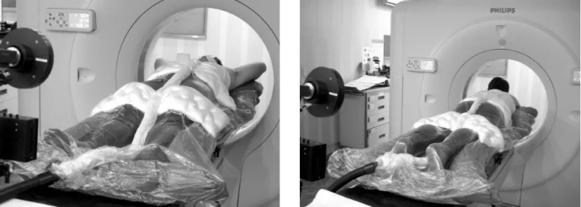
환자가 자유로운 호흡은 가능하면서 최대한 편한 자세를 유지하고 환자 자세에 따른 간의 움직임을 최소화하기 위해 전신 진공 쿠션(Whole body vac-lock, Medical intelligence, Germany)을 사용하였다. 또한 호흡에 의한 움직임을 제한 하기 위해 Body-fix(Vacuum pump, Medical intelligence, Germany)를 이용하여 80 mbar의 압력으로 공기를 제거한 뒤 2가지 방법 즉 바로 누운 자세(Supine position)와 엎드린 자세(Prone position)로 환자 고정기구를 제작하였다(Fig 1).
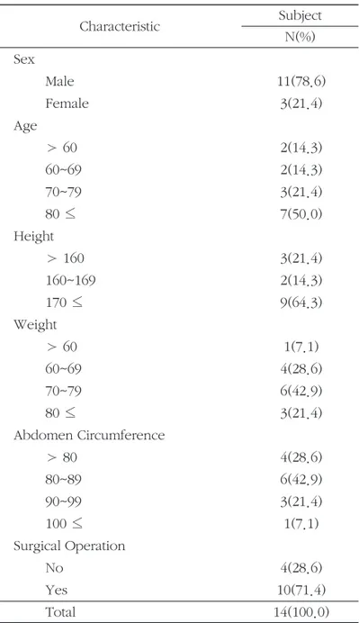
Table 1. Patient’s characteristics
Sex
Male 11(78.6)
Female 3(21.4)
Age
≻ 60 2(14.3)
60~69 2(14.3)
70~79 3(21.4)
80 ≤ 7(50.0)
Height
≻ 160 3(21.4)
160~169 2(14.3)
170 ≤ 9(64.3)
Weight
≻ 60 1(7.1)
60~69 4(28.6)
70~79 6(42.9)
80 ≤ 3(21.4)
Abdomen Circumference
≻ 80 4(28.6)
80~89 6(42.9)
90~99 3(21.4)
100 ≤ 1(7.1)
Surgical Operation
No 4(28.6)
Yes 10(71.4)
Total 14(100.0)
Subject Characteristic N(%)
Fig 1. Simulation process to immobilize using the Vac-lock with Body-fix in supine and prone positions.
Fig 2. The contouring target and critical organs are shown on planning CT imaging in supine and prone position.
2) 영상획득 및 윤곽
호흡에 의한 움직임을 제한한 바로 누운 자세(Supine position)와 엎드린 자세(Prone position)의 종양 용적 평가 를 위하여 전산화단층 모의치료기(CT-simulator, Philips, Holland)와 RPM respiratory gating system 1.7 (S/W)을 통 해 횡격막 상연에서 장골상연(Iliac crest)까지 3mm 슬라이 스 두께로 4차원컴퓨터단층촬영(4DCT, 4 Dimension Computed Tomography)영상을 획득하였고, 획득한 영상 을 치료계획 프로그램인 Pinnacle 3.0을 이용하여 각각의
정상조직과 종양조직의 용적을 그렸다. 이때 종양조직의 용 적은 육안적 종양체적(GTV, Gross tumor volume), 임상적 표적체적 (CTV, Clinical target volume), 내부 표적체적 (ITV, Internal target volume)을 이용하여 치료계획용적 (PTV, Planning target volume)을 설정하였다(Fig 2).
비교를 위해 정상 간(Normal liver) 용적은 전체 간에서 치료계획용적(PTV, Planning target volume)을 뺀 용적으 로 정의하였다.
3) 계획 및 선량 체적 히스토그램(DVH, Dose volume histogram)
설정된 치료 용적은 Tomotherapy planning system V4.2(Tomotherapy, Inc. Madison, WI, USA)로 전달하여 Pitch 0.172, Field Width 1.0 cm, Modulation factor(MF) 2.0을 적용하여 처방선량 50Gy (95% prescription line)로 움직임을 제한한 바로 누운 자세(Supine position)와 엎드 린 자세(Prone position)에서 최적화(Optimization)시켰다.
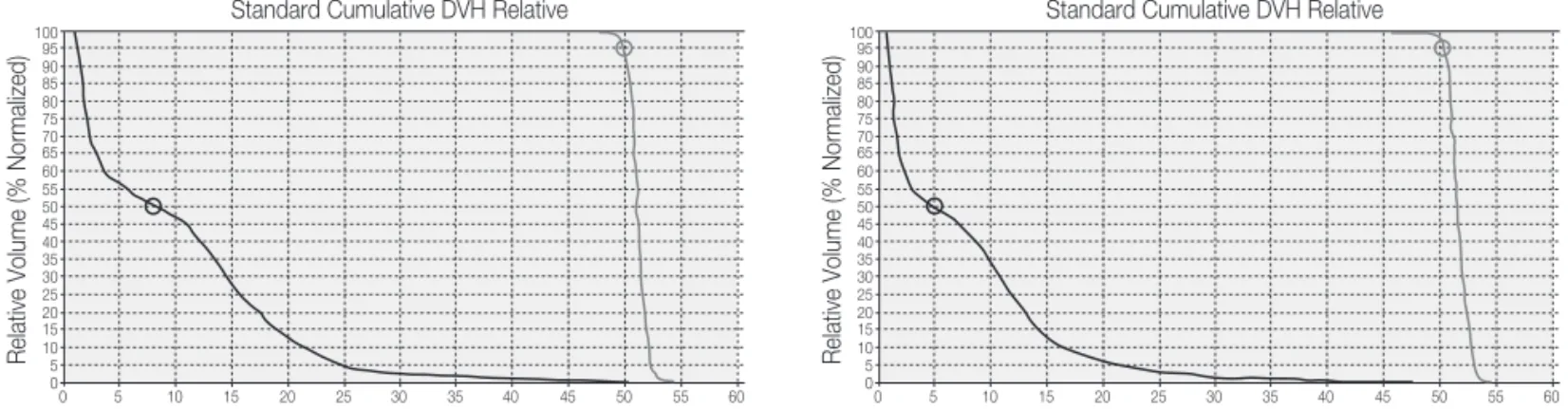
최적화 시킨 후 바로 누운 자세(Supine position)와 엎드린 자세(Prone position)에서 종양 선량과 정상 간 선량을 선 량 체적 히스토그램(DVH, Dose volume histogram)을 통 해 같은 용적에서 선량 분포를 비교하였다.(Fig 3).
4) 분석방법
본 연구에 수집된 자료는 SPSS 13.0 for Windows를 사용 하여 분석하였다.
각 환자에 대한 바로 누운 자세(Supine position)와 엎드 린 자세(Prone position)에서의 표적 움직임, 종양용적 (CTV, Clinical target volume, PTV, Planning target volume)과 정상 간의 용적, 선량 체적 히스토그램(DVH,
Dose volume histogram)을 t-test분석을 통해 평균과 표준 편차를 분석하고 유의성을 평가하였으며 유의수준은 <0.05 이었다.
결 과
1. 표적(Target) 움직임 평가
① 호흡에 의한 움직임을 제한한 바로 누운 자세(Supine position)
호흡에 의한 움직임을 제한한 바로 누운 자세(Supine position)에서 표적의 왼쪽-오른쪽(LR, Left-right) 움직임은 평균 2.76±1.25 mm이고, 앞-뒤(AP, Anterior-posterior)와 상하(SI. Superior-inferior) 방향의 움직임은 각각 4.02±
1.63 mm, 11.56±3.08 mm이었다.
② 호흡에 의한 움직임을 제한한 엎드린 자세(Prone position)
호 흡 에 의 한 움 직 임 을 제 한 한 엎 드 린 자 세 (Prone position)에서는 표적의 상하(SI. Superior-inferior) 방향에 서 7.45±1.96 mm로 가장 크게 움직였으며, 왼쪽-오른쪽 Fig 3. DVH(Dose volume histogram) of the PTV(Planning target volume) and critical organs.
Table 2. Intrafractional motion of the liver in the supine and prone positions Patient position Supine
(mean±SD,mm)
Prone (mean±SD,mm) Characteristic
Left-right
Anterior-posterior Superior-inferior
2.76±1.25 4.02±1.63 11.56±3.08
2.21±0.93 3.36±1.17 7.45±1.96
4.418 4.027 7.487
0.001 0.001 0.000
t p-value
Data presented as mean±standard deviation.
p-Value from SPSS 13.0 for Windows.
(LR, Left-right)와 앞-뒤(AP, Anterior-posterior)의 평균 움 직임은 각각 2.21±0.93 mm, 3.36±1.17 mm 이었다.
호흡의 의한 움직임을 제한한 바로 누운 자세(Supine position)와 엎드린 자세(Prone position)간의 간 종양의 움 직 임 은 3방 향 모 두 에 서 유 의 한 차 이 를 나 타 내 었 다 (p=0.001, 0.001, 0.000)(Table 2).
2. 표적(Target) 용적(Volume) 분석
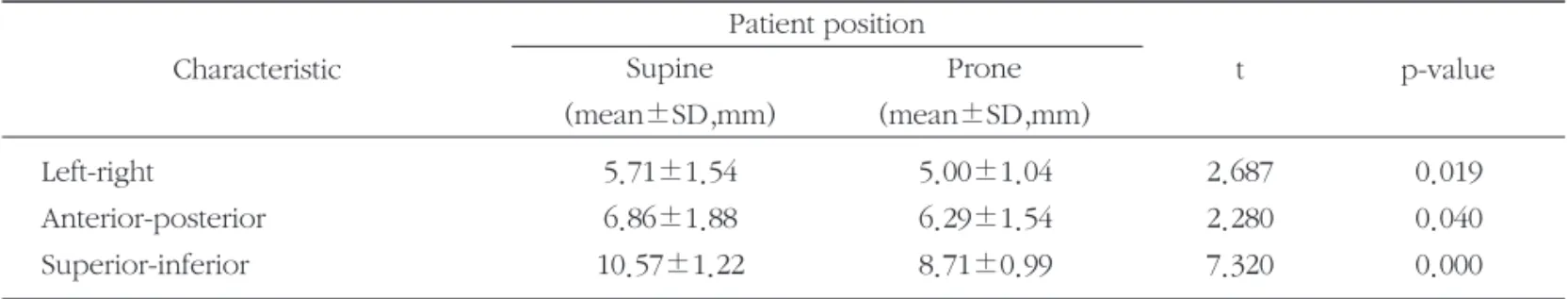
호흡에 따른 종양조직과 정상조직 윤곽의 여백(Margin) 은 바로 누운 자세(Supine position)에서 왼쪽-오른쪽(LR, Left-right) 평균은 5.71±1.54 mm, 앞-뒤(AP, Anterior- posterior) 6.86±1.88 mm, 상하(SI. Superior-inferior) 방향 10.57±1.22 mm 였으며, 엎드린 자세(Prone position)에 서는 왼쪽-오른쪽(LR, Left-right) 5.00±1.04 mm, 앞-뒤 (AP, Anterior-posterior) 6.29± 1.54 mm, 상 하 (SI.
Superior-inferior) 방향 8.71±0.99 mm로 각 방향에서의 여 백은 유의한 차이를 나타내었다(각각 p=0.019, 0.040, 0.000)(Table 3).
① 호흡에 의한 움직임을 제한한 바로 누운 자세(Supine position)
호흡에 의한 움직임을 제한한 바로 누운 자세(Supine position)에서의 평균 용적은 CTV(Clinical target volume) 187.12±210.84 cm3이었고, PTV(Planning target volume) 279.48±253.59 cm3이었으며 정상 간(Normal liver)에서는 1003±212.32 cm3이었다.
② 호흡에 의한 움직임을 제한한 엎드린 자세(Prone position)
호 흡 에 의 한 움 직 임 을 제 한 한 엎 드 린 자 세 (Prone position)용적은 CTV(Clinical target volume)의 평균 용적 은 187.00±210.26 cm3이었고, PTV(Planning target volume) 평균 용적은 251.51±230.86 cm3이었으며 정상 간 (Normal liver)의 평균 용적은 1033.29±196.71 cm3로 각각 분석되었다. PTV(Planning target volume)용적과 정상 간 의 용적에서 유의한 차이를 보였다(p=0.002, 0.001). 그러 나 CTV(Clinical target volume)용적은 통계학적으로 유의 한 차이를 보이지 않았다(p>0.05)(Table 4).
3. 선량 체적 히스토그램(DVH, Dose volume histogram)
완성된 계획을 움직임을 제한한 바로 누운 자세(Supine position)와 엎드린 자세(Prone position)에서 PTV(Planning Table 3. CTV margin of the liver target in the supine and prone positions
Patient position Supine
(mean±SD,mm)
Prone (mean±SD,mm) Characteristic
Left-right
Anterior-posterior Superior-inferior
5.71±1.54 6.86±1.88 10.57±1.22
5.00±1.04 6.29±1.54 8.71±0.99
2.687 2.280 7.320
0.019 0.040 0.000
t p-value
Data presented as mean±standard deviation.
p-Value from SPSS 13.0 for Windows.
Table 4. Comparison of the volume in the supine and prone positions Patient position Supine
(mean±SD,cm3)
Prone (mean±SD,cm3) Characteristic
CTV PTV
Normal Liver
187.12±210.84 279.48±253.59 1003.30±212.32
187.00±210.26 251.51±230.86 1033.29±196.71
0.272 3.768 -4.203
0.790 0.002 0.001
t p-value
Data presented as mean±standard deviation.
target volume) 용적 최고 선량(Maximum Dose) 53.07±
3.79 Gy, 53.88±3.63 Gy로 종양 용적에 선량이 전달되어 유 의한 차이를 나타냈다(p=0.025). 그러나 PTV(Planning target volume)의 평균(Mean) 용적 선량은 51.73±3.59 Gy, 51.83±3.39 Gy로 유의한 차이를 나타내지는 않았다 (p>0.05).
움직임을 제한한 바로 누운 자세(Supine position)에서의 정상 간 용적은 최고는 52.56±3.62 Gy, 용적(Volume) V5 39.83±10.03 Gy, V10 34.68±10.74 Gy, V20 27.44±9.16 Gy, V50 15.38± 6.31 Gy이 고 , 엎 드 린 자 세 (Prone position)에서는 정상 간 용적은 최고는 49..56±3.55 Gy, 용적(Volume)은 V5 36.74±10.07 Gy, V10 31.52±10.88 Gy, V20 25.06±10.18 Gy, V50 12.87±5.41 Gy로 치료자 세의 변화가 용적선량의 변화에 통계적 유의성을 보인 다.(p=0.001, 0.008, 0.011, 0.010, 0.001). 정상 간의 평균 (Mean) 용적 선량은 환자 자세 변화에 따른 유의한 차이를 보이지 않았다(p>0.05)(Table 5).
결 론
간에 대한 방사선 치료 시 매번 발생할 수 있는 위치 잡이 오차와 호흡에 의한 간의 상하움직임을 정량적으로 분석하 는데 있어서 전자포탈영상전송장치(EPID, Electronic Portal Imaging Devices)와 투시장치, 그리고 최근에는 RPM system을 이용한 4차원 전산화 단층 치료 계획 장치
(4DCT-Sim, 4 Dimensional Computed Tomography Simulator)의 역할이 결정적이라 할 수 있다. 4차원 전산화 단층 치료 계획 장치를 사용한 장기 움직임은 호흡주기 당 위상(Phase) 과 자유 호흡동안을 평가할 수 있다. 위상과 위 상의 재구성과 재 손실 때문에 4차원 전산화단층 치료계획 은 실질적인 호흡 주기를 반영하고 호흡 주기를 통해 복부 장기 움직임을 정확하게 측정할 수 있다.2,7,8,9,10,11,14,15,16)
더욱이 호흡으로 인한 움직임의 영향이 큰 간은 움직임 이 없는 타 종양에 비교해 볼 때 움직임에 대한 종양위치 변화율의 양상에 대한 분석이 필수적이다. 간이 호흡에 의 해 움직인다는 점이 방사선치료에 있어서 매우 중요한 요 소임은 널리 알려져 있고, 이에 대한 극복 방안들도 연구되 고 있다.2,17,18,19)
특히 Kitamura14)등은 간암의 경우 종양의 부위, 수술의 기왕력, 간경변증 여부에 따라 4 ~ 9 mm의 움직임이 보였 다고 보고하고 있다.
이전에 보고된 Yong Seok Kim 등의 연구에 의하면 간 첨부의 움직임은 바로 누운 자세와 엎드린 자세에서 17.3 mm, 13.0 mm, 간 아래쪽 끝은 14.4 mm, 11.0 mm로 본 연구와 비교분석해보면 바로 누운 자세 환자들의 정상 호 흡 동안 평균 간의 움직임은 11.0 mm 그리고 깊은 호흡에 서는 12.0-75.0 mm의 범위로 다소 적은 움직임이 관찰되
었다.2,20)우리는 4DCT를 이용하여 바로 누운 자세와 엎드
린 자세에서의 움직임의 평균은 상하(Superior-Inferior) 방향으로 각각 11.56±3.08 mm, 7.45±1.96 mm로 측정 되었다. 이 결과는 상하방향의 내부 여백을 줄여 내부 종 Table 5. Comparison of the Dose in the DVH(Dose volume histogram)
Patient position Supine
(mean±SD,Gy)
Prone (mean±SD,Gy) Characteristic
PTV
Mean 51.73±3.59 51.83±3.39 -0.497 0.628
Max 53.07±3.79 53.88±3.63 -2.532 0.025
Normal Liver
Mean 15.44±6.42 14.97±5.99 0.817 0.429
Max 52.56±3.62 49.51±3.55 4.296 0.001
V5 39.83±10.03 36.74±10.07 3.097 0.008
V10 34.68±10.74 31.52±10.88 2.965 0.011
V20 27.44±9.16 25.06±10.18 3.004 0.010
V50 15.38±6.31 12.87±5.41 4.267 0.001
t p-value
Data presented as mean±standard deviation.
p-Value from SPSS 13.0 for Windows.
양 체척(ITV)의 감소를 시킬 수 있으나 이러한 위치변화가 치료 중(Intra-fraction)에 발생하게 되면 장기 움직임에서 큰 차이가 발생하여 정상조직 손상을 일으키기 때문에 신 중해야 한다.
이에 호흡으로 인한 움직임의 감소를 확인하고 여백을 줄임으로서 실질적인 종양과 정상조직의 용적을 줄일 수 있었다. 호흡에 의한 움직임을 제한한 바로 누운 자세 (Supine position)와 엎드린 자세(Prone position)의 간 종 양 용적의 평균은 각각 279.48±253.59 cm3, 251.51±
230.86 cm3였으며, 정상 간 용적은 1003.30±212.32 cm3, 1033.29±196.71 cm3로 간 종양 용적을 줄여 정상 간 용 적을 증가 시킬 수 있었다. 이는 엎드린 자세에서의 환자 호흡에 의한 종양의 움직임을 보다 적게 할 수 있었으며 표 적 용적을 감소시켜 정상 간의 용적에 방사선 방어를 확보 할 수 있는 것으로 해당용적의 방사선 독성으로부터 보호 할 수 있다는 것을 의미한다.
즉, 환자의 체위는 바로 누운 자세보다 엎드린 자세가 상 하 방향에서 간의 움직임을 상당히 감소시켰으며 종양 용적 을 줄이고 정상 간 용적을 상대적으로 증가시켜 적합한 선 량분포를 나타낼 수 있었다. 이는 엎드린 자세가 바로 누운 자세보다 높은 치료 효과비를 획득하였다고 할 수 있다.2) 또한 Body Fix를 이용하여 복부압박을 시행하여 호흡을 제 어함으로써 표적체적(target volume)의 경계여유를 감소시 킬 수 있었다.
결과적으로 엎드린(Prone) 자세와 Body-fix 사용은 간의 움직임을 감소 시켜주고 있으며, 이를 통해 PTV 경계여유 를 감소시킬 수 있다. 이는 움직임이 감소 될 때 치료계획과 동일한 선량이 실제 치료에 전달 될 것이다. 따라서 간 내 종양과 손상위험장기의 거리가 유지된다면 정위적 방사선 치료를 진행함에 있어서 호흡에 의한 움직임을 제한한 엎드 린 자세(prone position)가 도움이 될 수 있을 것으로 생각 된다.
본 연구의 제한점을 살펴보자면 첫째, 실험에 참여한 환 자의 수가 적었다는 것이다. 비록 장기 움직임의 정도를 다른 연구와 비교 하였지만 본 연구에서의 시행한 환자의 수는 통계적으로 유의성을 확보하기에는 다소 부족하였
다.2,21,22) 둘째, 4차원 전산화단층치료 모의치료에 필요한
평균 진행 시간은 30분 정도로 일반 전산화단층 모의치료 의 진행시간인 5분과 비교해 보았을 때 많은 소요시간이 필요하며 이는 일반적인 모의 치료절차에 적용하기 적절 치 못한 점이 있다.
환자의 개인적인 특성상 체중의 압박으로 인하여 간의 변 형이 생기면 종양의 위치 구별이 용이하지 않을 수 있으며 치료계획용 모의치료영상 획득 시 환자의 호흡주기에 따라
획득된 영상이 달라질 수 있다. 이러한 경우, 추가적으로 체 중과 Body-fix의 사용 그리고 동시에 움직였던 횡격막의 거 리를 관찰하여 영상 융합과정(Image-Guide Process)을 거 쳐야만 오차를 최소한으로 줄일 수 있을 것이라 생각된다.
이러한 제한점은 호흡주기를 정밀하게 분석할 수 있는 장비 가 개발되거나 호흡조절 방사선치료의 발전된 치료법이 개 발된다면 극복할 수 있을 것으로 생각되며 이러한 제한점이 극복된다면 간 내 종양의 방사선 치료 시 우수한 치료성과 를 얻을 수 있을 것으로 사료된다.
참고문헌
1. Lee S, Seong JS, Kim YB, et al. Use of respiratory motion reduction device(RRD) in treatment of hepatoma. J Korean Soc Ther Radiol Oncol 2001;
19(4): 319-326
2. Kim YS, Park SH, Ahn SD, et al. Differences in abdominal organ movement between supine and prone positions measured using four-dimensional computed tomograpy. Radiother Oncol 2007; 85: 424- 428
3. International Commission on Radiation Units and Measurements, Report No. 62: Prescribing, recording and reporting photon beam therapy (supplement to ICRU report 50). Bethesda: ICRU. 1999
4. Kvinnsland Y, Muren LP. The impact of organ motion on intestine doses and complication probabilities in radiotherapy of bladder cancer. Radiother Oncol 2005;
76: 43-7
5. Li XA, Qi XS, Pitterle M, et al. Interfractional variation in patient setup and anatomic change assessed by daily tomotherapy. Int J Radiat Oncol Biol Phys 2007; 68:
581-91
6. Rardall KT, James MB, Lon HM, et al. Potential benefits of eliminating planning target volume expansions for patient breathing in the treatment of liver tumor. Int J Radiat Oncol Biol Phys 1997; 38: 613-617
7. John H. Heinzerling, John F. Anderson, Lech Papiez, et al. Four-dimensional computed tomography scan analysis of tumor and organ motion at varying levels of abdominal compression during stereotactic treatment of lung and liver. Int J Radiat Oncol Biol Phys 2008; 70:
1517-1578
8. Mian Xi, Meng-Zhong Liu, Li Zhang, et al. How many sets of 4DCT images are sufficient to determine internal target volume for liver radiotherapy?.
Radiother Oncol 2009; 92: 255-259
9. David P. Gierga, George T. Y. Chen, Jong H. Kung, et al. Quantification of respiration-induced abdominal tumor motion and its impact on IMRT dose distribution. Int J Radiat Oncol Biol Phys 2004; 58:
1584-1595
10. Eike Rietzel, George T. Y. Chen, Noah C. Choi, et al.
Four-dimensional image-based treatment planning:
Target volume segmentation and dose calculation in the presence of respiratory motion. Int J Radiat Oncol Biol Phys 2005; 61: 1535-1550
11. Takashi Aruga, Jun Itami, Moriyo Aruga, et al. Target volume definition for upper abdominal irradiation using CT scans obtained during inhale and exhale phases. Int J Radiat Oncol Biol Phys 2000; 48: 465-469 12. Bradley JD, Nofal AN, El Naqa IM, et al. Comparison of helical, maximum intensity projection (MIP), and averaged intensity(AI) 4D CT imaging for stereotactic body radiation therapy(SBRT) planning in lung cancer. Radiother Oncol 2006; 81: 264-8
13. Guckenberger M, Wilbert J, Meyer J, et al. Is a single respiratory correlated 4D-CT study sufficient for evaluation of breathing motion? Int J Radiat Oncol Biol Phys 2007; 67: 1352-9
14. De Ne ve W, Va n de l He uve l F, De Be uke le e r M, e t a l. Routine clinical on- line portal imaging followed by immediate field adjustment using a tele- controlled patient couch. Radiother Oncol 1992; 24: 45- 54 15. Ha nle y J, Lumley M, Mage ras G, et a l.
Measurements of patient positioning errors in three- dimensional conformal radiotherapy of the prostate.
Int J Radiat Oncol Biol Phys 1997; 37: 435-444 16. Edward D. Brandner, Andrew Wu, Hungcheng Chen,
et al. Abdominal organ motion measured using 4D CT. Int J Radiat Oncol Biol Phys 2006; 65: 554-560 17. James D. Brierley, Laura A. Dawson, Elliott Sampson,
et al. Rectal motion in patients receiving preoperative radiotherapy for carcinoma of the rectum. Int J Radiation Oncology Biol Phys 2011; 80(1): 97-102 18. Amish P. Shah, Patrick A. Kupelian, Twyla R.
Willoughby, et al. An evaluation of intrafraction
motion of the prostate in the prone and supine positions using electromagnetic tracking. Radiother Oncol 2011; 99: 37-43
19. Capocaccia R, Sant M, Berrino F, et al. Hepatocellular carcinoma: trends of Incidence and survival in Europe and United State at the end of the 20thcentury.
AmJGastroenterol2007;102:1661-1670
20. Weiss PH, Baker JM, Potchen EJ. Assessment of hepatic respiratory excursion. J Nucl Med 1972; 13:
758-9
21. Balter JM, Ten Haken RK, Lawrence TS, et al.
Uncertainties in CT-based radiation therapy treatment planning associated with patient breathing. Int J Radiat Oncol Biol Phys 1996; 36: 167?-74
22. Langen KM, Jones DT. Organ motion and its management. Int J Radiat Oncol Biol Phys 2001; 50:
265-78
Won Seok Jung, Ju Ho Kim, Young Jae Kim, Ryung Mi Shin, Jeong Hun Oh, Geon A Jeong, Jun Young Jo, Gi Chul Kim, Tae Kyu Choi
The effect of patient position on dose in radiation therapy of liver cancer
Department of Radiation Oncology, Kyunghee College Hospital, Seoul, Korea Department of Radiation Oncology, Yonsei Cancer Center, Seoul, Korea Department of Radiological Technology, Daegu Health College, Daegu, Korea
Purpose :To analyze tumor`s movement and volume change from changing position in order to minimize movement caused by breathing.
Materials and Methods : We conducted survey of 14 patients with HCC(Hepatocellular carcinoma). Patient immobilization device was made in two ways(Supine position, prone position) and from image acquisition, tumor`s movement, volume and dose are analyzed.
Results :The mean movement of target(LR, Left-right) in supine position and prone position was 2.76±1.25 mm, 2.21±0.93 mm. AP(Anterior-posterior) and SI(Superior-inferior) was 4.02±1.63 mm, 11.56±3.08 mm, and 3.36±1.17 mm, 7.45±1.96 mm.
Treatment volume was decreased and normal liver volume was increased in prone position.
Conclusion :We could reduce the margin of the treatment volume by minimizing the movement of liver caused by breathing.
Especially in prone position, it is considered to be able to decrease the movement of the liver and increase normal liver volume.
Abstract
Keyword :Supine position, Prone position, Tumor volume