서 론
어류는 개체 발생 동안 자어기에서 치어기로 변태하기 직전 에 형태적, 생리적, 행동적으로 매우 극심한 변화를 겪으므로 자세한 발생학적 지식은 발생학적 측면뿐만 아니라 어업생물 학적 및 양식업에서도 매우 중요하다
(Koumoundouros et al.,
1999).
또한 이 두 분야와 관련하여 해부학의 발달은 유어기의 형태발달 단계에서 기능적인 발달 경향과 환경에 대한 선 호도를 이해하는데 도움을 준다
(Fukuhara, 1992).
어류의 종 묘생산에 있어서 자치어기의 골격 발달과정에 관한 관심과 정확한 지식은 사육 초기에 골격 이상의 탐지와 제거를 위해 필 수적이며
(Koumoundouros et al., 1997a, b),
유어기의 계통 분류학적 기초 형질을 제공하고 있어 이 분야를 연구하는 열 기가 높다(Mook, 1977; Potthoff et al., 1988; Faustino and power, 1999; Liu, 2001; Sfakianakis et al., 2004).
점농어는 농어목
(Perciformes)
농어과(Lateolabracidae)
농 어속(Lateolabrax)
에 속하는 어종으로 최근에 별종으로 보고 되었다(Kim et al., 2001).
농어속어류는 우리나라를 비롯한 중국, 일본 등 동아시아 국가에서 상업적으로 매우 중요한 어 종으로써 인기가 많은 어류이며, 특히 점농어는 성장이 빨라 양식업에서 매우 선호되는 어종이다(Yokogawa and Tajima, 1996; Kim et al., 1998).
그러나 점농어에 관한 연구는 호르271
한국산 점농어(
Lateolabrax maculatus
) 자치어의 골격발달과 비늘형성강충배 . 명정구
1* . 김용억
2. 김형철
3Early Osteological Development and Squamation in the Spotted Sea Bass Lateolabrax maculates (Pisces: Lateolabracidae)
Korea Inter-University Institute of Ocean Science, Pukyong National University, Busan 608-737, Korea
1
Marine Living Resources Research Department, Korea Ocean Research & Development Institute, Ansan 426-744, Korea
2
Department of Marine Biology, Pukyong National University, Busan 608-737, Korea
3
Division of Environmental Research, National Fisheries Research & Development Institute, Busan 619-705, Korea Early osteological development of the skull, vertebral column, and fins, and squamation in the spotted sea bass, Lateo- labrax maculates, were studied under extensive larval rearing conditions. The first ossification during cephalic skeleton development began with the premaxillary, dentary, and parasphenoid at 6.4 mm Total length (Notochord length 6.1 mm) and was completed by 25.2 mm TL (Standard length 20.3 mm). Ossification of the cartilaginous caudal complex started simultaneously in the urostyle and two preural centra at 12.2 mm TL (10.4 mm) and it was completely ossified by 32.0 mm TL (26.4 mm). The principal caudal fin rays, with a count of 9/8, began to ossify at 11.6 mm TL (10.2 mm) and the procurrent caudal fin rays, with counts of 4 (upper) and 3 (lower), started to ossify by 12.6 mm TL (10.9 mm). Ossifica- tion of these parts was completed by 21.4 mm TL (17.5 mm). Ossification of the vertebral column was first observed in the first to fourth centra at 8.3 mm TL (7.5 mm) and was fully completed by 21.7-35.0 mm TL (17.8-29.3 mm). The pectoral girdle started to ossify by 5.6 mm TL (5.4 mm) and was completed by 26.8 mm TL (21.8 mm). Eight pectoral fin rays were ossified at 11.6 mm TL (10.2 mm) and 16-18 rays were fully ossified by 13.8 mm TL (12.0 mm). Also, the dorsal, anal, and pelvic fin rays started to ossify at 12.2 mm TL (10.4 mm) and were completed by 12.8 mm TL (11.2 mm), 23.8 mm TL (19.4 mm), and 13.8 mm TL (12.0 mm), respectively. Ossification of the anal and dorsal pterygi- ophores initially occurred by 12.6 mm TL (10.9 mm) and 14.3 mm TL (12.2 mm), and was completed by 21.4 mm TL (17.5 mm) and 19.3 mm TL (15.9 mm), respectively. Squamation started at three centers of differentiation: the middle region of the trunk, the anterior of the caudal peduncle, and on the caudal peduncle at 23.8 mm TL (19.4 mm). The body was covered with scales, except the snout, at 57.2-60.2 mm TL (SL 47.1-49.2 mm).
Key words: Lateolabrax maculatus, Osteological development, Squamation
Chung-Bae Kang, Jung-Goo Myoung1
*
, Yong Uk Kim2 and Hyongchol Kim3부경대학교 해양과학공동연구소, 1한국해양연구원, 2부경대학교, 3국립수산과학원
*
Corresponding author: [email protected]몬
(Baek et al., 2001; Kim et al., 1999),
초기생활사(Kim et al., 1998; Nakayama et al., 1996),
형태 및 유전학적 연구(Yokogawa and Seki, 1995; Park et al., 1996; Myoung et al., 1997; Liu et al., 2006)
등 일부 보고가 있을 뿐이며, 골 격 발달 및 비늘형성 과정에 관한 연구는 알려져 있지 않다.본 연구의 목적은 자치어 단계에서 농어속어류의 다른 종 과 형태적으로 매우 유사하여 분류와 동정이 어려운 점농어 를 대상으로 분류학적 연구의 일환에서 발육상태에 따른 내 부 골격발달 과정과 비늘형성에 관한 상세한 기본 패턴을 기 술하는데 있다.
재료 및 방법
점농어 자치어는
1995
년10
월30
일 전남 고흥에서 채집한 어미로부터 채란, 인공 수정시킨 후 양식장에서 사육시킨 것 을 대상으로 관찰하였다. 사육시 수생균의 발생을 억제시키 기 위하여1
주일 간격으로 약욕 처리하였으며, 먹이는 부화 후4
일째부터Rotifer (Branchionus rotundiformis)
를 공급하 였고,16
일부터는 미립자 배합사료를 함께 공급하였다. 해수 는 부화 후 7일째부터 지수식으로 관리하였고 이후 여과 해수 를 공급하여 유수시켰다. 사육 기간 중 수온은12.8-22.0
℃(
평균20.4
℃)
이었다. 부화 자어는 부화 후3
개월까지 사육하 면서30
일째까지는 매일 무작위로10-15
마리씩 추출하였고 그 이후로는5-7
마리씩4-5
일 간격으로 추출하여5
% 포르말 린에 고정하였다.자치어는
2
중 염색법(Kawamura and Hosoya, 1991)
으로염색한 후 입체해부현미경과
Drawing tube
를 이용하여 스케 치하였고 각 부위별 골화의 정확성을 확인하기 위해 성어 개 체에서 채골한 골격과 비교 검토하였다. 골격 각 부위 명칭은Okiyama (1988), Kendall et al. (1984)
에 따랐으며, 크기는0.01mm
까지 측정하였다결 과
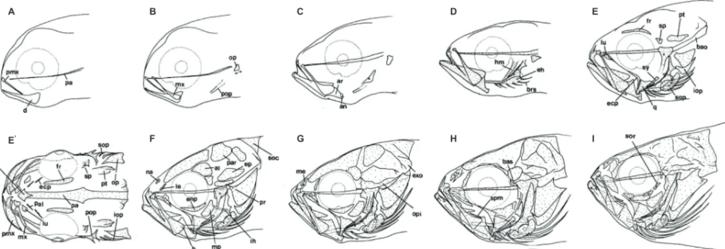
점농어 자치어의 발육단계에 따른 골격발달은 두개골
(cra- nium),
내장골(visceral skeleton),
척추(vertebral column),
미골(caudal skeleton),
견대골(pectoral girdle bone),
지느 러미(fin-rays)
와 담기골(pterygiophore)
및 비늘형성과정 (squamation)을 구분하여 관찰하였다.두부골격
두개골과 내장골로 이루어진 점농어의 두부 골격 발달 과정 을 나타내었다
(Fig. 1; Table 1).
전장6.4 mm(
체장6.1 mm)
의 자어는 처음으로 위턱을 구성하는 전상악골(premaxillary)
과 아래턱을 구성하는 치골(dentary)
및 두개골의 기저를 형 성하는 부설골(parasphenoid)
이 먼저 골화하였다(Fig. 1A).
전장
6.6 mm (6.4 mm)
의 후기자어는 주상악골(maxillary)
이 골화되어 위턱의 빠른 골화가 진행되었으며, 새개부를 형성 하는 전새개골(preopercle)
과 주새개골(opercle)
의 골화가 시 작되었다(Fig. 1B).
전장7.5 mm (7.2 mm)
에 달하면 위턱의 전상악골은 상행돌기와 관절돌기가 형성되고4-5
개의 이빨이Fig. 1. Development of the head skeleton in Lateolabreax maculatus. A-I, lateral view; E', dorsal view. A, 6.4 mm TL; B, 6.6 mm TL; C, 7.5 mm TL; D, 8.8 mm TL; E-E', 11.2 mm TL; F, 12.0 mm TL; G, 14.1 mm TL; H, 15.5 mm TL; I, 25.2 mm TL. al, alisphenoid; an, angular; ar, articular; bao, basioccipital; bas, basisphenoid; brs, branchiostegal rays; d, dentary; eh, epihyal; enp, endopterygoid; ecp, ectopterygoid; ep, epiotic; exo, exoccipital; fr, frontal; h, hypophyal; hm, hyomandibular;
ih, interhyal; iop, interopercle; le, lateral ethmoid; lu, lucryma; me, mes ethmoid; mp, metapterygoid; mx, maxillary; na, nasal;
op, opercle; opi, opisthotic; pa, parasphenoid; pal, palatine; par, parietal; pr, prootic; pmx, premaxillary; pop, preopercle; pt, pterotic; q, quadrate; soc, supraoccipital; sop, subopercle; sor, suborbital; sp, sphenotic; spm, supramaxillary; sy, symplectic.
Empty area, cartilage; stippled area, ossified
A
E'
B
F
C
G
D
H
E
I
Table 1. Development process of the head skeleton in Lateolabrax maculatus
Elements Total length (mm)
6.4 6.6 7.5 8.8 10.5 11.2 12.0 12.2 12.6 14.1 15.5 21.7 25.2
Cranium
Parasphenoid Basioccipital Exoccipital Supraoccipital Opisthotic Epiotic Alisphenoid Parietal Prootic Basisphenoid Pterotic Sphenotic Vomer Frontal Lateral ethmoid Mes ethmoid NasalOrbital region
Preorbital SuborbitalJaw bone
Maxillary Premaxillary Supramaxillary Dentary Articular AngularHyoid arch
Ceratohyal Epihyal Hypohyal Interhyal Glossohyal Branchiostegal rayPalate
Palatine Metapterygoid Ectopterygoid Endopterygoid Hyomandibular Symplectic Quadrate
Opercular
Opercle Preopercle Subopercle Interopercleblack bar, ossified state;●, initial ossification; →, , full complement state of elements;
형성되며, 아래턱은 관절골
(articular)
과 각골(angular)
이 골 화되었다. 설궁(hyoid arch)
은 각설골(ceratohyal)
의 골화가 먼저 시작되고 그 아래쪽에2
개의 새조골이 관절되어 골화가 시작되었다. 새개부의 전새개골과 주새개골은 더욱 골화가 진행되어 넓게 발달하며 전새개골에 하나의 구멍이 형성되었 다(Fig. 1C)
. 전장8.8 mm (8.5 mm)
로 성장하면 설궁에 상설 골(epihyal)
이 골화 직전에 이르며, 모두6
개의 새조골이 골 화되었다. 구개부는 가장 먼저 설악골(hyomandibular)
의 일 부분이 전새개골의 앞에서 골화가 시작되었다. 또한 주상악 골의 앞부분은 전상악골의 관 절돌기와 관절할 부분이 오목하 게 골화가 진행되었다(Fig. 1D)
. 전장11.2 mm (9.8 mm)
에 달하면 설악골은 더욱 골화가 진행되어 하나의 커다란 구멍 이 형성되며, 구개골(palatine)
, 외익상골(ectopterygoid)
, 방 골(quadrate)
, 접속골(sympletic)
등이 동시에 골화하고 구개 골에 구개치가 형성되었다. 새개부는 하새개골(subopercle)
과 간새개골(interopercle)
이 골화되어 새개부 구성요소의 골 화가 함께 진행되었으며, 부설골의 앞쪽에는 서골(vomer)
이, 뒤쪽에는 기저후두골(basioccipital)
이 골화되었다. 눈 의 위.아래쪽에 각각 액골(frontal)
과 안전골(preorbital =lu- crymal)
이 골화하며, 설악골의 위쪽에 설이골(sphenotic)
과 익이골(pterotic)
이 골화되었다(Fig. 1E-E').
전장12.0 mm (10.4 mm)
에 달하면 두개골 중에서 노정골(parietal),
상이 골(epiotic),
익설골(alisphenoid),
전이골(prootic),
상후두골(supraoccipital),
비골(nasal),
측사골(lateral ethmoid)
등이 골화되며, 구개부에 내익상골(endopterygoid)
과 후익상골(metapterygoid)
이 마지막으로 골화되었다. 설궁에는 하설골(hypohyal)
과 간설골(interhyal)
및7
개의 새조골이 골화되어 설궁의 모든 골격들의 골화가 완료되어 이 시기에 골화가 급 속히 진행되었다(Fig. 1F).
전장14.1 mm (12.0 mm)
에 달하 면 외후두골(exoccipital),
후이골(opisthotic)
및 중사골(mes ethmoid)
이 골화되었다(Fig. 1G).
전장15.5 mm (13.2 mm)
에 달하면 두개골 중 기저설골(basisphenoid)
이 최후로 골화 되며, 악골 중 상주상악골(supramaxillary)
이 가장 늦게 골화 되어 두개골 및 내장골의 골화가 완료 직전에 달하였다(Fig.
1H).
전장25.2 mm (20.3 mm)
에 달하면 안하골(suborbital)
이 골화되어 두부를 구성하는 모든 골격들의 골화가 최종 완 료되었다(Fig. 1I).
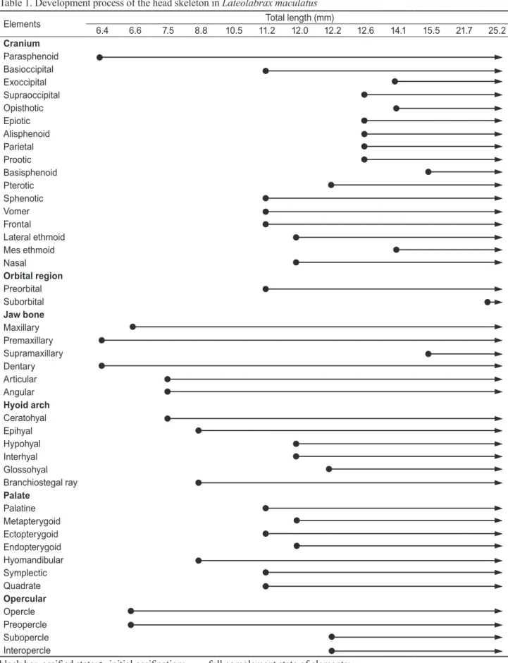
미골
대부분의 경골어류에서 꼬리지느러미를 지지하는 미골
(caudal skeleton)
은 적당히 변형된 몇 개의 추골과 골편으 로 이루어진 미골복합체(caudal complex)
로 구성된다(Fig.
2; Table 2).
미골은 전장
9.1 mm (8.7 mm)
까지는 전혀 골화가 일어나지 않고 미부봉상골(urostyle)
이45
°로 휘어져 있다(Fig. 2A).
전 장12.2 mm (10.4 mm)
로 성장하면 미부봉상골의 일부와 마 지막 미추골의 제3-4
번째 추체 및 하미축골이 형성될 뒤 가 장자리에 꼬리지느러미 상하를 구성하는 두 줄기가 골화되었 다(Fig. 2B).
전장12.6 mm (10.9 mm)
에 이르면 추체의 골화 가 완료되고, 신경극(neural spine)
과 혈관극(hemal spine)
이 골화되며,5
개의 하미축골(hypural bone)
과1
개의 준하 미축골(parhypural bone)
이 동시에 골화되고 꼬리지느러미Fig. 2. Development of the caudal skeleton in Lateolabrax maculatus. A, 9.1 mm TL; B, 12.2 mm TL; C, 12.6 mm TL; D, 14.1 mm TL; E, 14.9 mm TL; F, 21.7 mm TL; G, 24.8 mm TL; H, 32.0 mm TL. cbp, caudal bony plate; ep 1-3, epural 1-3; hs, hemal spine; hy 1-5, hypural 1-5; ns, neural spine; ph, parhypural; us, urostyle; uro, uroneural. Empty, cartilage; stippled, ossified.
A
E
B
F
C
G
D
H
의 위쪽과 아래쪽을 형성하는 줄기가 각각
5, 6
개가 골화되었 다(Fig. 2C).
전장14.1 mm (12.0 mm)
에 달하면 미골의 골 화 상태는 거의 변화가 없으나 신경극과 혈관극 및 꼬리지느 러미 줄기의 골화가 현저히 진행되었다(Fig. 2D).
전장14.9 mm (12.5 mm)
가 되면 첫 번째 상미축골(epural bone)
과 미 신경골(uroneural)
및 미골판(caudal bony plate)
의 골화가 시 작되었다(Fig. 2E).
전장21.7 mm (17.8 mm)
가 되면 첫 번 째 상미축골이 미부봉상골의 등쪽 면을 따라 길게 형성되며, 두 번째 상미축골이 골화되었다. 신경극과 혈관극은 더욱 길 게 골화가 진행되고 마지막 두 번째 혈관극의 기저부에 하나 의 구멍이 형성되었다(Fig. 2F).
전장24.8 mm (20.2 mm)
가 되면 미골의 마지막 구성요소인 세 번째 상미축골의 골화가 시작되며, 마지막2-3
번째 신경극의 기저부에 구멍이 형성되 었다(Fig. 2G).
전장32.0 mm (26.4 mm)
에 달하면 신경극의기저부 및 미골판에도 하나씩의 구멍이 형성되어 이 시기에 모든 골편들의 골화가 완성되어 성어의 형태를 갖추게 되었 다
(Fig. 2H).
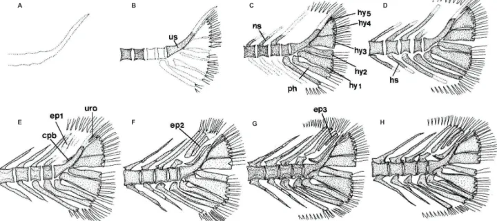
척추골
어류의 척추골은 몸의 중축을 이루는 골격으로 척수
(spinal cord)
와 혈관(blood vessel)
을 보호한다. 점농어 자치어의 척 추골은 두개골이나 내장골보다 늦게 골화가 시작되었고 척추 골은 복추골 앞쪽의 추체와 신경극의 골화가 먼저 시작되었 으나 신경극과 혈관극은 거의 동시에 골화가 완성되었다(Fig.
3; Table 2).
전장
8.3 mm (7.5 mm)
인 자어는 복추골에서1-4
번째 추체(centrum)
의 골화가 신경극(neural spine)
과 함께 골화가 시Table 2. The development process of pectoral girdle, caudal skeleton and vertebrae in Lateolabrax macualtus
Elements Total length (mm)
5.6 8.3 9.1 10.0 10.5 12.2 12.6 14.1 14.9 15.6 21.7 22.9 24.0 24.8 26.0 26.8 32.0 35.0 Pectoral girdle
Cleithrum
Dorsal post leithrum Ventral post leithrum Supracleithrum Posttemporal
Supraposttemporal 1st 2nd Coracoid
Scapula Actinost(radial)
Caudal skeleton
UrostyleHypural 1-5th
Parhypural
Epural 1st
2nd 3rd Uroneural
Caudal bony plate
Vertebrae
Neural spine Hemal spine Parapophysis Abdominal vertebra Caudal vertebrablack bar, ossified state; ●, initial osccification; →, full complement state of elements.
Fig. 3. Schematic diagram showing development of the vertebra column, caudal skeleton, fin support, and spinous and soft fin in Lateolabrax maculatus. A, 8.3 mm TL; B, 10.0-12.2 mm TL; C, 12,6-13.1 mm TL. D, 15.5 mm TL; E, 16.6 mm TL; F, 21.7 mm TL; G, 35.0 mm TL. Scales are vertebrae numbers. Dotted, cartilage; solid, ossified.
A
B
C
D
E
F
G
작되었다
(Fig. 3A).
이후 전장9.1 mm (8.7 mm)
로 성장하 면 복추골의 추체와 신경극은 모두 골화가 완료되고 측돌기(parapophysis)
의 골화가 시작되었다. 미추골은 앞쪽에서부 터 골화가 시작되었으며, 동시에 혈관극(hemal spine)
과 신 경극의 골화가 꼬리쪽으로 진행되었다. 전장10.0-12.2 mm (8.9-10.4 mm)
에 달하면2-4
개의 추체가 골화되지 않은 상태 에서 미부봉상골(urostyle bone)
의 일부가 골화하였다. 또한 신경극과 혈관극의 끝 부분은 아직 완전히 골화되지 않아서 끝 부분이 뾰족하지 않았으며, 제1 미추골의 혈관극이 오목 하게 분지되어 혈관간극(interhemal spine)
이 삽입될 수 있도 록 변형되었다(Fig. 3B).
전장12.6-13.1 mm (10.9-11.3 mm)
에 달하면 모든 추체의 골화가 완료되며, 신경극과 혈관극에 는 신경궁문(neural arch)
과 혈도궁문(hemal arch)
이 골화되었다.
3-7
번째 추체에 늑골(rib)
이 골화하여 관절되었다(Fig.
3C).
전장15.5-16.6 mm (13.1-14.1 mm)
에 달하면 먼저 미 추골의 등쪽과 배쪽에 각각 전관절돌기와 후관절돌기가 발 달하고, 이후 복추골의 등쪽에도 전관절돌기와 후관절돌기 가 발달하였다. 또한 미추골의 신경극 기부에 구멍이 형성되 기 시작하였다. 늑골은7-9
개로 증가하였고 상늑골이1-4
번 째 추체 상에서 골화되었다(Fig. 3D, E)
. 전장21.7-35.0 mm (17.8-29.3 mm)
에 달하면14
개의 늑골이 모두 골화되어 각 측돌기에 관절되며, 각각의 신경극에 하나씩의 구멍이 형성 되어 모든 척추골을 형성하는 골격이 완성되었다(Fig. 3F, G).
지느러미 및 담기골
각 지느러미 및 등지느러미를 지지하는 신경간극과 뒷지
Table 3. Development process of fin ray and pterygiophore in Lateolabrax maculatus
Total length (mm)
Counts
Fin-Rays Pterygiophores
D A PT PL
C
FIS D A
Upper
procurrent Principal Lower procurrent
9.4 0 0 0 0 0 0 0 0 0 0
11.6 0 0 8 0 0 9+8 0 0 0 0
12.2 0-0, 11 II, 7 12 3 0 9+8 0 0 0 0
12.6 XII-I, 9 II, 7 14 3 4 9+8 3 0 0 1
12.8 XII-I, 12 II, 7 14 4 5 9+8 4 0 0 1
13.0 XII-I, 14 II, 9 14 4 5 9+8 5 0 (12) 1(1)
13.1 XIII-I, 14 II, 8 14 5 6 9+8 6 0 (12) 1(2)
13.8 XIII-I, 13 II, 8 17 1, 5 7 9+8 6 0 (12) 1(1)
14.3 XII-I, 13 II, 7 16 1, 5 8 9+8 7 0 3(15) 1(3)
14.4 XII-I, 14 II, 9 16 1, 5 7 9+8 6 0 5(13) 1(3)
15.6 XII-I, 14 II, 7 18 1, 5 9 9+8 8 0 6(16) 1(1)
15.6 XII-I, 13 II, 9 18 1, 5 10 9+8 9 0 19(2) 1(2)
16.0 XIII-I, 13 II, 9 18 1, 5 9 9+8 8 0 21 5(2)
16.6 XIII-I, 13 II, 9 18 1, 5 9 9+8 8 3 21 5(2)
19.3 XII-I, 14 II, 9 18 1, 5 11 9+8 11 3 24 5(3)
21.4 XIII-I, 13 II, 9 18 1, 5 14 9+8 13 3 24 9
21.7 XIII-I, 13 II, 9 18 1, 5 14 9+8 13 3 24 9
22.8 XII-I, 14 II, 9 19 1, 5 14 9+8 13 3 24 9
23.8 XII-I, 13 III, 8 17 1, 5 15 9+8 14 3 25 9
26.8 XII-I, 13 II, 8 19 1, 5 14 9+8 13 3 25 8
28.6 XII-I, 14 II, 8 19 1, 5 14 9+8 13 3 25 9
30.4 XII-I, 13 III, 9 18 1, 5 14 9+8 13 3 25 10
33.8 XII-I, 13 III, 8 18 1, 5 13 9+8 13 3 25 10
35.7 XIII-I, 13 III, 8 18 1, 5 14 9+8 14 3 27 10
( ), present before ossification; A, anal; C, caudal; D, dorsal; FIS, free interneural spine; PL, pelvic; PT, pectoral.
상측두골
(supraposttemporal)
에1
개의 가시가 형성되었다.견갑골은 거의 사각형으로, 오훼골은 망치모양으로 골화가 진행되며 각각에 구멍이 형성되었다
(Fig. 5F).
전장26.8 mm (21.8 mm)
에 달하면 후측두골의 중간 부분에1
개의 가시가 추가로 형성되고, 상쇄골과 관절 부위에3-7
개의 가시가 형성 되었다. 또한 쇄골에도 최대3
개의 가시가 형성되었다. 후측 두골의 위쪽 가지에 마지막 상측두골 하나가 더 골화되어 쇄 골을 형성하는 모든 골격이 완성되었다(Fig. 4G).
비늘형성
비늘의 형성은 주로 비늘 열의 수와 비늘에 의해 몸 표면에 덮이는 부분의 범위에 따라서 다음과 같이 진행되었다.
전장
23.8 mm (19.4 mm)
에 달하면 비늘은 몸 정중앙 부 위를 따라서 최초로 형성되는데 그 위치는 몸통중앙부에1
열, 꼬리자루 시작부분에 2열 및 꼬리자루 후방에 2열의 비늘 이 불연속적으로 형성되었다(Fig. 5A).
전장24.2 mm (19.8 mm)
에 달하면 몸통 중앙에서 꼬리자루에 걸쳐서 불연속적으 로 형성된 비늘이 서로 연속되어 출현하며, 비늘 열도 앞쪽(
몸통 중앙)은2-4
열에서 점점 꼬리자루로 향하면서4-5
열로 증가하였다(Fig. 5B).
전장26.8 mm (21.8 mm)
에 달하면 몸 통 중앙부의 비늘은 두부 쪽으로 확장되어 새개부의 후방까지 닿으며, 뒤쪽은 꼬리지느러미의 기저부를 제외하고 확장되었 다. 비늘 열은 새개부 바로 뒤쪽에서2-3
열, 몸통 중앙부위에 서4-5
열, 꼬리자루 부분에서6-7
열로 증가하여 뒤쪽으로 갈 느러미를 지지하는 혈관간극 즉, 담기골(pterygiophore)
의골화 과정을 나타내었다
(Table 3; Fig. 3).
지느러미 중에서 꼬리지느러미가 부화 후 약30
일경인 전장11.6 mm (10.2 mm)
에 주 줄기 수는 이미 정수에 달하여 골화되었으며, 전장12.6 mm (10.9 mm)
에 달하면 꼬리지느러미 부속줄기(pro- current)
의 골화가 시작하고 전장21.4 mm (17.5 mm)
에 이 르면 그 수가 위쪽이14-15
개, 아래쪽이13-14
개로 정수에 달하여 골화가 완료되었다. 가슴지느러미는 전장11.6 mm (10.2 mm)
에 최초로8
개의 줄기가 골화되며, 전장13.8 mm (12.0 mm)
에16-19
개로 골화가 완료되었다. 등지느러미는 전장12.2 mm (10.4 mm)
에서 최초로11
개의 줄기가 골화되 기 시작하여13.0mm (11.3mm)
에 이르면 이미 지느러미의 수는 정수에 달하였다. 그러나, 등지느러미의 경우 두 번째 등지느러미의 가시(극)이 성어와 닮은 완전한 형태를 갖춘 것 은14.30-15.60 mm (12.2-13.3 mm)
에서 나타났다. 배지느 러미는 전장12.2 mm (10.4 mm)
에 최초로 3개의 줄기가 골 화되고, 전장13.8 mm (12.0 mm)
에 달하면 그 수가 정수에 달하여 골화가 완료되었다. 뒷지느러미는 등지느러미, 배지 느러미와 같이 전장12.2 mm (10.4 mm)
에서 골화가 진행됨 과 동시에 그 수가 정수에 달하여 있었으나, 완전한3
개의 가 시 형태를 갖춘 것은 약23.8-30.4 mm (19.4-24.6 mm)
전후 였으며, 지느러미 중에서 가장 늦게 골화되었다.담기골의 골화는 전장
12.6 mm (10.9 mm)
에서 첫 번째 혈 관간극이 뒷지느러미의 2개의 가시와 관절되어 가장 먼저 골 화가 시작되며, 신경간극은 전장14.3 mm (12.2 mm)
에서 등 지느러미의 2개의 가시와 관절되어 골화가 시작되었다. 그러 나 골화의 진행은 신경간극이 다소 빨리 진행되어 전장19.3 mm (15.9 mm)
에서, 혈관간극은21.4 mm (17.5 mm)
에서 골화가 완료되는 개체가 출현하였다. 유리신경간극은 전장16.6 mm (14.1 mm)
에서 골화가 시작되어 전장21.7 mm ( 17.8 mm)
에서 완료되었다.견대골
가슴지느러미를 지지하는 견대골
(shoulder girdle bone)
은 수 개의 막골이 부속되며, 후측두골(posttemporal)
에 의해 두 개골에 관절되었다(Fig. 4; Table 2).
전장
5.6 mm (5.4 mm)
의 자어는 쇄골(cleithrum)
이 골화 되기 시작하였고 전장8.3 mm (7.5 mm)
에는 쇄골의 위 부 분에 상쇄골(supracleithrum)
이 골화하였다(Fig. 4A, B).
전 장10.5 mm (9.2 mm)
에는 후측두골이 골화되어 상쇄골과 관절되며,2
개의 후쇄골(postcleithrum)
이 긴 막대모양으로 골화되었다(Fig. 4C).
전장12.6 mm (10.9 mm)
에 달하면 후 측두골에2
개, 상쇄골에1
개의 가시가 형성되고 가슴지느러 미 줄기가 골화되었다(Fig. 4D).
전장15.6 mm (13.3 mm)
에 달하면 후측두골의 아래쪽이 보다 넓게 발달하며, 오훼골(coracoid),
견갑골(scapula),
사출골(actinost=radial)
이 골화 하였다(Fig. 4E).
전장22.9 mm (18.9 mm)
에 달하면 쇄골과Fig. 4. Development of the pectoral girdle in Lateolabrax maculatus. A, 5.6 mm TL; B, 8.3 mm TL; C, 10.5 mm TL;
D, 12.6 mm TL; E, 15.6 mm TL; F, 22.9 mm TL; G, 26.8 mm TL. cl, cleithrum; cr, coracoid; dpcl, dorsal post cleithrum;
pt, posttemporal; r, radial; sc, scapula; scl, supracleithrum;
spt 1-2, supraposttemporal 1-2; vpcl, ventral post cleithrum.
A
E
B
F C
G D
느러미 기저부 부근에 형성되었다
(Fig. 5I, J).
전장57.2-60.2 mm (47.1-49.2 mm)
에 달하면 주둥이 부분을 제외한 모든 부 위에 비늘이 형성되었다(Fig. 5K, L).
고 찰
일반적으로 경골어류는 부화 시에 골격발달에 있어서 주목 할 만한 변화를 나타낸다고 알려져 있다(
Koumoundouros et al., 2001).
농어목어류(Perciformes)
는 두개골과 지느러미의 발달이 부화 이후에 관찰되나(Matsuoka, 1985; Koumoun- douros et al., 1997b, 2001a, 2001b; Faustion and Power, 1999; Sfakianakis et al., 2004, 2005),
연어과어류(Salmoni- dae)
는 부화 이전에 두개골과 지느러미의 골격 발달이 시작 한다(Kendall et al., 1984).
점농어는Table 1
과2
에서 나타난 바와 같이 부화 이후에 골화가 진행되었으며, 전장5.6 mm (5.4 mm)
에 가장 먼저 쇄골의 골화가 진행되고 이어서 전장6.4 mm (6.1mm)
에 이르면 두개골을 형성하는 골격 중 전상 악골, 치골 및 부설골의 골화가 진행되었다. 쏨뱅이목어류(Han et al., 2001; Kim et al., 1997)
는 쇄골과 함께 악골, 부 설골이 동시에 골화가 진행되며, 청베도라치과에 속하는 저 울베도라치Entomacrodus stellifer lighti (Kim et al., 1992)
는 쇄골과 부설골이 먼저 골화하고 이후 악골의 골화가 이어 졌으며, 복어목에 속하는 졸복Takifugu pardalis (Han et al.,
2005)
은 쇄골과 부설골 및 악골이 동시에 골화가 진행되어 분류군별로 차이를 나타내었다. 하지만 이들 종 모두 턱을 구 성하는 악골이 비교적 빠른 속도로 골화하는 특징을 나타내 었다. 이것은 두개골이 섭이와 호흡을 위한 중요한 필요 골격 요소로써 골화가 우선적으로 일어나는 부위이며
(Vandewalle et al., 1997),
이 시기에 입의 개구와 섭이 기능에sternohyoi- deus muscle
을 지지하는 쇄골이 중요한 역할을 하기 때문이 다(Wagemans and Vandewalle, 1999).
한편 점농어와 유사 속인Morone
속(Fritzsche and Johnson, 1980)
어류는 전상 악골, 주상악골, 치골 및 쇄골이 체장4.6-6.0 mm
사이에 골 화가 진행되어 점농어와 비슷한 크기에서 골화하였다.자치어기의 형태 발달에 따른 내부 골격발달, 특히 두부 골 격의 가시의 형성은 개체 발육 상태 및 발육 환경에 따라서 다 소 차이는 있으나 두정부, 새개부, 완와 및 견대부 등에 형성 되는 가시는 유어기의 매우 중요한 분류형질이다
(Okiyama,
1988).
점농어는 견대부의 주 골격인 쇄골에 가시가 출현하였으나,
Nakayama et al. (1996)
의 경우 특별한 언급이 없 어 가시가 출현하지 않는 것으로 판단된다. 그러나 농어속 어류 성어의 견대부 골격을 채골하여 쇄골의 형태를 살펴보 면 농어, 점농어, 넙치농어3
종 모두 견대부의 등쪽 가장자 리가 톱니모양을 형성하고 있는데 이 견대부에 형성된 톱니 모양이 어린 자치어기 때 쇄골에 형성되는 가시가 그 기원으 로 판단된다. 점농어는 전장22.9-26.8 mm (18.9-21.8 mm)
사이에서1-3
개의 가시가 형성되어 이것을 뒷받침하였다. 그 수록 비늘열 수가 증가하였다. 또한 새개부 바로 뒤에서 배지느러미 쪽으로 경사를 이루는
3-4
개의 비늘이1
열로 형성되 었다(Fig. 5C).
전장28.3 mm (23.2 mm)
에 달하면 새개부 뒤 쪽의 비늘이4-6
개로 증가하고, 몸체에 형성된 비늘이 증가 하여 몸통부위에서 미부까지 형성된 비늘 열은10
줄로 증가 하며, 전장29.4 mm (24.6 mm)
에 달하면 미부에 형성된 비 늘은 등쪽과 배쪽으로 확장되어 완전히 덮이기 직전에 달하 였다(Fig. 5D, E).
전장34.2 mm (27.5 mm)
에 달하면 새개부 뒤쪽 가장자리를 따라서 형성된 비늘이 몸통에 형성된 비늘 열과 서로 연결되면서 몸통 중앙 부위의 비늘이 등쪽과 배쪽 으로 상하 확장되고 미부는 거의 대부분이 비늘로 완전히 덮 였으며, 새개부의 위쪽과 배지느러미 전후에 새롭게 비늘이 출현하였다(Fig. 5F).
전장34.7 mm (28.0 mm)
에 달하면 배 지느러미와 뒷지느러미 사이에 형성된 비늘이 가슴지느러미 부근에서 몸체에 형성된 비늘과 서로 연결되어 범위가 확장되 었다(Fig. 5G).
전장39.3 mm (32.3 mm)
에 달하면 가슴지느 러미와 배지느러 미 사이의 일부 및 등지느러미 가시부의 기 저 부분을 제외하고선 새개부 뒤쪽에서 꼬리까지 비늘로 덮 여있다(Fig. 5H).
전장46.8-49.0 mm (38.4-40.1 mm)
사이에 달하면 비늘은 눈의 후방(안하골 부위), 주전개골 및 가슴지Fig. 5. Development of the squamation in Lateolabrax maculatus. A, 23.8 mm TL; B, 24.2 mm TL; C, 26.8 mm TL; D, 28.3 mm TL; E, 29.4 mm TL; F, 34.2 mm TL; G, 34.7 mm TL; H, 39.3 mm TL; I, 46.8 mm TL; J, 49.0 mm TL; K, 57.2 mm TL; L, 60.2 mm TL. Solid area, scales.
A G
H
I
J
K
L B
C
D
E
F
러나
Nakayama et al. (1996)
의 경우 최대관찰 표본의 크기 가 체장17.2 mm
로 점농어와 많은 차이를 보였는데, 이는 관 찰 표본의 크기 때문에 가시가 출현하지 않은 것으로 생각되 나 향후 보다 큰 개체를 대상으로 정밀한 조사가 필요하다고 생각된다. 한편 가슴지느러미를 지지하는 골격 중 견갑골은 다소 큰foramen
이 형성되는데 이는 농어목어류의 전형적인 형질이다(Koumoundouros et al., 2001b).
점농어의 경우 견 갑골에 둥근 구멍이 뚫린foramen
이 형성되며, 특이하게 전 장22.9mm
에 이르면 오훼골에도foramen
이 형성되었다가 이후 전장26.8 mm (21.8mm)
에 달하면 사라지는 특징을 보 였다. 견갑골에foramen
가 형성되는 종은 황돔Dentex den- tex (Koumoundoures et al., 2001b), Lutjanus campechanus (Potthoff et al., 1988),
자리돔류(Emery, 1973)
등 많은 농어 목어류를 비롯하여 쏨뱅이목어류(Kim et al., 1997; Han et al., 2001; Kim et al., 1997)
에서도 형성되어 이는 어류의 일 반적인 형질로 보여진다.척추골의 골화
(Table 2; Fig. 3)
와 관련해서 점농어는 전장8.3 mm (7.5 mm)
에서 복추골의 제1-4
번째 추체가 신경극 과 함께 골화가 진행되어 점차 미추골 쪽으로 골화가 진행되 며, 미부의 추체가 거의 골화된 이후에 미부봉상골이 골화 하였는데, 저울베도라치(Han et al., 1992)
도 이와 같은 골 화 순서를 나타내었다. 반면에 졸복(Han et al., 2005), Di- plodus sargus (Koumoundouros et al., 2001a),
붉은쏨뱅이(Han et al., 2001)
은 복추골 쪽의 신경극이 먼저 골화가 일어 난 후 추체의 골화가 진행되는 차이를 보였다. 또한 척추골 의 골화는 후기자어기 부터 진행되는데,Polochthys mauli (Ozawa, 1976)
는 척추골의 골화가 몸 중앙에서 먼저 골화 한 후 앞, 뒤쪽으로 골화가 진행되는 종류도 있으나, 점농어 는 날치Prognichthys agoo (Park and Kim, 1987), Diplodus sargus (Koumoundouros et al., 2001a),
붉은쏨뱅이(Han et al., 2001)
등과 마찬가지로 복추골에서 골화가 먼저 진행된 후 미추골 쪽으로 골화가 진행되었으며, 미부봉상골은 미골 부위의 추체보다 먼저 골화하였다.일반적으로 농어목어류에서 후방의 등지느러미와 뒷지느러 미의 발달은 전방의 등지느러미가 발달하기 이전에 먼저 발 달하기 시작하는 것이 일반적이다
(Johnson, 1984; Faustino and Power, 1999).
점농어의 경우 등지느러미와 뒤지느러미 의 발달은 일반 농어목어류와 같은 경향을 나타내었으나 내 부 골화의 진행은 전, 후방의 등지느러미와 뒷지느러미는 전 장12.2-12.6 mm (10.4-10.9 mm )
사이에 거의 동시에 진행 되었으며, 이들의 지지골격인 담기골의 골화는 신경간극이 전장12.6 mm (10.9 mm)
에서, 혈관간극은 전장14.3 mm
(12.2 mm)
에서 진행되어 다소 차이가 있으나 골화의 진행방향은 공통적으로 전방에서 후방으로 진행되는 특징을 나타내 었다. 반면에
Diplodus puntazzo (Sfakianakis et al., 2005)
와Diplodus sargus (Komoundouros et al., 2001a)
는 신경 간극과 혈관간극의 골화는 전, 후방에서 거의 동시에 골화가이루어 지며,
Pagellus erythrinus (Sfakianakis et al., 2004)
는 신경간극의 후방에서 골화가 먼저 진행된 이후 신경간극 과 혈관간극의 골화가 전 후방에서 동시에 진행되었고 참돔(Matsuoka, 1985)
은 신경간극과 혈관간극의 골화가 동시에 진행되나 신경간극의 경우 전, 후방에서 골화가 먼저 진행된 이후 점점 중앙쪽으로 골화가 진행되어 종 간에 다양한 골화 과정을 나타내었다.비늘형성은 자연상태에서 서식처의 변화에 대한 반응과 더 불어 방어기작과 밀접한 관계가 있으므로 매우 중요하다
(Fu- kuhara, 1992).
점농어의 비늘발달은 초기에 발생지점과 열 에 있어서 매우 다양한 변화를 나타내었는데, 몸통 중앙과 꼬리자루 앞부분 및 꼬리자루 중앙부분 등 세 곳에서 불연 속적으로 형성된 후 서로 전후로 확장되어 곧 바로 연결되는 특징을 나타내었다. 비늘형성에 있어 다른 종들을 살펴보면 황돔(Koumoundouros, 2001b),
넙치Paralichthys olivaceus (Seikai, 1980),
농어Lateolabrax japonicas (Fukuhara and Fushimi, 1982), Tilapia sparrmani (Fujita, 1971)
등은 꼬 리자루에서, 참돔(Fukuhara, 1976),
감성돔Mylio macro- cephalus (Fukuhara, 1977),
붉돔Evynnis japonica (Fuku- hara and Fushimi, 1981)
은 몸통 중앙에서, 황새치Xiphias gladius (Potthoff and Kelley, 1982)
는 몸통 중앙 복부 부위에 서 가장 먼저 비늘이 형성되며, 돌돔Oplegnathus fasciatus (Fukuhara and Ito, 1987)
과 쥐노래미Hexagrammos otakii (Fukuhara and Fushimi, 1984)
는 몸통 앞 부위와 중앙부위 두 곳에서,Rivulus marmoratus (Park and Lee, 1988)
은 두 정부에서, 용치놀래기Halichoeres poecilopterus (Kimura and Kiriyama, 1993)
는 몸통 앞 부위의 측선이 시작되는 부 위에서 처음으로 출현하여 종마다 매우 다양한 양상을 보였 다.Sire and Arnulf (1990)
은 문헌 조사를 토대로 대부분의 경골어류에서 비늘이 형성되는 부위를7
곳으로 구분하였는 데, 조사어종의85
종 가운데61
종이 처음 비늘이 발생하는 부위는 꼬리자루 부분이며, 나머지24
종은 다른 부위에서 비 늘이 출현하였다고 밝혔다. 점농어는 비늘이 측선부위를 따 라 동시에 세 곳에서 발생하였고 이후 곧바로 확장 연결되었 다. 같은 속의 농어(Fukuhara and Fhshimi., 1982)
는 꼬리자 루에서만 비늘이 출현하여 점농어와 뚜렷한 차이를 나타내 었다. 또한 비늘의 첫 출현시기는 점농어는 전장24.0 mm (19.5mm)
전후에서 농어는 체장19.0 mm
로 비슷하였으나, 두부를 제외한 몸 전체에 비늘이 최종 형성되는 시기는 점농 어가 전장46.8 mm (38.4mm)
이나 농어는 체장35 mm
로 두 종 간에 다소 차이를 나타내었다. 그러나 이러한 비늘형성 시 기와 완료 시기는 추후 자연산과의 비교 검토가 필요한 부분 이라고 생각된다.Mook (1997)
에 의하면 어류의 골격형성은 그들의 생활 방 식에 의해 골화가 통제되며 이러한 생활방식의 차이는 골화 되는 정도와 순서에 변화를 초래하여 자치어의 골결 발달에 다양한 변화를 줄 수 있다고 지적하였는데, 자치어의 골격 연구는 종 동정뿐만 아니라 성어의 골격 이해 및 계통의 추정에 도움을 줌으로 골격발달에 대한 보다 구체적인 연구가 필요 하다고 본다.
사 사
본 연구를 위하여 수고해주신 한국해양연구원 통영해양생 물연구보존센터의 박용주님과 현장 직원들, 세보수산 직원 들에게 감사의 뜻을 표합니다.
참고문헌
Baek H-J, An C-M and Kim H-B. 2001. Biosynthesis of C21- steroids in spotted sea bass (Lateolabrax maculatus) ova- ries. J Korean Fish Soc 34, 638-642.
Emery AR. 1973. Ecology and functional osteology damselfish (Pisces; Pomacentridae) at Alligator Reef, Florida Keys.
Bull Mar Sci 23, 649-770.
Faustino M and Power DM. 1999. Development of the pectoral, pelvic, dorsal and anal fins in cultured sea bream. J Fish Biol 54, 1094-1110.
Fritzsche A and Johnson GD. 1980. Early osteological devel- opment of white perch and striped bass with emphasis on identification of their larvae. American Fish Soc 109, 387- Fujita K. 1971. Early development of the squamation in Tilapia 406.
sparrmani. Japan J Ichthyol 18, 90-93.
Fukuhara O and Ito K. 1987. On the formation of the fins and squamation in the Japanese parrot fish, Oplegnathus fascia-
tus reared in the laboratory. Bull Nansei Reg Fish Lab, 9-17.
Fukuhara O and Fushimi T. 1981. Observations of morphol- ogy and squamation in Evynnis japonica Tanaka (Sparidae) reared in the laboratory. Bull Nansei Reg Fish Reg Lab, 1-8.
Fukuhara O and Fushimi T. 1982. Development of fins and squamation in the percichthyid fish, Lateolabrax japonicas.
Japan J Ichthyol 29, 173-178.
Fukuhara O and Fushimi T. 1984. Squamation of larval green- ling Hexagrammos otakii (Pisces: Hexagrammidae) reared in the laboratory. Bull Japan Soc Sci Fish 50, 759-761.
Fukuhara O. 1976. Morphological studies of larva of red sea bream. II. Early development squamation. Bull Nansei Reg Fish Res Lab, 13-18.
Fukuhara O. 1977. Some morphological observation on larvae and juveniles of the kurodai, Mylio macrocephalus (Spari- dae, Teleostei) reared in the laboratory. Bull Nansei Reg Fish Res Lab, 1-16.
Fukuhara O. 1992. Study on the development of functional morphology and behavior of the larvae of eight commer- cially valuable teleost fishes. Contr Fish Res Jpn Sea Block 25, 1-122.
Han K-H, Cho J-K, Lee S-H, Hwang S-Y, Yoon S-M, Seo W-I and Kim C-C. 2005. Osteological development of the lar-
vae and juveniles of Takifugu pardalis (Teleostei: Tetra- odontodae). Korean J Ichthyol 17, 29-35.
Han K-H, Lim S-K, Kim K-S, Kim C-W and Yoo D-J. 2001.
Osteological development of the larvae and juveniles of
Sebastiscus tertius (Barsukov et Chen) in Korea. Korean J
Ichthyol 13, 63-68.Johnson GD. 1984. Percoidei: development and relationship.
In: Moser HG, Richards WJ, Cohen DM, Fahay MP, AW Kendall, SL Richardson (eds). Ontogeny and systematics of fishes. American Society of Ichthyologists and Herpetolo- gists, special publication no 1, Allen Lawrence KS, 464- Kawamura K and Hosoya K. 1991. A modified double staining 498.
technique for making a transparent fish-skeletal specimen.
Bull Nat’l Res Inst Aquaculture 20, 11-18.
Kendall AW, Ahlstrom EH and Moser HG. 1984. Early life his- tory stages of fishes and their characters. In: Moser HG, Richards WJ, Cohen DM, Fahay MP, Kendall AW, Richard- son SL (eds). Ontogeny and systematics of fishes. American Society of Ichthyologists and Herpetologists, special publi- cation no 1. Allen Lawrence KS, 11-12.
Kim CH, Jun JC and Kim SU. 1998. Early life history of Kore- an temperate sea bass Lateolabrax sp. (Pisces: Moronidae).
Bull Net Fish Res Dev Agency 54, 79-85.
Kim J-W, Lee W-K, Yang S-W, Jeong K-S, Cho Y-C, Rho Y-G, Bang I-C, Kim K-S, Lim S-K, Yoo M-S and Kwon H-B.
1999. Immunohistochemical identification of the two froms of gonadotripin releasing hormones (sGnRH, cGnRH-II) in spotted sea bass (Lateolabrax sp.) brain. J Korean Fish Soc 32, 266-270.
Kim YU, Han K-H and Kang C-B. 1992. Morphological and skeletal development of larvae and juveniles of Entomacro-
dus stellifer (Herre). Korean J Ichthyol 4, 31-43.
Kim YU, Han K-H, Kang C-B, Kim JK and Byun S-K. 1997.
The early life history of the rockfish, Sebastiscus marmora-
tus 2. Morphology and skeletal development of larvae and
juvenile. Korean J Ichthyol 9, 186-194.Kim YU, Myoung J-G, Kim Y-S, Han K-H, Kang C-B and Kim J-G. 2001. The marines fishes of Korea. Hanguel, Busan, Korea, 382.
Kimura S and Kiriyama T. 1993. Development of eggs, larvae and juveniles of the labrid fish, Halichoeres poecilopterus, reared in the laboratory. Japan J Ichthyol 39, 371-377.
Koumoundouros G, Gagliardi F, Divanach P, Boglione C, Cataudella S and Kentouri M. 1997a. Normal and abnormal osteological development of caudal fin in Sparus aurata L.
fry. Aquaculture 149, 215-226.
Koumoundouros G, Oran G, Divanach P, Stefanakis S and Kentouri M. 1997b. The opercular complex deformity in intensive gilthead sea bream (Spartus aurata L.) larvicul- ture. Moment of apparition and description. Aquaculture 156, 165-177.
Koumoundouros G, Sfakianakis DG, Maingot E, Divanach P
and Kentouri M. 2001a. Osteological development of the vertebral column and of the fins in Diplodus sargus (Tele- ostei: Perciformes: Sparidae). Mar Bio 139, 853-862.
Koumoundouros G, Divanach P and Kentouri M. 1999. Os- teological development of the vertebral column and of the caudal complex in Dentex dentex. J Fish Bio 54, 424-436.
Koumoundouros G, Divanach P and Kentouri M. 2001b. Osteo- logical development of Dentex dentex (Osteichthyes: Spari- dae): dorsal, anal, paired fins and squamation. Mar Bio1 38, 399-406.
Liu C-H. 2001. Early osteological development of the yellow tail Seriola dumerili (Pisces: Carangidae). Zool Stud 40, 289-298.
Liu J-X, Gao T-X, Yokogawa K and Zhang Y-P. 2006. Differen- tial population structuring and demographic history of two closely related fish species, Japanese sea bass (Lateolabrax
japonicas) and spotted sea bass (Lateolabrax maculatus) in
Northwestern Pacific. Mol Phylogenet Evol 39, 799-811.Matsuoka M. 1985. Osteological development in the red sea bream, Pagrus major. Japanese J Ichthyol 32, 35-51.
Mook D. 1977. Larval and osteological development of the sheepshead, Archosargus probatocephalus (Pisces: Spari- dae). Copeia 1977(1), 126-133.
Myoung J-G, Park C-W, Kim M-S, Kim J-M, Kang C-B and Kim YU. 1997. Morphological changes during starvation in early development stages of spotted sea bass, Lateolabrax sp. I. Post-larval stage. Korean J Ichthyol 9, 15-21.
Nakayama K, Kinoshita I, Seikai T, Nakabo T and Tanaka M.
1996. Morphological comparisons during early stage rear- ing of Chinese and Japanese forms of the temperate bass,
Lateolabrax japonicas .Japan J Ichthyol 43, 13-20.
Okiyama M. 1988. An Atlas of the Early State Fishes in Japan.
Tokai Univ. Press. 1157pp.
Ozawa T. 1976. Early life history of the gonostomatid fish, Pol-
lichtys mauli in the oceanic region off southern. Japan J
Ichthyol 23, 43-54.Park E-H and Lee S-H. 1988. Scale growth and squamation chronology for the laboratory-reared Hermaphroditic fish
Rivulus marmoratus (Cyprinodontidae). Japan J Ichthyol
34, 476-482.Park J-Y, Kim K-K and Kim Y. 1996. Genetic characterization of two types of sea bass, Lateolabrax japonicas in Korea by isozyme analysis. J Aquaculture 9, 437-445.
Park YS and Kim YU. 1987. Studies on the larvae and juveniles of flying fish, Prognichthysa goo (Temminck and Schlegel) (Pisces, Exocoetidae) .II. Osteological development of lar- vae and juveniles. Bull Korean Fish Soc 20, 447-456.
Potthoff T, Kelley S and Collins LA. 1988. Osteological devel- opment of the red snapper, Lutjanu scampechanus (Lut- janidae). Bull Mar Sci 43, 1-40.
Pottoff T and Kelly S. 1982. Development of the vertebral col- umn, fins and fin supports, branchiostegal rays, and squa- mation in the swordfish, Xiphias gladius. Fish Bull 80, 161- 186.
Seikai T. 1980. Early development of squamation in relation to color anomalies in hatchery-reared flounder, Paralichthys
olivaceus .Japan J Ichthyol 27, 249-254.
Sfakianakis DG, Doxa CK, Kouttouki S, Koumoundouros G, Maingot E, Divanach P and Kentouri M. 2005. Osteologi- cal development of the vertebral column and of the fins in
Diplodus puntazzo (Cetti, 1777). Auqaculture 250, 36-46.
Sfakianakis DG, Koumoundouros G, Divanach P and Kentouri M. 2004. Osteological development of the vertebral column and of the fins in Pagellus erythrinus (L. 1758). Tempera- ture effedt on the developmental plasticity and morpho-an- atomical abnormalities. Auqaculture 232, 407-424.
Sire JY and Arnulf I. 1990. The development of squamation in four teleostean fishes with a survey of the literature. Japan J Ichthyol 37, 133-143.
Vandewalle P, Gluckmann I, Baras E, Huriaux F and Focant B.
1997. Postembryonic development of the cephalic region in
Heterobranchus longifilis. J Fish Bio 50, 227-253.
Wagemans F and Vandewalle P. 1999. Development of the car- tilaginous skull in Solea solea: trends on Pleuronectiforms.
Ann Sci Nat 1, 39-52.
Yokogawa K and Tajima T. 1996. Morphological and genetic characters of artificially propagated sea bass in Taiwan.
Fisheries Sci 62, 361-366.
Yokogawa K and Seki S. 1995. Morphological and genetic dif- ference between Japanese and Chinese sea bass of the genus