Introduction
Since total knee arthroplasty (TKA) is considered a high risk factor for venous thromboembolism (VTE), thromboprophylaxis is commonly recommended
1). However, the actual incidence of
VTE and the need for routine thromboprophylaxis after TKA in Asians are controversial. A recent metaanalysis showed that the incidences of deep vein thrombosis (DVT) and symptomatic pul
monary embolism (PE) were low (40.4% and 0.01%, respectively) in Asians without chemoprophylaxis, despite the westernized life
style and aging of the population
2). However, since the incidence of DVT proven by venography is reported to be 50%–76.5% and that of PE proven by lung perfusion scan is 17.1% in cases with
out prophylaxis, routine thromboprophylaxis should be reconsid
ered
3,4). In contrast to Western studies, which typically describe the incidence of VTE with chemoprophylaxis, most Asian re
ports have investigated the incidence without chemoprophylaxis.
Thus, due to insufficient data, the need for prophylaxis after TKA in Asians remains debatable.
Recently, rivaroxaban (factor Xa inhibitor) has shown superior
Rivaroxaban and Acetylsalicylic Acid for Prevention of Venous Thromboembolism Following Total Knee Arthroplasty in Korean Patients
Kyu Sung Chung, MD 1 , Tae Yang Shin, MD 2 , Sang Hoon Park, MD 3 , Hyuck Kim, MD 4 , and Choong Hyeok Choi, MD 3
1
Department of Orthopedic Surgery and Sports Medical Center and Sports Medical Research Institute, Seoul Paik Hospital, College of Medicine, Inje University, Seoul;
2Department of Orthopaedic Surgery, Jeonju Korea Hospital, Jeonju;
3Department of Orthopaedic Surgery, College of Medicine, Hanyang University, Seoul;
4
Department of Thoracic and Cardiovascular Surgery, College of Medicine, Hanyang University, Seoul, Korea
Purpose: To investigate the incidence of venous thromboembolism (VTE) after total knee arthroplasty (TKA) with chemoprophylaxis using acetylsalicylic acid (AA) or rivaroxaban in Korean patients.
Materials and Methods: Between May 2011 and November 2013, 268 TKA patients (330 cases) were randomly allocated to 3 groups (group A:
subcutaneous injection of 5,000 IU lowmolecularweight heparin for 2 days followed by oral administration of 100 mg AA for 5 days; group X7: oral administration of 10mg rivaroxaban for 7 days; and group X10: oral administration of 10 mg rivaroxaban for 10 days). Multidetectorrow computed tomography (MDCT) was performed at 10 days and 3 months postoperatively to evaluate VTE changes.
Results: The VTE incidence was 38.2%, 20.0%, and 10.0% in groups A, X7, and X10, respectively (p<0.001). Pulmonary embolism (PE) was identified in 19.1%, 10.0%, and 2.7% in groups A, X7, and X10, respectively (p<0.001). Proximal or symptomatic deep vein thrombosis (DVT) occurred primarily in group A, but the incidence was not significantly different among groups. On followup MDCT, PE was resolved completely with treatment in 29/30 (96.7%), and so was asymptomatic distal DVT in 24/27 (88.8%) without treatment.
Conclusions: Rivaroxaban had a lower incidence of overall VTE than AA, but no difference was observed in symptomatic VTE. The 10day course of rivaroxaban had a lower incidence of overall VTE than the 7day course.
Keywords: Knee, Arthroplasty, Venous thromboembolism, Rivaroxaban, Acetylsalicylic acid pISSN 2234-0726 · eISSN 2234-2451
Knee Surgery & Related Research
Received November 24, 2017; Revised February 11, 2018;
Accepted March 29, 2018
Correspondence to: Choong Hyeok Choi, MD
Department of Orthopaedic Surgery, College of Medicine, Hanyang University, 222 Wangsimniro, Seongdonggu, Seoul 04763, Korea Tel: +82222908483, Fax: +82222993774
Email: [email protected]
247
This is an Open Access article distributed under the terms of the Creative Commons Attribution NonCommercial License (http://creativecommons.org/licenses/bync/4.0/) which permits unrestricted noncommercial use, distribution, and reproduction in any medium, provided the original work is properly cited.
Copyright © 2018 KOREAN KNEE SOCIETY www.jksrr.org
results compared with lowmolecularweight heparin (LMWH)
5). However, there has been no report on the VTE incidence after TKA with chemoprophylaxis using rivaroxaban or acetylsalicylic acid (AA) in Asian patients. Traditionally, the VTE prevalence is believed to be lower in Asians than in Caucasians
6), and recent reports confirmed this belief
2), and shorter prophylactic regimens tend to be favored in Asian patients; however, no supporting evi
dence has been found thus far.
In the present study, we investigated the actual incidence of VTE and hemorrhagic outcome after TKA with currently avail
able chemical thromboprophylaxis in Korean individuals. We also studied whether a shorter prophylactic duration has a similar prophylactic effect compared with the currently recommended duration in Korean patients.
Materials and Methods
The protocol was approved by the Institutional Review Board (HYUH IRB no. 2011R37), and all patients provided informed consent. The inclusion criteria for this prospective, randomized and controlled study were patients who underwent primary TKA, including unilateral or staged bilateral TKA with a 2week interval, between May 2011 and November 2013. A total of 278 patients (341 cases) who met the inclusion criteria were allocated into 3 groups according to the different prophylactic regimens.
We assessed the patients for VTE using a 64channel multide
tectorrow computed tomography (MDCT) indirect venography system (Brilliance 64; Philips, Eindhoven, The Netherlands) at 10 days postoperatively. The exclusion criteria included renal insuf
ficiency, contrast allergy, simultaneous bilateral TKA, MDCT conducted before 10 days postoperatively due to suspicious VTE symptoms
7,8), a previous VTE history and varicose veins. After
Excluded (29 patients, 46 cases)
Simultaneous bilateral TKA (17 patients, 34 cases) Chronic kidney disease (2 patients, 2 cases) Hypersensitivity to contrast media (1 patient, 1 case) Previous VTE history (5 patients, 5 cases)
Varicose vein (4 patients, 4 cases)
Allocation to group A (94 patients, 114 cases)
Discontinued intervention
1. MDCT before 10 days postoperatively due to suspicious VTE (4 patients, 4 cases)
Group A (90 patients, 110 cases)
Allocation to group X7 (95 patients, 114 cases)
Assessed for eligibility (307 patients, 387 cases)
Randomized (278 patients, 341 cases)
Discontinued intervention
1. MDCT before 10 days postoperatively due to suspicious VTE (2 patients, 2 cases) 2. Acute renal failure
(1 patient, 2 cases)
Group X7 (92 patients, 110 cases)
Allocation to group X10 (89 patients, 113 cases)
Discontinued intervention
1. MDCT before 10 days postoperatively due to suspicious VTE (3 patients, 3 cases)
Group X10 (86 patients, 110 cases)
Fig. 1. Flow diagram of patients included in the present study. TKA: total knee arthroplasty, VTE: venous thromboembolism, MDCT: multidetector
row computed tomography.
applying the exclusion criteria, 268 patients were ultimately included in the study with 110 cases allocated in each group ac
cording to the computergenerated randomization code (Fig.
1). In the cases of staged bilateral TKA, the same prophylactic regimen was used for the first and second operations. Baseline demographics and clinical characteristics including Charlson co
morbidity index were similar among 3 groups (Table 1).
1. Thromboprophylactic Regimens
Patients were randomly allocated into 3 groups according to the different prophylactic regimens. In group A (90 patients, 110 cases), subcutaneous injection of 5,000 international units (IU) of
Fragmin (dalteparin, LMWH; Pfizer, New York, USA) was given daily for 2 postoperative days, which was followed by 5 days of 100 mg aspirin (AA; Bayer, Leverkusen, Germany) therapy. In group X7 (92 patients, 110 cases), a shorter regimen of 10 mg Xarelto (rivaroxaban, factor Xainhibitor, Bayer) was given orally for 7 days postoperatively. In group X10 (86 patients, 110 cases), 10 mg Xarelto was given orally for 10 days postoperatively. Pro
phylaxis was started at 6 hours after the end of surgery. For all patients, intermittent pneumatic compression was applied imme
diately after surgery and maintained for 7 days. However, a com
pression stocking was not applied. A continuous passive motion machine was used at 1 day postoperatively.
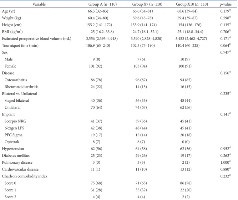
Table 1. Baseline Demographics and Clinical Characteristics
Variable Group A (n=110) Group X7 (n=110) Group X10 (n=110) pvalue
Age (yr) 66.5 (32–83) 66.6 (34–81) 68.6 (39–84) 0.179
a)Weight (kg) 60.4 (34–80) 59.8 (45–78) 59.4 (39–87) 0.598
a)Height (cm) 155.2 (141–172) 155.9 (141–174) 154 (136–176) 0.135
a)BMI (kg/m
2) 25 (16.2–33.8) 24.7 (16.1–32.1) 25.1 (18.8–34.4) 0.706
b)Estimated preoperative blood volume (mL) 3,556 (2,393–4,918) 3,540 (2,828–4,820) 3,453 (2,462–4,727) 0.171
a)Tourniquet time (min) 106.9 (65–240) 102.3 (75–190) 110.4 (60–225) 0.064
b)Sex 0.747
c)Male 9 (8) 7 (6) 10 (9)
Female 101 (92) 103 (94) 100 (91)
Disease 0.156
c)Osteoarthritis 86 (78) 96 (87) 94 (85)
Rheumatoid arthritis 24 (22) 14 (13) 16 (15)
Bilateral vs. Unilateral 0.235
c)Staged bilateral 40 (36) 36 (33) 48 (44)
Unilateral 70 (64) 74 (67) 62 (56)
Implant 0.141
c)Scorpio NRG 41 (37) 39 (36) 45 (41)
Nexgen LPS 42 (38) 48 (44) 45 (41)
PFC Sigma 19 (17) 15 (14) 20 (18)
Optetrak 8 (7) 8 (7) 0 (0)
Hypertension 62 (56) 64 (58) 62 (56) 0.952
c)Diabetes mellitus 25 (23) 29 (26) 19 (17) 0.263
c)Pulmonary disease 3 (3) 3 (3) 2 (2) 1.000
d)Cardiovascular disease 11 (1) 11 (10) 13 (12) 0.880
c)Charlson comorbidity index 0.232
c)Score 0 75 (68) 71 (65) 86 (78)
Score 1 31 (28) 35 (32) 22 (20)
Score 2 4 (4) 4 (4) 2 (2)
Values are presented as mean (range) or number of cases (%).
a)
KruskalWallis test.
b)Oneway analysis of variance test.
c)Chisquare test.
d)Fisher exact test.
2. Evaluation of VTE
We assessed the occurrence of a VTE event using MDCT at 10 days postoperatively. Six seconds after the signal density level reached 100 hounsfield units (HU), a computed tomography (CT) scan was performed from the costophrenic angle to the lung apex. After the injection of contrast media (Ultravist 370, Iopromide, Bayer) thorough the antecubital vein, indirect ve
nography was performed to obtain contrastenhanced venous phase images. Based on the literature, the estimated radiation dose received per MDCT scan of this study is approximately 7 mSv for PE scan and 8 mSv for DVT scan
9,10). A single radiologist evaluated the CT images in a blinded manner. Symptomatic PE was defined as PE with additional symptoms, such as dyspnea, pleuritic chest pain, cough, hemoptysis, tachypnea, rales, or tachycardia
7). Symptomatic DVT was defined as DVT involving discomfort of the calf or thigh, a Homan’s sign, swelling, localized hotness, skin discoloration, tenderness, or prominence of the superficial veins
8). If the patient presented with severe symptoms, we performed MDCT before 10 days postoperatively. The VTE cases were divided into PE and DVT, and the latter was further subcategorized into proximal DVT (occurring in the popliteal vein and above) and distal DVT (occurring below the popliteal vein).
If a patient had asymptomatic distal DVT, the patient was only observed conservatively without any treatment. We only treated patients with PE, symptomatic DVT, or proximal DVT, and a pulmonologist or a chest surgeon was consulted for the manage
ment of these patients with anticoagulants. At 3 months post
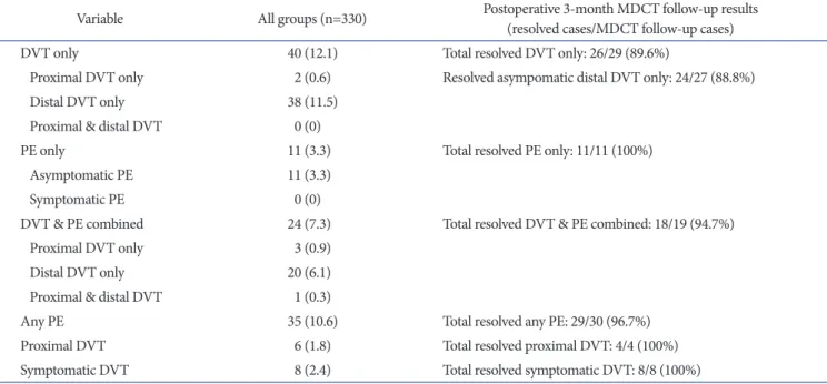
operatively, patients who had VTE events underwent followup MDCT to evaluate the change in VTE.
3. Evaluation of Hemorrhagic Outcome
The bleeding amount was checked by drain removed routinely after 48 hours. Bleeding events were classified as major or minor.
Major bleeding comprised intracranial, intraocular, retroperi
toneal, intraspinal or pericardial bleeding
11). Minor bleeding in
cluded any unexpected hematoma (>25 cm
2), threatened wound hematoma, nasal, gingival, rectal or vaginal bleeding, macroscop
ic hematuria, coughing or vomiting blood
12). Transfusion volume was compared among groups. Additionally, the transfusion volume of staged bilateral TKA patients was compared to that of unilateral TKA patients in each group.
4. Statistical Analysis
The sample size and power calculation was based on the ex
pected incidence of VTE that was approximately 50%
13)in group
A, 20%
14)in group X7, and 7%
12)in group X10. The minimal expected difference in VTE incidence was noted between groups X7 and X10, which was 13%. Statistical significance was set at 5%
(p<0.05) with a power of 80%; thus, the minimum sample size for each group was set as 110 cases by the chisquare test, consid
ering a dropout rate of 10%. Statistical analysis was performed with SPSS ver. 18.0 (SPSS Inc., Chicago, IL, USA). For compari
son of the categorical and proportional variables, the Chisquare test was used. If more than 20% of the expected frequencies were less than 5, the Fisher exact test was performed. Oneway analysis of variance was used to compare the results of numerical con
tinuous variables. However, for some variables without normal distribution, the KruskalWallis test was used.
Results
The incidence of VTE is described in Table 2. Compared to group A, the incidence was significantly lower in groups X7 and X10. The incidence was significantly lower in group X10 than that in group X7 (p=0.038). Group X10 had a significantly lower incidence compared with groups A and X7 in terms of PE and PE plus proximal DVT plus symptomatic DVT. In contrast, no significant differences were observed between groups A and X7 (p=0.056 and p=0.074, respectively). The incidences of proximal DVT and symptomatic DVT were similar among the 3 groups, and symptomatic PE was not observed in any of the groups. All DVT events occurred on the operated side.
The results of followup MDCT are summarized in Table 3. In the cases of asymptomatic distal DVT only, 24/27 (88.8%) dem
onstrated complete resolution, whereas the remaining 3 cases showed a reduced amount of thrombosis without symptoms. All the proximal and symptomatic DVT cases resolved completely, except for 4 patients who refused to take followup MDCT with
out suspicious VTE symptoms. In cases of PE, 96.7% showed complete resolution (29/30); only 1 patient, in group X7, did not show resolution of PE, which then progressed to chronic PE.
However, complete resolution of this patient’s PE was confirmed by followup MDCT after 6 months of anticoagulation therapy.
The mean bleeding amount was similar among the 3 groups
(692 mL in group A, 705 mL in group X7, and 700 mL in group
X10; p=0.889) and there was no observable major or minor
bleeding event. The transfusion volume was similar among 3
groups (908 mL in group A, 1,014 mL in group X7, and 1,062
mL in group X10; p=0.072). The transfusion volume of staged
bilateral TKA patients (909 mL in group A, 1,011 mL in group
X7, and 1,067 mL in group X10) was similar to that of unilateral
Table 2. Incidence of Venous Thromboembolism (VTE) according to Different Chemical Prophylaxis Regimens
Variable No. of
cases
Each group
pvalue
a)pvalue
a)A X7 X10 A vs. X7 A vs. X10 X7 vs. X10
No VTE 255 68 (71.8) 88 (80.0) 99 (90.0)
VTE 75 42 (38.2) 22 (20.0) 11 (10.0) <0.001 0.003 <0.001 0.038
DVT only
Proximal only 2 2 (1.8) 0 (0) 0 (0) 0.331
b)0.498
b)0.498
b)
Distal only 38 19 (17.3) 11 (10.0) 8 (7.3) 0.056 0.116 0.024 0.471
Proximal & distal 0 0 (0) 0 (0) 0 (0)
PE only
Asymptomatic 11 3 (2.7) 5 (4.5) 3 (2.7) 0.798
b)0.721
b)1.000
b)0.721
b)Symptomatic 0 0 (0) 0 (0) 0 (0)
DVT combined PE 24 18 (16.4) 6 (5.5) 0 (0) <0.001 0.009 <0.001 0.029
b)Proximal DVT only 3 3 (2.7) 0 (0) 0 (0) 0.109
b)0.247
b)0.247
b)
Distal DVT only 20 15 (13.6) 5 (4.5) 0 (0) <0.001 0.019 <0.001 0.060
b)Proximal & distal DVT 1 0 (0) 1 (0.9) 0 (0) 1.000
b)1.000
b) 1.000
b)Any DVT 64 39 (35.5) 17 (15.5) 8 (7.3) <0.001 0.001 <0.001 0.056
Any PE 35 21 (19.1) 11 (10.0) 3 (2.7) <0.001 0.056 <0.001 0.027
Any proximal DVT 6 5 (4.5) 1 (0.9) 0 (0) 0.051
b)0.212
b)0.060
b)1.000
b)Symptomatic DVT 8 5 (4.5) 3 (2.7) 0 (0) 0.105
b)0.721
b)0.060
b)0.247
b)P E+symptomatic DVT
+proximal DVT 41 24 (21.8) 14 (12.7) 3 (2.7) <0.001 0.074 <0.001 0.005
Values are presented as number (%).
DVT: deep vein thrombosis, PE: pulmonary embolism.
a)