Is Routine Thromboprophylaxis Needed in Korean Patients Undergoing Unicompartmental Knee Arthroplasty?
This study was undertaken to determine the prevalence and the natural course of venous thromboembolism (VTE) without thromboprophylaxis to ascertain whether routine thromboprophylaxis is necessary following unicompartmental knee arthroplasty (UKA) in Korean patients. The medical records and multidetector row computed tomography (MDCT) imaging of the consecutive 77 UKAs in 70 patients were reviewed. In all patients, MDCTs were undertaken preoperatively and at 1-week after surgery, and VTE symptoms were evaluated. At postoperative 6-months, follow-up MDCTs were undertaken in all patients in whom VTEs were newly detected after surgery. VTE lesions were newly detected in 18 (26%) of the 70 patients. However, none of the patients complained of VTE-related symptoms and MDCT demonstrated that all VTEs were small and involved limited portion without lower leg edema or pleuroparenchymal complication. At the 6-month follow up MDCT, all types of VTEs were shown to be completely resolved, regardless of their location. All of the VTE lesions maintained an asymptomatic status for 6-month after surgery. VTE following UKA in Korean patients who do not receive thromboprophylaxis seems to occur frequently, but all of the VTEs are clinically insignificant and all VTEs are spontaneously regressed.
Routine thromboprophylaxis or thrombolytic treatment in Korean patients undergoing UKA may not be necessary.
Keywords: Venous Thromboembolism; Natural History; Arthroplasty; Replacement; Knee;
Thromboprophylaxis In Jun Koh,1,2 Ju Hwan Kim,3 Man Soo Kim,1
Sung Won Jang,1 Chulkyu Kim,1 and Yong In1,2
1Department of Orthopaedic Surgery, The Catholic University of Korea, Seoul St. Mary’s Hospital, Seoul, Korea; 2Department of Orthopaedic Surgery, The Catholic University of Korea College of Medicine, Seoul, Korea; 3Gangseo Himchan Hospital, Seoul, Korea
Received: 13 September 2015 Accepted: 10 December 2015 Address for Correspondence:
Yong In, MD
Department of Orthopaedic Surgery, The Catholic University of Korea, Seoul St. Mary’s Hospital, 222 Banpo-daero, Seocho-gu, Seoul 06591, Korea
E-mail: [email protected]
http://dx.doi.org/10.3346/jkms.2016.31.3.443 • J Korean Med Sci 2016; 31: 443-448
INTRODUCTION
Venous thromboembolism (VTE), either deep vein thrombosis (DVT) or pulmonary embolism (PE) is one of the most common complications following total knee arthroplasty (TKA) (1,2). Al- though the clinical relevance and natural course of asymptom- atic DVT remains unclear, symptomatic DVT is considered to have substantial risk for morbidity and fatal PE (1,3-5). Thus, routine thromboprophylaxis for preventing VTE is recommend- ed in all patients undergoing TKA, except in the case of high bleeding risk (1,4,5). On the other hand, unicompartmental knee arthroplasty (UKA) is less invasive and has a faster recov- ery time compared to TKA, and patients undergoing UKA are generally younger and more active compared with patients un- dergoing TKA. Indeed, the prevalence of symptomatic VTE fol- lowing UKA in patients receiving routine pharmacological throm- boprophylaxis is remarkably lower than that of TKA (6-9). How- ever, no gold standard strategy for preventing VTE in patients undergoing UKA has been established, and the optimal aggres- siveness of thromboprophylaxis in UKA patients remains a chal- lenging issue (8).
Recent studies have reported the prevalence of VTE follow- ing TKA was lower in the Korean compared to Western popula-
tion (10-14), and all VTE following TKA in Korean patients were completely resolved without pharmacological thrombolytic treatment (15,16). Thus, taking into account for surgical inva- siveness and patients demographics of UKA, the prevalence of VTE following UKA in the Korean population is estimated to be lower than that of Western populations with mild clinical course.
However, equivocal results, regarding the prevalence of VTE fol- lowing TKA between the Asian and Western population, have reported (17,18). Moreover, most previous studies have tended focus on the prevalence of VTE following major orthopaedic surgery, such as total hip arthroplasty (THA) or TKA (19,20).
Therefore, the incidence of both asymptomatic and symptom- atic VTE and the clinical features of VTE following UKA in the Korean population remain to be determined.
This study was conducted to determine the prevalence of VTE following UKA in Korean patients who do not receive thrombo- prophylxis; and the natural course of VTE following UKA in pa- tients who do not receive therapeutic thrombolytic treatment.
MATERIALS AND METHODS Subjected patients
The medical records of 71 consecutive patients who underwent Musculoskeletal Disorders
78 primary UKAs (unilateral or same-day bilateral) between March 2013 and March 2014 at single institution were reviewed using a prospectively collected database. After approved by in- stitutional review board of our institution, only those patients who underwent UKA and had known clinical and radiographic outcomes with a minimum follow-up period of 1 year were in- cluded. Exclusion criteria included a history or current treat- ment of VTE or; newly diagnosed VTE at the preoperative eval- uation. One patient was excluded because of a newly detected PE at the preoperative evaluation. Consequently, 77 knees of 70 patients, consisting of 60 female patients (67 knees) and 10 male patients (10 knees), were selected for final analyses. The mean age of the patients was 61 years (standard deviation [SD] 6.7, range 40-82). The mean body mass index (BMI) was 26.1 kg/m2 (SD 3.4, range 17.5-33.7).
All surgeries were performed by a single surgeon (I.Y.) under general anesthesia in a standard fashion. Oxford (Biomet, War- saw, IN, USA) mobile bearing UKAs were used in 42 knees of 40 patients and Zimmer® Unicompartmental High-Flex Knee (Zim- mer, Warsaw, IN, USA) fixed bearing UKAs were used in 35 knees of 30 patients using standardized cementation technique. A pneu- matic tourniquet that inflated to 300 mmHg was applied. Pati- ents were encouraged to start their ambulation from the opera- tion day and gradually increasing range-of-motion exercises.
None of the patients received either pharmacological or me- chanical thromboprophylaxis for VTE prevention. The mean hospital stay was 8 days (SD, 1.6; range, 7-16 days).
VTE were diagnosed based upon the findings of multidetec- tor-row computed tomography (MDCT) venography (Soma- tome definition AS+, Siemens, Munich, Germany) which was read by a single radiologist. The presence of VTE was defined as any types of VTE, either DVT or PE detected on MDCT, regard- less of symptom. In all patients, MDCTs were undertaken pre- operatively and at postoperative 7 days. All patients who had newly diagnosed postoperative VTE were consulted to special- ized internists in thrombolytic therapy whether to perform ther- apeutic thrombolytic treatment. The specialized internists agreed that all patients were followed up without thrombolytic treat- ment, because all of the VTEs were asymptomatic without ra- diographically pleuroparenchymal complications. A clinical in- vestigator (K.J.H.) explained the findings of MDCT and the man- agement plan to all patients who had newly detected VTE. Fi- nally, follow up MDCT venography was performed at 6-months after surgery in all patients in whom VTE was diagnosed post- operatively.
A research assistant (K.M.S.) collected all clinical informa- tion. The clinical data included demographic information (age, gender, height, weight, and BMI). The indications for UKA were classified as 1) primary degenerative arthritis, 2) osteonecrosis, or 3) other causes. Pre-existing medical comorbidities included 1) cardiovascular conditions (coronary artery disease, conges-
tive heart failure, valvular insufficiency, and arrhythmia), 2) pul- monary conditions (chronic obstructive pulmonary disease and asthma), 3) metabolic-endocrine disorder, 4) neurologic conditions (stroke, Parkinson’s disease, and seizure disorder]), 5) history of malignancy or chemotherapy, 6) history of varicose vein with phlebitis and history of VTE (DVT or PE) or familial history of VTE. VTE-related symptoms were classified into 1) asymptomatic, 2) DVT-related symptoms (pain and edema of the limb, venous distension or enlargement, or the Homan’s sign [21]), 3) PE-related symptoms (chest pain or discomfort, dyspnea, tachycardia, or hemoptysis), or 4) both DVT- and PE- related symptoms. Type of UKA was classified as 1) unilateral UKA, or 2) same-day bilateral UKA. Tourniquet time was also recorded. VTE lesions were classified into 1) No VTE, 2) distal DVT alone, defined as a thrombosis in the calf veins, 3) proxi- mal DVT alone, defined as a thrombus in the femoral and/or popliteal veins, 4) PE with distal DVT, 5) PE with proximal DVT, or 6) PE alone. Each of established VTE lesions was monitored and each finding from MDCT venography was recorded sepa- rately.
Statistical analysis
Statistical analyses were performed using SPSS® for Windows (Version 18.0; SPSS Inc., Chicago, IL, USA). Descriptive statis- tics were used to report the prevalence of each VTE lesion. Chi- square tests were used to determine the statistical significance between differences for each categorical variable, and the Wil- coxon sign ed-rank test was used to evaluate the significance of differences between continuous variables. To determine the rates of VTEs, the proportion of each VTE lesion was calculated separately. To determine the clinical features of VTE, the pro- portion of each VTE-related symptom was calculated separate- ly. To determine the natural course of VTE, each VTE patient was categorized as 1) complete resolution; or 2) residual lesion;
and the prevalence according to the time period was assessed.
P values < 0.05 were considered significant.
Ethics statement
The present study was approved by institutional review board of Seoul St. Mary’s Hospital (IRB No. KC14RISI0254). The inform- ed consents were obtained from all patients.
RESULTS
Although VTE lesions were newly detected in 18 (26%) out of the 70 patients, all of VTEs, either DVT or PE was asymptomat- ic. The most common type of VTE was the isolated distal DVT (16%) followed by PE combined with distal DVT (7%) and iso- lated proximal DVT (3%) (Fig. 1). In addition, none of the pa- tients complained of VTE-related symptoms; and MDCT ve- nography demonstrated that all VTEs were small and involved
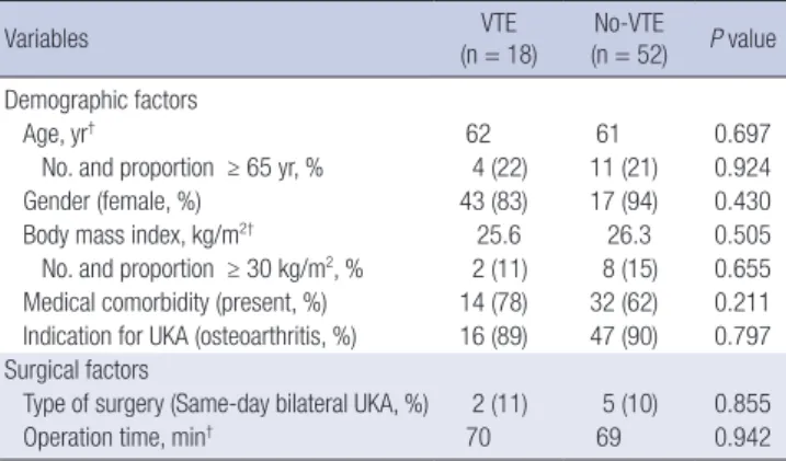
Table 1. Comparison of demographic and surgical factors between VTE and No-VTE groups*
Variables VTE
(n = 18) No-VTE (n = 52) P value Demographic factors
Age, yr† 62 61 0.697
No. and proportion ≥ 65 yr, % 4 (22) 11 (21) 0.924
Gender (female, %) 43 (83) 17 (94) 0.430
Body mass index, kg/m2† 25.6 26.3 0.505
No. and proportion ≥ 30 kg/m2, % 2 (11) 8 (15) 0.655 Medical comorbidity (present, %) 14 (78) 32 (62) 0.211 Indication for UKA (osteoarthritis, %) 16 (89) 47 (90) 0.797 Surgical factors
Type of surgery (Same-day bilateral UKA, %) 2 (11) 5 (10) 0.855
Operation time, min† 70 69 0.942
*Data are presented as numbers of patients (percentages); †Data are presented as mean values. VTE, venous thromboembolism; UKA, unicompartmental knee arthro- plasty.
Fig. 1. Prevalence of VTE following UKA in patients who did not receive thrombopro- phylaxis. The most common VTE lesion was isolated distal DVT (16%) followed by PE with concomitant distal DVT (7%) and isolated proximal DVT (3%).
No VTE n = 52 (74%) Distal DVT
n = 11 (16%)
Proximal DVT n = 2 (3%) PE with distal DVT
n = 5 (7%)
Fig. 2. Serial MDCT venography images showing the natural course of postoperative- ly developed proximal DVT following unilateral UKA in a 71-year old female patient.
Normal venous flows are observed at both proximal thighs preoperatively. Arrow indi- cates the left poplieal vein (A). Mild engorgement of the popliteal vein (arrow) is noted in the left proximal thigh at postoperative 1-week (B). At postoperative 6-month, the DVT lesion is completely regressed without thrombolytic treatment. Arrow indicates the left popliteal vein (C).
A
B
C
limited portion of calf vein or proximal tibioperoneal vein with neither lower leg edema nor pleuroparenchymal complication.
There were no differences in demographic and surgical risk fac- tors for VTE occurrence between the VTE and no-VTE groups (Table 1).
At the 6-month follow up MDCT, all types of VTEs were shown to be completely resolved, regardless of their location (Fig. 2 and 3). All of the VTE lesions maintained an asymptomatic status for 6 months after the surgery.
DISCUSSION
This study found that although VTE in Korean patients under- going UKA occurred frequently (26%), all VTEs were clinically asymptomatic and radiographically insignificant. In addition, they were completely resolved without any treatment.
In this study, the prevalence of VTE after UKA was 26%; how- ever all of the VTEs were asymptomatic. The prevalence of VTE in this study seems to be higher than that of recent studies, which reported very low VTE prevalence following UKA, ranging from 0% to 5% (6-8,22,23) (Table 2). However, it is difficult to compare this study with previous studies because previous studies evalu- ated only symptomatic VTE patients who received routine phar- macological thromboprophylaxis. As there was no symptomat- ic patient in this study, if we had performed MDCT in sympto- matic patients, the prevalence of VTE in this study would be comparable to those of previous studies. These findings concur with previous studies reporting lower prevalence of VTE, even after TKA in the Korean compared with Western population (10-12,15,16) and indicate that appropriate strategies for pre- venting VTE following UKA reflecting prevalence and clinical features of this study should be established for the Korean pop-
ulation.
One interesting finding in this study was that all of the PEs occurred irrespective of proximal DVTs. In this study, MDCT venography revealed all of the PEs (7%) occurred concomitant-
ly with distal DVT. However, all of the proximal DVTs (3%) were localized within the femoral vein without pulmonary involve- ment. These findings concur with previous studies reporting no significant association of proximal DVT and PE. These findings also asked question the mechanical propagation theory, which a proximal DVT was more likely to propagate and lead to PE (24,25). These findings suggest that further studies are neces- sary to determine whether routine thromboprophylaxis for pre- venting DVT would reduce the incidence of PE in patients un- dergoing UKA.
This study found that all of the VTEs, either DVT or PE nei- ther progress nor result in sequelae. In this study, all patients who had newly detected VTE followed up without receiving thrombolytic treatment. Follow up MDCT venography at post- operative 6-month showed that all VTE lesions resolved com- pletely. These findings agree with previous studies, which re- ported complete resolution of VTEs without thrombolysis even in patients who underwent TKA, regardless of size or location (15,16). The results of this study, together with those of previous studies, suggest that therapeutic thrombolytic treatment might be unnecessary in patients who had only radiological, clinically asymptomatic, VTEs following UKA.
This study has several limitations. First, all study participants were Korean and most were women (86%). Thus, the findings of this study may not be widely generalizable because the prev- alence of VTE following UKA may be manifested differently among various ethnic populations and by gender. The female predominance among Korean patients undergoing knee arthro- plasty has been well documented previously (26,27). Second, we performed MDCT at postoperative 7 days in all patients and 6 months after surgery in patients who had newly diagnosed postoperative VTE. Therefore, asymptomatic VTEs which oc- curred 7 days after surgery might be missed. However, no gold standard period for monitoring of postoperative VTE has been established. Recent clinical practice guidelines recommend the minimum period of thromboprophylaxis as at least 7 to 10 days after major orthopaedic surgery (1,3,4). Third, as routine throm- Table 2. Summary of previous studies reporting prevalence of VTE following UKA
Author (year) Country No. of cases Prophylaxis Evaluation Prevalence Comments
Current study (2015) Korea 70 No All patients Overall 26%
DVT 26%/PE 7% All PE developed with distal DVT.
All VTEs were asymptomatic and resolved sponta- neously.
Lombardi et al. (2007) [7] US 423 Chemoprophylaxis Symptomatic patient Overall 0% -
Chan et al. (2009) [22] UK 239 Mechanical (No routine
Chemoprophylaxis) Symptomatic patient Overall 5%
DVT 3%/PE 3% 1 patient died due to PE.
VTE occurred 6% of SD BUKA and 4% of St BUKA.
Berend et al. (2010) [6] US 828 Chemoprophylaxis Symptomatic patient Overall 0.1%
DVT 0.1%/PE 0% -
Willis-Owen et al. (2011) [8] UK 1,080 Chemoprophylaxis Symptomatic patient Overall 0.3%
DVT 0.3%/PE 0%
Overall VTE prevalence after TKA was 2.2%
Chen et al. (2013) [23] Singapore 171 Chemoprophylaxis Symptomatic patient Overall 4%
DVT 2%/PE 1% VTE occurred 2% of SD BUKA and 4% of StBUKA.
VTE, venous thromboembolism; DVT, deep vein thrombosis; PE, pulmonary embolism; SD BUKA, same-day bilateral UKA; St BUKA, staged bilateral UKA.
Fig. 3. Serial MDCT scans showing the natural course of postoperatively developed PE following unilateral UKA in a 58-year old female patient. Preoperative finding. Ar- row indicates the posterior basal segmental artery of right lower lobe (A). Newly de- veloped PEs are noted on bifurcation of the right middle and lower lobe at postopera- tive 1-week. Arrow indicates the posterior basal segmental artery of right lower lobe (B). These lesions are completely regressed at postoperative 6-month without treat- ment. Arrow indicates the posterior basal segmental artery of right lower lobe (C).
A
B
C
boprophylaxis rather than VTE screening test is recommended in all patients undergoing TKA (1,3,4), routine MDCT for VTE screening is not generally accepted in real clinical practice. How- ever, asymptomatic VTE is common and symptoms of VTE are known to be nonspecific. Thus, routine preoperative and post- operative MDCTs were only way to investigate the accurate prev- alence and natural history of VTEs following UKA. Fourth, al- though angiography is considered to be the traditional standard for detecting VTE, MDCT venography was used in this study.
MDCT can evaluate DVT and PE simultaneously and previous studies validated its usefulness for detecting VTE in patients following knee arthroplasty (13,28-30). Fifth, the strategy for PE management used in this study may not be generally accepted because prompt initiation of anticoagulation therapy is recom- mended after diagnosis of acute PE to prevent thrombus exten- sion and recurrent fatal PE. However, imaging tests for PE diag- nosis are recommended after clinical probability assessment based on risk stratification and clinical signs and symptoms (31-34). Thus, no patient who had newly diagnosed postopera- tive VTE in this study would be indicated to perform MDCT be- cause all VTEs were clinically asymptomatic with a low mortal- ity risk. Moreover, asymptomatic VTE reported to be resolved spontaneously without any long-term sequelae, even after ma- jor orthopaedic surgery (15,35,36). We discussed these issues with specialized internists in thrombolytic therapy before re- search commencement.Finally, the effect of the small sample size should be considered. This study is underpowered and sub- ject to a type II error to detect the difference between the VTE and no-VTE group and to demonstrate the natural course of VTE. Other issues related to MDCT, such as cost and radiation hazard make it difficult to extend the patient enrolment time period. Further detailed prospective studies with sufficient sam- ple size are needed to determine between group differences and the natural course of VTE and to identify the risk factors for VTE after UKA. Despite these limitations, this study is the first to report the prevalence and natural course of VTE following UKA in Korean patients who receive neither thromboprophy- laxis nor therapeutic thrombolytic treatment.
In conclusion, this study demonstrates that VTE following UKA in Korean patients who do not receive thromboprophy- laxis seems to occur frequently, but all of the VTEs are clinically asymptomatic. Moreover, all VTEs are spontaneously regressed within 6 months after UKA without long-term sequelae. The re- sults of this study suggest that routine thromboprophylaxis or thrombolytic treatment in Korean patients undergoing UKA may not be necessary.
ACKNOWLEDGMENT
We would like to thank Ho Jong Chun MD, PhD and Hera Kang MD, Department of Radiology, Seoul St. Mary’s Hospital, Seoul,
Korea, for their assistance with interpretation of all radiograph- ic images.
DISCLOSURE
The authors declare that they have no potential conflicts of in- terest.
AUTHOR CONTRIBUTION
Study concept and design: In Y. Data collection: Kim JH, Jang SW, Kim MS, Kim C. Data interpretation and statistical analysis:
Koh IJ. Drafting the first manuscript: Koh IJ. Critical review: Koh IJ, In Y. Approval of the final manuscript: all authors.
ORCID
In Jun Koh http://orcid.org/0000-0002-7941-1587 Ju Hwan Kim http://orcid.org/0000-0002-8288-8853 Man Soo Kim http://orcid.org/0000-0002-1374-9964 Sung Won Jang http://orcid.org/0000-0003-1276-1141 Chulkyu Kim http://orcid.org/0000-0001-7220-6938 Yong In http://orcid.org/0000-0002-5932-3934
REFERENCES
1. Falck-Ytter Y, Francis CW, Johanson NA, Curley C, Dahl OE, Schulman S, Ortel TL, Pauker SG, Colwell CW Jr; American College of Chest Physicians.
Prevention of VTE in orthopedic surgery patients: Antithrombotic Thera- py and Prevention of Thrombosis, 9th ed: American College of Chest Phy- sicians Evidence-Based Clinical Practice Guidelines. Chest 2012; 141:
e278S-325S.
2. Haas SB, Barrack RL, Westrich G. Venous thromboembolic disease after total hip and knee arthroplasty. Instr Course Lect 2009; 58: 781-93.
3. Geerts WH, Bergqvist D, Pineo GF, Heit JA, Samama CM, Lassen MR, Col- well CW; American College of Chest Physicians. Prevention of venous thromboembolism: American College of Chest Physicians Evidence-Bas- ed Clinical Practice Guidelines (8th Edition). Chest 2008; 133: 381S-453S.
4. Johanson NA, Lachiewicz PF, Lieberman JR, Lotke PA, Parvizi J, Pellegrini V, Stringer TA, Tornetta P 3rd, Haralson RH 3rd, Watters WC 3rd. Ameri- can academy of orthopaedic surgeons clinical practice guideline on. Pre- vention of symptomatic pulmonary embolism in patients undergoing to- tal hip or knee arthroplasty. J Bone Joint Surg Am 2009; 91: 1756-7.
5. Lieberman JR, Hsu WK. Prevention of venous thromboembolic disease after total hip and knee arthroplasty. J Bone Joint Surg Am 2005; 87: 2097- 112.
6. Berend KR, Morris MJ, Lombardi AV Jr. Unicompartmental knee arthro- plasty: incidence of transfusion and symptomatic thromboembolic dis- ease. Orthopedics 2010; 33: 8-10.
7. Lombardi AV Jr, Berend KR, Tucker TL. The incidence and prevention of symptomatic thromboembolic disease following unicompartmental knee arthroplasty. Orthopedics 2007; 30: 46-8.
8. Willis-Owen CA, Sarraf KM, Martin AE, Martin DK. Are current thrombo-
embolic prophylaxis guidelines applicable to unicompartmental knee replacement? J Bone Joint Surg Br 2011; 93: 1617-20.
9. Duchman KR, Gao Y, Pugely AJ, Martin CT, Callaghan JJ. Differences in short-term complications between unicompartmental and total knee ar- throplasty: a propensity score matched analysis. J Bone Joint Surg Am 2014; 96: 1387-94.
10. Cho KY, Kim KI, Khurana S, Bae DK, Jin W. Is routine chemoprophylaxis necessary for prevention of venous thromboembolism following knee arthroplasty in a low incidence population? Arch Orthop Trauma Surg 2013; 133: 551-9.
11. Kim KI, Cho KY, Jin W, Khurana SS, Bae DK. Recent Korean perspective of deep vein thrombosis after total knee arthroplasty. J Arthroplasty 2011;
26: 1112-6.
12. Lee WS, Kim KI, Lee HJ, Kyung HS, Seo SS. The incidence of pulmonary embolism and deep vein thrombosis after knee arthroplasty in Asians re- mains low: a meta-analysis. Clin Orthop Relat Res 2013; 471: 1523-32.
13. Park KH, Cheon SH, Lee JH, Kyung HS. Incidence of venous thromboem- bolism using 64 channel multidetector row computed tomography-indi- rect venography and anti-coagulation therapy after total knee arthroplas- ty in Korea. Knee Surg Relat Res 2012; 24: 19-24.
14. Cha SI, Lee SY, Kim CH, Park JY, Jung TH, Yi JH, Lee J, Huh S, Lee HJ, Kim SY. Venous thromboembolism in Korean patients undergoing major or- thopedic surgery: a prospective observational study using computed to- mographic (CT) pulmonary angiography and indirect CT venography. J Korean Med Sci 2010; 25: 28-34.
15. Kim YH, Kim JS. Incidence and natural history of deep-vein thrombosis after total knee arthroplasty. A prospective, randomised study. J Bone Joint Surg Br 2002; 84: 566-70.
16. Kim YH, Yoo JH, Kim JS. Factors leading to decreased rates of deep vein thrombosis and pulmonary embolism after total knee arthroplasty. J Ar- throplasty 2007; 22: 974-80.
17. Leizorovicz A; SMART Venography Study Steering Committee. Epidemi- ology of post-operative venous thromboembolism in Asian patients. Re- sults of the SMART venography study. Haematologica 2007; 92: 1194-200.
18. Piovella F, Wang CJ, Lu H, Lee K, Lee LH, Lee WC, Turpie AG, Gallus AS, Planès A, Passera R, et al. Deep-vein thrombosis rates after major ortho- pedic surgery in Asia. An epidemiological study based on postoperative screening with centrally adjudicated bilateral venography. J Thromb Hae- most 2005; 3: 2664-70.
19. Chung LH, Chen WM, Chen CF, Chen TH, Liu CL. Deep vein thrombosis after total knee arthroplasty in Asian patients without prophylactic anti- coagulation. Orthopedics 2011; 34: 15.
20. Lee SY, Ro H, Chung CY, Lee KM, Kwon SS, Sung KH, Park MS. Incidence of deep vein thrombosis after major lower limb orthopedic surgery: anal- ysis of a nationwide claim registry. Yonsei Med J 2015; 56: 139-45.
21. Crawford ES. The seventh John Homans Lecture: heroes in vascular sur- gery. J Vasc Surg 1992; 15: 417-23.
22. Chan WC, Musonda P, Cooper AS, Glasgow MM, Donell ST, Walton NP.
One-stage versus two-stage bilateral unicompartmental knee replace- ment: a comparison of immediate post-operative complications. J Bone Joint Surg Br 2009; 91: 1305-9.
23. Chen JY, Lo NN, Jiang L, Chong HC, Tay DK, Chin PL, Chia SL, Yeo SJ. Si- multaneous versus staged bilateral unicompartmental knee replacement.
Bone Joint J 2013; 95-B: 788-92.
24. Parvizi J, Jacovides CL, Bican O, Purtill JJ, Sharkey PF, Hozack WJ, Roth- man RH. Is deep vein thrombosis a good proxy for pulmonary embolus?
J Arthroplasty 2010; 25: 138-44.
25. Parvizi J, Parmar R, Raphael IJ, Restrepo C, Rothman RH. Proximal deep venous thrombosis and pulmonary embolus following total joint arthro- plasty. J Arthroplasty 2014; 29: 1846-8.
26. Koh IJ, Kim TK, Chang CB, Cho HJ, In Y. Trends in use of total knee arthro- plasty in Korea from 2001 to 2010. Clin Orthop Relat Res 2013; 471: 1441- 50.
27. Koh IJ, Kim MW, Kim JH, Han SY, In Y. Trends in high tibial osteotomy and knee arthroplasty utilizations and demographics in Korea from 2009 to 2013. J Arthroplasty 2015; 30: 939-44.
28. Watanabe H, Sekiya H, Kariya Y, Hoshino Y, Sugimoto H, Hayasaka S. The incidence of venous thromboembolism before and after total knee ar- throplasty using 16-row multidetector computed tomography. J Arthro- plasty 2011; 26: 1488-93.
29. Miyagi J, Funabashi N, Suzuki M, Asano M, Kuriyama T, Komuro I, Mori- ya H. Predictive indicators of deep venous thrombosis and pulmonary arterial thromboembolism in 54 subjects after total knee arthroplasty us- ing multislice computed tomography in logistic regression models. Int J Cardiol 2007; 119: 90-4.
30. Song EK, Seon JK, Park SJ, Cho SB, Choi MS. Diagnosis of the deep vein thrombosis with multidetector-row computed tomographic venography after totalknee arthroplasty. J Korean Orthop Assoc 2008; 43: 294-300.
31. van der Hulle T, Dronkers CE, Klok FA, Huisman MV. Recent developments in the diagnosis and treatment of pulmonary embolism. J Intern Med 2016; 279: 16-29.
32. Quadery R, Elliot CA, Hurdman J, Kiely DG, Maclean RM, Sabroe I, van Veen JJ, Condliffe R. Management of acute pulmonary embolism. Br J Hosp Med (Lond) 2015; 76: C150-5.
33. Limbrey R, Howard L. Developments in the management and treatment of pulmonary embolism. Eur Respir Rev 2015; 24: 484-97.
34. Konstantinides SV, Torbicki A, Agnelli G, Danchin N, Fitzmaurice D, Galiè N, Gibbs JS, Huisman MV, Humbert M, Kucher N, et al. 2014 ESC guide- lines on the diagnosis and management of acute pulmonary embolism.
Eur Heart J 2014; 35: 3033-69.
35. Kim YH, Oh SH, Kim JS. Incidence and natural history of deep-vein throm- bosis after total hip arthroplasty. A prospective and randomised clinical study. J Bone Joint Surg Br 2003; 85: 661-5.
36. Ginsberg JS, Turkstra F, Buller HR, MacKinnon B, Magier D, Hirsh J. Post- thrombotic syndrome after hip or knee arthroplasty: a cross-sectional study. Arch Intern Med 2000; 160: 669-72.