INTRODUCTION
For useful biological resources, the interest in their use and the researches for them have focused on plants such as natural medicine and the interest in use and development of useful animals have been very low around the world. How- ever, diversity and usefulness of animals is currently retho- ught and development of technologies in the agricultural and biological industries leads to confirmation of high possibility
of using amphibians as a biological resource (Erspamer et al. 1986; Rinaldi 2002; Lu et al. 2008; Gomes et al. 2011;
Wang et al. 2012). The use as a biological resource empha- sizes the importance of gene pool and the use for productive, medical and dietary purposes. In particular, the use of amphi- bians for a medical purpose has been well known in both of Oriental and Western countries and in Korea it also has been utilized as a medicinal animal in traditional oriental medi- cine and fork remedies (Mor et al. 1994; Batista et al. 1999;
Je et al. 2007; Qian et al. 2008; Cho et al. 2009; Jin et al.
2009). However, its effects and usage as a material for tradi- tional oriental medicine shown in references and antient books were induced from experiences, fork remedies and
─
─ 157 ──
Antioxidative Activity and Anti-inflammatory Effects on the Murine Macrophages of Methanol Extracts
of Amphibians
Sang-Bum Kim, Min-Ho Chang1, Sang-Hyun Han2and Hong-Shik Oh2,*
Ohyun Middle School, Jeju 690-061, Korea
1National Park Research Institute, Korea National Park Service, Namwon 590-811, Korea
2Department of Science Education, Jeju National University, Jeju 690-756, Korea
Abstract -- Oxidative stress has been reported to be one of causes of neuritis. This study examined antioxidative activities of methanol extracts of six amphibian species known to be medicinal animals (Rana catesbeiana, R. coreana, R. rugosa, R. dybowskii, R. nigromaculata, and Hyla japonica) and investigated their effects of inhibiting nitric oxide (NO) production and cytotoxicity on the murine macrophage RAW264.7 cells. As inflammation is closely associated with reactive oxygen species, assays on 1,1-diphenyl-2-picrylhydrazyl (DPPH) radical scavenging activity, xanthine oxidase inhi- bitory activity, superoxide anion radical scavenging activity and NO scavenging activity of the extracts of the six species were performed to investigate their antioxidative activity. The results obtained were as follows; All extracts showed antioxidative activity, and the activity of R. dybowskii was the highest in comparison among those. Anti-inflammatory effects of the extracts were also examined, the five extracts except that of R. rugosa did not show cytotoxicity for RAW264.7 cells at the maximal concentration (1,000μμg mL--1). Selectivity index, meaning NO scavenging activity com- pared to cytotoxicity, showed the highest level in the extract of R. dybowskii. These results will be very useful basic data for future studies on prevention and treatment of human diseases to under- stand the biological roles of amphibian extracts throughout the antioxidative or anti-inflammatory pathways.
Key words : antioxidative activity, cytotoxicity, anti-inflammatory effect, Korean amphibians
* Corresponding author: Hong-Shik Oh, Tel. 064-754-3283, Fax. 064-725-4902, E-mail. [email protected]
traditional oriental medicine without a scientific analysis (Park and Lee 1998; Park et al. 2005).
Amphibians are usually called as ‘wa’ in traditional orien- tal medicine because it croaks well. In [Donguibogam] its cold characteristic was reported to control fissure and foods of children and in [Pen-Tsao-Kang-Mu] it was written to relieve diarrhea and pathology of fever. In traditional orien- tal medicine, it is used for nephropathy, diuresis, nutrition and flatulence, dried powder in warm honey water or boiled amphibian bodies by itself is eaten as a special efficient medi- cine of pulmonary tuberculosis, roborant and asthma. This study was conducted to provide necessary data for researches on antioxidants and anti-inflammatory materials and on their isolation and mechanism to prevent and cure diseases by in- vestigating biological activity such as antioxidant activity and anti-inflammatory effect of methanol (MeOH) extracts of six amphibian species collected in Korea, which has been rarely studied and has been reported to be used as a fork remedy.
MATERIALS AND METHODS
1. Animals and preparation of extracts
Six amphibian species (R. catesbeiana, R. coreana, R.
rugosa, R. dybowskii, R. nigromaculata, and H. japonica) were used for this study obtained from American Bullfrog Capture Operation Division (Jeongeup, Jeollabuk-do, Repub- lic of Korea) and Frog Village (Muju, Jeollabuk-do, Repub- lic of Korea). After lyophilization of animal specimens, samples were subsequently grinded, deposited on 500 mL 80% MeOH, and extracted three times by using a sonicator.
And then, the supernatant was isolated and evaporated and after frozen drying it was used with diluted with 100 mg mL-11 : 1 ethanol (EtOH) : phosphate-buffered saline (PBS) solution.
2. Cell culture
Murine macrophage cell line RAW264.7 was obtained from Korean Cell Line Bank (Seoul, Republic of Korea) and was incubated at 37�C with 5% CO2conditions using Dulbecco’s modified Eagle’s medium (DMEM) including 100 units mL-1 penicillin-streptomycin and 10% fetal bovine serum (FBS).
Subcultures were conducted every 3~4 days.
3. DPPH radical scavenging activity assay
To examine antioxidant activity of each sample, the Blois (1958) method measuring radical scavenging effect with DPPH (Sigma, USA) was used. DPPH solution was made by dissolving around 2 mg DPPH in 15 mL EtOH. After adding 6.25 mL dimethylsulfoxide (DMSO) to 12 mL of the solution, it was diluted with EtOH for absorbance of the con- trol to be 0.94~0.97 at 517 nm wavelength and was shaken for 10 sec. In addition, 100μL samples of each concentra- tion dissolved in MeOH were put on 96 well plate and a same amount of 0.4 mM DPPH was added. After 10 min incubation at room temperature, absorbance was measured at 517 nm.
4. Xanthine oxidase inhibitory activity assay
The production of uric acid caused by xanthine/xanthine oxidase was measured with the increased absorbance at 290 nm (Cheng et al. 1998) and allopurinol (Sigma, USA) was used as the control. For the mixture, each samples of various concentrations, 0.5 mM xanthine and 1 mM EDTA were pre- pared in 200 mM phosphate buffer (pH 7.5) and 50 unitsμL-1 xanthine oxidase was added to induce production of uric acid. Xanthine oxidase inhibitory activity was presented with the decreased rate of absorbance of the produced uric acid.
5. Superoxide anion scavenging activity assay
The amount of superoxide anion formed by using phena- zine methosulfate (PMS)/NADH system was measured at 517 nm with nitroblue tetrazolium reduction method (Frido- vich 1970; Nishikimi et al. 1972; Liu et al. 1997). The mix- ture was prepared with each sample, 125μM NADH and 63μM NBT in 200 μL PBS (pH 8.4) and 8 μM PMS was added to provoke production of superoxide. Superoxide anion scavenging activity was shown with the decreased rate of absorbance of the produced superoxide.
6. NO scavenging activity assay
NO scavenging activity was analyzed by using sodium nitroprusside (SNP) forming naturally NO (Green et al. 1982;
Marcocci et al. 1994). Each samples of various concentra- tions were added to 10 mM SNP and was incubated at 25�C for 3 hr. After the reaction, Griess solution [1% (w/v) sul-
fanilamide, 0.1% N-1-naphylethylen diamine in 2.5% (v/v) phosphoric acid] of a same amount with the mixture was added. It was at room temperature for 10 min and its absor- bance was measured at 540 nm. NO scavenging activity was calculated with the amount of residual nitrite. The activity was presented with % of the scavenging activity at 500μg mL-1.
7. Cytotoxicity assay
RAW264.7 cells were put into 96 well microplates with 2
×105cells well-1by using the DMEM and were incubated for 18 hr. After each samples of different concentrations and 100 ng mL-1lipopolysaccharide (LPS) (Sigma, USA) were added and incubated for 24 hr. 2 mg mL-13-(4,5-dimehtyl- thiazol)-2,5-diphenyl-tetrazolium bromide (MTT) was added and was incubated for 1 hr and the media was removed. After the formazan sediment produced by reduction of MTT by adding 200μL DMSO was dissolved and absorbance was measured at 540 nm with a microplate reader (Biotek, USA).
By comparing the absorbance of each sample at each con- centration with that of the untreated sample, cytotoxicity of the sample to RAW264.7 cells was evaluated.
8. NO production inhibition assay
RAW264.7 cells (2×10⁵cells well-1) were put into 96 well plates and each samples of various concentrations were treat- ed. After adding LPS (100 ng mL-1), it was incubated for 24 hr. After mixing 100μL supernatant of the media with 100μL Griess solution they were incubated on 96 well plates for 10 min and absorbance was measured at 530 nm. The amount of NO was compared with that of sodium nitrite as a standard.
9. Statistical analysis
The results were presented with a mean and standard devia- tion and a statistical significance was analyzed with Student’s t-test.
RESULTS AND DISCUSSION
1. Antioxidative activity of amphibian extracts
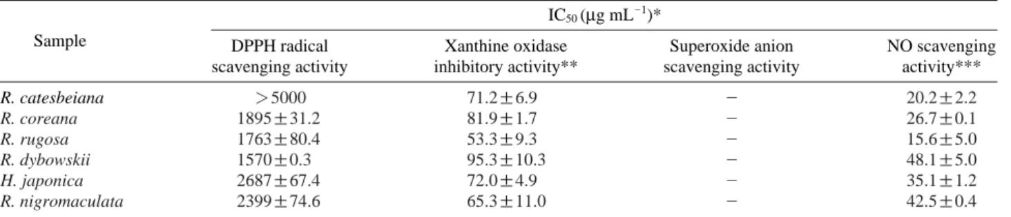
The antioxidant activities of MeOH extracts of R. cates- beiana, R. coreana, R. rugosa, R. dybowskii, R. nigromacu- lata, and H. japonica were presented in Table 1 and Fig. 1.
DPPH free radical scavenging activity was increased with depending on the treated concentrations (Fig. 1A) and the IC50values showing a concentration with 50% scavenging activity of each sample was the lowest in R. dybowskii by recording 1,570μg mL-1and the highest in R. catesbeiana (Table 1). Xanthine oxidase inhibitory activity of the amphi- bian MeOH extracts was measured with xanthine/xanthine oxidase system. Xanthine oxidase inhibitory effect at 625 μg mL-1showed the highest level, 95.3% in R. dybowskii and the lowest level, 53.3% in R. rugosa (Table 1, Fig. 1).
Superoxide anion radical scavenging activity of the extracts showed low levels in all of the samples by recording less than 5% (Table 1). NO is an active species with a strong cytotoxicity and the production of much NO provokes indirect effects including nitrosation and nitration and oxidation to induce harmful effects. NO scavenging activity of the MeOH extracts of the six species of Amphibia was measured with the amount of nitrite by using SNP forming NO. At 2.5 mg mL-1each sample showed 48~15% scavenging activity
Table 1. Comparison of antioxidative potential of MeOH extracts of amphibians
IC50(μg mL-1)*
Sample DPPH radical Xanthine oxidase Superoxide anion NO scavenging
scavenging activity inhibitory activity** scavenging activity activity***
R. catesbeiana ¤5000 71.2±6.9 - 20.2±2.2
R. coreana 1895±31.2 81.9±1.7 - 26.7±0.1
R. rugosa 1763±80.4 53.3±9.3 - 15.6±5.0
R. dybowskii 1570±0.3 95.3±10.3 - 48.1±5.0
H. japonica 2687±67.4 72.0±4.9 - 35.1±1.2
R. nigromaculata 2399±74.6 65.3±11.0 - 42.5±0.4
*IC50values were calculated from regression lines using seven different concentration in triplicate experiments.
**1.25 mg mL-1scavenging activity (% control).
***2.5 mg mL-1scavenging activity (% control).
- indicates ⁄5% radical scavenging activity in maximum concentration.
and the scavenging effect of R. dybowskii, R. nigromaculata and H. japonica was found to be good by recording 48.1, 42.5 and 35.1%, respectively while that of R. coreana, R.
catesbeiana and R. rugosa was relatively low by recording 26.7, 20.2 and 15.6%, respectively. Like previous studies on antioxidant activity of medicinal insect extracts revealing that the extracts of Anomala albopilosa, Sympetrum eroticum and Anax parthenope had high antioxidant activity (Kim et al. 2004; Yoon et al. 2007), this study also found that amphib- ians produced a superior antioxidant activity.
2. Anti-inflammatory activity of amphibian extracts
For vertebrates, infection and damage of capillary vessels of skin tissues provoke proliferation of synovial cells and increase of fibroblasts and macrophages along with satura-
tion of lymphocytes inside of connective tissues. Interlukin secreted from the lymphocytes by these changes, actives effector lymphocytes and promotes various enzyme such as hyaluronidase, elastase and collagenase and inflammatory mediators like prostaglandin to trigger inflammation destroy- ing connective tissues (Deby 1988; Shimizu and Wolfe 1990).
NO is a free radial produced from L-arginine with nitric oxide synthases (NOSs) (Palmer et al. 1988) and it shows many biological functions including body defence, signal transduction and a function as a secondary messenger of vasodilation (Monacada et al. 1991; Knowles and Mocada 1992; Nathan 1992). Constitutive NOS (cNOS) contains neuronal- and endothelial-NOS expressed in neurons and endothelial cells, respectively. The production of NO caused by cNOSs play a key role in managing homeostasis of a body (Kawamata et al. 2000). However, contrary to cNOSs, indu- cible NOS (iNOS) is presented on macrophages, vascular
Concentration (μg mL-1) Concentration 625μg mL-1
78.1 156.3 312.5 625.0 1250.0 2500.0 5000.0 0
20 40 60 80 100
A B C D E F
A B C D E F DPPH radical scavenging activity (% control)
Concentration (mg mL-1)
0.08 0.16 0.31 0.63 1.25 2.50 -10
0 10 20 30 40 50
Nitric oxide scavenging activity (% control)
0 20 40 60 80 100
Xanthine oxidase inhibitory activity (% control)
A B C D E F
(A)
(C)
(B)
Fig. 1. Dose-dependent scavenging effects on DPPH radical, NO and xanthine oxidase inhibitory activity by amphibian extracts. The data indicate the mean±S.D. of triplicate experiments. A: R. catesbeiana; B: R. coreana; C: R. rugosa; D: R. dybowskii; E: H. japonica;
F: R. nigromaculata.
smooth muscle cells, endothelial cells, hepatocytes and myo- cardial cells by following the stimuli of LPS, interferon-g, interleukin (IL)-1b and tumor necrosis factor (TNF)-α (Lee et al. 2000). The iNOS expressed on these tissues leads to inflammation, tumor (Nathan 1992), damage of tissues, gene variation and damage of nerves by producing much NO for a long time (Stuehr et al. 1991; Weisz et al. 1996). Like this, inflammation-related NO and prostaglandin E2are formed
by iNOS and cyclooxygenase-2.
Macrophage is known to be related with homeostasis by affecting many host responses such as acquired immunity as well as innate immunity and it is very critical for body def- ense in the early infection by making NO and cytokine dur- ing inflammatory reaction (Higuchi et al. 1990). LPS exist- ing on the outer membrane of gram positive bacteremia is reported to be an endotoxin and trigger sepsis and shock with
0 20 40 60 80 100 120 140
0 20 40 60 80 100
MTT reduction(% control) NO inhibition(% control)
0 20 40 60 80 100 120 140
0 20 40 60 80 100
MTT reduction(% control) NO inhibition(% control)
0 20 40 60 80 100 120 140
0 20 40 60 80 100
MTT reduction(% control) NO inhibition(% control)
0 20 40 60 80 100 120 140
0 20 40 60 80 100
MTT reduction(% control) NO inhibition(% control)
0 20 40 60 80 100 120 140
0 20 40 60 80 100
MTT reduction(% control) NO inhibition(% control)
0 20 40 60 80 100 120 140
0 20 40 60 80 100
MTT reduction(% control) NO inhibition(% control)
0.0 62.5 125.0 250.0 500.0 1000.0 Concentration (μg mL-1)
0.0 62.5 125.0 250.0 500.0 1000.0 Concentration (μg mL-1)
0.0 62.5 125.0 250.0 500.0 1000.0 Concentration (μg mL-1)
0.0 62.5 125.0 250.0 500.0 1000.0 Concentration (μg mL-1)
0.0 62.5 125.0 250.0 500.0 1000.0 Concentration (μg mL-1)
0.0 62.5 125.0 250.0 500.0 1000.0 Concentration (μg mL-1) MTT
NO
(A) (B)
(C) (D)
(E) (F)
Fig. 2. Effects of MeOH extracts on the NO production in LPS-stimulated RAW264.7 cells. Cells were treated with LPS (100 ng mL-1) alone or LPS plus the indicated concentrations of MeOH extracts for 24 hr. A: R. catesbeiana; B: R. coreana; C: R. rugosa; D: R. dybowskii;
E: H. japonica; F: R. nigromaculata.
released from the outer membrane when bacteria die. LPS increases synthesis of pro-inflammatory cytokine such as TNF-α, IL-1b and IL-6 on macrophages like RAW264.7 cells or mononuclear cells, and particularly TNF-α and IL-1b induce expression of iNOS (Higuchi et al. 1990; Willeaume et al. 1995; McDaniel et al. 1996).
According to the results, MeOH extracts of R. catesbeiana, R. coreana, R. dybowskii, R. nigromaculata, and H. japonica did not show cytotoxicity to the maximal concentration, 1,000 μg mL-1and TC50of R. rugosa showing 50% cytotoxicity was observed to be 333.6μg mL-1. Selectivity index meaning NO scavenging activity compared to cytotoxicity was the highest in R. dybowskii by recording 6.96 and those of R.
rugosa, R. coreana, R. catesbeiana, R. nigromaculata and H. japonica recorded 4.63, 3.46, 3.29, 2.92 and 2.51, respec- tively (Table 2, Fig. 2).
In conclusion, all MeOH extracts of the six species had antioxidant activity, and in comparison among those the activity of R. dybowskii was found to be the best by consider- ing DPPH radical scavenging activity, xanthine oxidase inhibitory activity and NO scavenging activity. In addition, as R. dybowskii did not trigger cytotoxicity to 1,000μg mL-1 and its selectivity index was also the highest (6.96) and its efficiency was observed to be the highest among the extracts.
These results of this study are considered to be important basic data for studies on antioxidants and anti-inflammatory components through extraction of active ingredients from amphibians and on isolation and mechanism of active com- ponents to prevent or to treat diseases. In the future, in vitro and in vivo biological functions of sequential fractions of amphibians are needed to be examined and additional resear- ches on possibility of an industrial use of EtOH or spirit extracts are also considered to be necessary.
REFERENCES
Batista CVF, LR Da Silva, A Sebben, A Scaloni, L Ferrara, GR Paiva, T Olamendi-Portugal, LD Possani and C Bloch Jr.
1999. Antimicrobial peptides from Brazilian frog Phyllome- dusa distincta. Peptides 20:679-686.
Blois MS. 1958. Antioxidant determinations by the use of a stable free radical. Nature 181:1198-1200.
Cheng ZJ, SC Kuo, SC Chan, FN Ko and CM Teng. 1998.
Antioxidant properties of butein isolated from Dalbergia odorifera. Biochem. Biophys. Acta 1392:291-299.
Cho JH, BH Sung and SC Kim. 2009. Buforins: Histone H2A- derived antimicrobial peptides from toad stomach. Biochim.
Biophys. Acta-Biomembr. 1788:1564-1569.
Deby C. 1988. Metabolism of polyunsaturated fatty acids, pre- cursors of eicosanoids. pp.11-36. In Prostaglandins: Bio- logy and chemistry of prostaglandins and related eicosanoids (Curtis-Prior PB ed.). Churchill Livingstone, Edinburgh.
Erspamer V, FG Erspamer and JM Cei. 1986. Active peptides in the skins of two hundred and thirty American amphibian species. Comp. Biochem. Physiol. C 85:125-137.
Fridovich I. 1970. Quantitative aspects of the production of superoxide anion radical by milk xanthine oxidase. J. Biol.
Chem. 245:4053-4057.
Gomes A, B Giri, A Alam, S Mukherjee, P Bhattacharjee and A Gomes. 2011. Anticancer activity of a low immunogenic protein toxin (BMP1) from Indian toad (Bufo melanosticua, Schneider) skin extract. Toxincon 58:85-92.
Green LC, DA Wagner, J Glogowski, PL Skipper, JS Wishnok and SR Tannenbaum. 1982. Analysis of nitrate, nitrite, and [15N]nitrate in biological fluids. Anal. Biochem. 126:131- 136.
Higuchi M, N Hisgahi, H Taki and T Osawa. 1990. Cytolytic mechanism of activated macrophage. Tumor necrosis fac- tor and L-arginine dependent mechanism acts as synergisti- cally as the major cytolytic mechanism of activated macro- phages. J. Immunol. 144:1425-1431.
Je J, Z Qian and S Kim. 2007. Antioxidant peptide isolated from muscle protein of bullfrog, Rana catebeiana Shaw. J. Med.
Food 10:401-407.
Jin LL, SS Song, Q Li, YH Chen, QY Wang and ST Hou. 2009.
Identification and characterization of a novel antimicrobial polypeptide from the skin secretion of a Chinese frog (Rana chensinensis). Int. J. Antimicrob. Ag. 33:538-542.
Kawamata H, H Ochiai, N Mantani and K Terasawa. 2000.
Enhanced expression of inducible NO synthase by Junen- taiho-to in LPS-activated RAW264.7 cells, a murine macro- phage cell line. Am. J. Chin. Med. 28:217-226.
Kim SB, SY Park, SH Kang, SY Choi, SJ Kim and WT Kim.
Table 2. Cell toxicity and the effects on LPS-induced NO produc- tion of the MeOH extracts in RAW264.7 cells
Sample TC50* IC50** Selectivity
(mg mL-1) (mg mL-1) index***
R. catesbeiana ¤1,000 341.8±0.1 2.92~
R. coreana ¤1,000 288.9±7.6 3.46~
R. rugosa 333.6±7.6 71.9±5.9 4.63
R. dybowskii ¤1,000 143.6±10.1 6.96~
H. japonica ¤1,000 303.5±14.5 3.29~
R. nigromaculata ¤1,000 397.8±20.0 2.51~
*TC50is the concentration producing 50% toxicity in RAW264.7 cells.
**IC50is the concentration producing 50% inhibition of NO production in RAW264.7 cells.
***Selectivity Index==TC50/IC50.
─
─ 163 ── 2004. Antioxidant activity of Dragofly’s extracts. Cheju J.
Life Science 7:35-51.
Knowles RG and S Mocada. 1992. Nitric oxide as signal in blood vessels. TIBS 17:399-402.
Lee BG, SH Kim, OP Zee, KR Lee, KY Lee, JW Han and HW Lee. 2000. Suppression of inducible nitric oxide synthase expression in RAW264.7 macrophages by two-carboline alkaloids extracted from Melia azedarach. Eur. J. Pharma- col. 406:301-309.
Liu F, VEC Ooi and ST Chang. 1997. Free radical scavenging activities of mushroom polysaccharide extracts. Life Sci.
60:763-771.
Lu CX, KJ Nan and Y Lei. 2008. Agents from amphibians with anticancer properties. Anticancer Drugs 19:931-939.
McDaniel ML, GH Kwon, CA Marshall and JA Corbett. 1996.
Cytokinins and nitric oxides in islet inflammation and dia- betes. Proc. Soc. Exp. Biol. Med. 211:24-32.
Marcocci L, JJ Maguire, MT Droylefaix and L Packer. 1994.
The nitric oxide-scavenging properties of Ginkgo biloba extract EGb 761, Biochem. Biophys. Res. Commun. 201:
748-755.
Monacada S, RM Palmer and EA Higgs. 1991. Nitric oxide:
physiology, pathophysiology, and pharmacology. Pharma- col. Rev. 43:109-142.
Mor A, K Hani and P Nicolas. 1994. The vertebrate peptide antibiotics dermaseptin have overlapping structural features but target specific microorganisms. J. Biol. Chem. 269:
31635-31641.
Nathan C. 1992. Nitric oxide as a secretory product of mam- malian cells. FASEB J. 6:3051-3064.
Nishikimi M, NA Roa and K Yagi. 1972. The occurrence of superoxide anion in the reaction of reduced phenazine me- thosulfate and molecular oxygen. Biochem. Biophys. Res.
Commun. 46:849-854.
Palmer RM, DS Ashton and S Moncada. 1988. Vascular endo- thelial cells synthesize nitric oxide from L-arginine. Nature 333:664-666.
Park JY, JC Heo, SM An, EY Yun, SM Han, JS Hwang, SW Kang, CY Yun and SH Lee. 2005. High throughput compa- tible screening of anti-oxidative substances by insect extract library. Korean J. Food Preserv. 12:482-488.
Park KT and JS Lee. 1998. Review on insect resources for medi- cal use in Kangwon province. Kor. J. Apiculture 13:79-92.
Qian Z, W Jung and S Kim. 2008. Free radical scavenging activ- ity of a novel antioxidative peptide purified from hydroly- sate of bullfrog skin, Rana catesbeiana Shaw. Bioresource Technol. 99:1690-1698.
Rinaldi AC. 2002. Antimicrobial peptides from amphibian skin:
an expanding scenario. Curr. Opin. Chem. Biol. 6:799-804.
Shimizu T and LS Wolfe. 1990. Arachidonic acid cascade and signal transduction. J. Neurochem. 55:1.
Stuehr HJ, NS Kwon, M Weise and C Nathan. 1991. Purifica- tion of the cytokine-induced macrophage nitric oxide syn- thase: and FAD- and FMN-containing flavoprotein. Proc.
Nat’l. Acad. Sci. USA 88:7773-7777.
Wang C, H Li, S Li, L Tian and D Shang. 2012. Antitumor effects and cell selectivity of temporin-1CEa, an antimi- crobial peptide from the skin secretions of the Cjinese brown frog (Rana chensinensis). Biochimie 94:434-441.
Weisz A, L Cicatielio and H Esumi. 1996. Regulation of the mouse inducible-type nitric oxide synthase gene promoter by interferon-gamma Bacterial lipopolysaccharide and NG- monomethyl-L-arginine. Biochem. J. 316:209-215.
Willeaume V, V Kruys, T Mijatovic and G Huez. 1995. Tumor necrosis factor-alpha production induced by viruses and by lipopolysaccharides in macrophages: similarities and dif- ferences. J. Inflamm. 46:1-12.
Yoon WJ, JA Lee, JY Kim, SB Kim and SY Park. 2007. Antioxi- dant activity and physiological function of the Anomala albopilosa extracts. J. Korean Soc. Food Sci. Nutr. 36:670- 677.
Received: 11 June 2012 Revised: 6 July 2012 Revision accepted: 9 July 2012