http://dx.doi.org/10.15279/kpba.2015.20.4.190
내시경 역행성 담췌관조영술 시행 후 발생하는 급성 췌장염의 예방: 내시경 시술적 관점
계명대학교 의과대학 내과학교실 이윤석·조광범
INTRODUCTION
Endoscopic retrograde cholangiopancreatography (ERCP) has become a standard procedure for the treatment of many pancreatobiliary diseases, even though magnetic resonance cholangiopancreatography (MRCP) and endoscopic ultra- sonography (EUS) have widely replaced the diagnostic as- pect of ERCP. However, even in experienced hands, ERCP could be challenging and procedure-related complications might be provoked such as pancreatitis, bleeding, infection
and duodenal perforation.1 Among those complications, post-ERCP pancreatitis (PEP) is one of the most problemat- ic complications.2 Then, many procedure-related or phar- macological interventions have been proposed to reduce the PEP. However, several pharmacologic agents which have been proposed so far, such as corticosteroid, gabexate me- sylate, nafamostat mesylate, octreotide, and somatostatin, have turned out to be non-effective or equivocal except for rectal non-steroidal anti-inflammatory drug (NSAID).3,4 Since rectal NSAID has not been available in South Korea
Prevention of Post-endoscopic Retrograde Cholangiopancreatography Pancreatitis: An Endoscopic Perspective
Yoon Suk Lee, Kwang Bum Cho
Department of Internal Medicine, Keimyung University School of Medicine, Daegu, Korea
Post-endoscopic retrograde cholangiopancreatography pancreatitis (PEP) remains to be the most common adverse event, even in experienced hands. While most of the PEP has a mild clinical course, it could be severe pancreatitis or into mortality.
Recently, several endoscopic techniques, such as pancreatic stent placement, guidewire-assisted cannulation, or early precut cannulation, have been suggested as a possible techniques for the PEP prophylaxis. Since several pharmacologic agents are turned out to be non-effective or equivocal except for the rectal non-steroidal anti-inflammatory drugs which are not available in Korea, this paper will describe the general aspects of PEP and focus on the endoscopy-techniques for PEP prophylaxis.
Korean J Pancreatobiliary 2015;20(4):190-197
Keywords: Endoscopic retrograde cholangiopancreatography, ERCP, Pancreatitis, Prophylaxis
Received Oct. 1, 2015 Revised Oct. 22, 2015 Accepted Oct. 27, 2015
Corresponding author : Kwang Bum Cho Department of Internal Medicine, Keimyung University School of Medicine, 56 Dalseong-ro, Jung-gu, Deagu 41931, Korea
Tel. +82-53-250-7088 Fax. +82-53-250-7088 E-mail; [email protected]
This is an Open Access article distributed under the terms of the Creative Commons Attribution Non-Commercial License (http://
creativecommons.org/licenses/by-nc/3.0/) which permits unrestricted non-commercial use, distribution, and reproduction in any medium, provided the original work is properly cited.
Copyright © 2015 by The Korean Journal of Pancreas and Biliary Tract
until now, this paper will describe the general aspects of PEP and focus on the endoscopy-techniques for PEP prophylaxis.
1. Incidence of post-ERCP pancreatitis
Post-ERCP hyperamylasemia is quiet common. However, such a transient elevation of serum amylase does not always indicate PEP. By using the PEP definition proposed by Cot- ton et al.5 in 1991, which is the presence of new pancreatic- type abdominal pain and 3 or more times the upper limits of normal occurring 24 hours after the procedure that re- quires at least 2 days-hospitalization, the incidence of PEP is reported to be 1-10%.5-8 In a recently published systematic review of 108 studies including only randomized and con- trolled trials (RCTs), the incidence of PEP was reported to be 9.7%.9 Furthermore, when PEP was classified into mild, moderate, and severe based on the length of hospitalization;
mild (prolongation of planned hospitalization to 2-3 days), moderate (to 4-10 days), and severe (to more than 10 days, or hemorrhagic pancreatitis, phlegmon or pseudocyst, or intervention),5 the mild, moderate, and severe pancreatitis was reported to be 5.7%, 2.6%, and 0.5%, respectively.9 However, in high-risk patients, the incidence of PEP was in- creased up to 14.7% and mild, moderate, and severe pancre- atitis was reported to be 8.6%, 3.9%, and 0.8%, respectively.9
2. Mechanisms of post-ERCP pancreatitis
There are several mechanisms regarding the development of PEP.10 First, mechanical injury to the papilla from multi- ple cannulation trials may lead to papillary edema and swell- ing as well as spasm of sphincter of Oddi, then resulting in obstruction to outflow of pancreatic juice and PEP develop- ment eventually. Second, hydrostatic injury from contrast dye or saline injection into the pancreatic duct during inad- vertent pancreatic duct cannulation or sphincter manome- try. Cheng et al.11 conducted a prospective multicenter study with 15 United States centers and 1115 patients and con- cluded that two or more contrast injections into the pancre- atic duct significantly increased the occurring of PEP. Third, thermal injury from electrosurgical current during endo- scopic sphincterotomy, thermal coagulation for bleeding
control, and endoscopic papillectomy.12 This is also related to mechanical injury resulting in papillary edema from ther- mal burn. Lastly, translocation of intestinal flora or bacteria from contaminated duodenoscopy or accessories might lead to infection and have a role in the development of PEP.12 Whatever the mechanism may cause to PEP, once activated, the inflammatory cascade are similar to other pancreatitis from alcohol, biliary tract disease and so on. Therefore, many strategies for each part of the mechanisms have been provided to avert PEP.
3. Risk factors for post-ERCP pancreatitis
The identification of high risk patients for PEP is impor- tant because preventive intervention could be conducted in advance, such as pancreatic duct stenting or pharmacologic prophylaxis.
Based on previous large-scale studies,13-15 the risk factors are subdivided into three categories: 1. operator-related fac- tors; inadequate training, lack of experience, 2. patient-re- lated factors; younger age, female sex, normal serum biliru- bin, recurrent pancreatitis, prior ERCP-induced pancreatitis, sphincter of Oddi dysfunction (SOD), allergy to contrast media, pancreas divisum, 3. procedure-related factors; diffi- cult cannulation, sphincter of Oddi manometry, precut sphincterotomy, pancreatic sphincterotomy, biliary balloon sphincteroplasty, ampullectomy, failed cannulation, cannu- lation time > 10 minutes, at least one pancreatic deep wire pass, two or more injections of contrast agent into the pan- creatic duct, minor papilla sphincterotomy. The definition of difficult cannulation was very heterogeneous in each study. European society of gastrointestinal endoscopy (ESGE) guideline updated in 2014 specifically defines the difficult cannulation as follows: duration of > 5 minutes, >
5 attempts, or 2 pancreatic guidewire passages.16
PREVENTION OF POST-ERCP PANCREATITIS
1. Guidewire-assisted cannulation
Mechanical injury from repeated cannulation attempts
have been considered to be important mechanism for PEP development,12,17 since this could lead to papillary edema and obstruction of pancreatic ductal flow. Moreover, acci- dental contrast injection into pancreatic duct may lead to chemical and hydrostatic injuries as well. Therefore, a guidewire-assisted cannulation technique has been postulat- ed to improve biliary cannulation and prevent PEP by re- ducing inadvertent contrast injection. Although the results of many studies are conflicting and inconclusive,18-20 several meta-analysis suggest that the guidewire-assisted cannula- tion reduce the risk of PEP,21,22 which has been recommend- ed by European guideline provided by ESGE.6 Recently in 2013, a meta-analysis of 12 randomized trials with 3450 pa- tients also reported that the guidewire-assisted cannulation improved the biliary cannulation rate (84% vs. 77%), de- creased the risk of PEP (3.5% vs 6.7%),23 while Nakai et al.24 reported that unintentional guidewire insertion into pancre- atic duct and a small common bile duct (diameter < 9 mm) were risk factors for PEP with the use of guidewire-assisted cannulation.
Regarding pancreatic guidewire-assisted cannulation (double guidewire technique) for selective bile duct cannu- lation by straightening the papilla, outcomes were inconclu- sive and conflicting so far. However, the outcomes of two RCTs regarding the comparison of double guidewire tech- nique and precut technique were recently reported,25,26 which showed that successful biliary cannulation were simi- lar but, the double guidewire technique had a higher inci- dence of PEP (38% vs. 11%, p = 0.01).26 These results were reflected in the 2014 updated version of ESGE guideline, and which recommend that if this method is used, a pro- phylactic pancreatic stent should be placed.16
2. Early precut sphincterotomy
Selective biliary cannulation is a pivotal element during ERCP, whereas it is unsuccessful in 5% to 10% cases with standard cannulation techniques.27 Therefore, in such a dif- ficult cannulation cases, precut sphincterotomy is usually performed as a rescue method and it is the essential compo- nent as expert endoscopist. However, in most cases, precut
sphincterotomy was reserved for the last salvage technique when all other standard techniques have failed,28 since it has been regarded as challenging technique and independent risk factor for PEP.29,30
However, it remains to be elusive whether the PEP is in- creased by the precut itself or the prolonged cannulation tri- als, in which multiple attempts and inadvertent pancreatic duct cannulations might be act as confounding factors.31 ESGE guideline also comment that prolonged cannulation attempts using standard techniques may impart a risk for PEP greater than the precut sphincterotomy itself.6 Several RCTs have conducted for comparing the early implementa- tion of precut sphincterotomy and the repeated attempts with a standard technique so far and those studies conclud- ed that the early implementation of precut sphincterotomy during difficult cannulation dose not increase the risk of PEP.32-35 Moreover, in 2009, multicenter, prospective-RCT by Manes et al.36 concluded that early precut implementa- tion was associated with lower PEP. A recently published meta-analysis by Sundaralingam et al.37 in 2015 reported that early implementation of precut sphincterotomy did not increase the risk of PEP compared with standard approach.
Furthermore, they concluded that the risk of PEP could be reduced when it is performed by experienced expert endos- copists although further well-designed studies are necessary to confirm these findings.37
3. Prophylactic pancreatic duct stenting
There are many prospective studies and meta-analyses suggesting pancreatic duct stent placement could be effec- tive for the PEP prophylaxis.38-47 As a possible explanation for this, it have been proposed that pancreatic duct stent may reduce pancreatic ductal pressure caused by papillary edema or spasm of the sphincter of Oddi.48 A recent meta- analysis with fourteen RCTs by Mazaki et al.47 also con- firmed that prophylactic pancreatic stenting could be effec- tive for PEP prevention after ERCP as compared with control group with relative risk (RR) of 0.39, 95% confiden- tial interval (CI) 0.29-0.53. Furthermore, subgroup analysis according to the severity of PEP from the meta-analysis re-
vealed that mild to moderate PEP as well as severe PEP have beneficial effect for PEP prophylaxis (RR 0.45; 95% CI 0.32- 0.62 vs. RR 0.26; 95% CI 0.09-0.76).47 Therefore, there might be no dissent from the conclusion of prophylactic ef- fect of pancreatic duct stent.
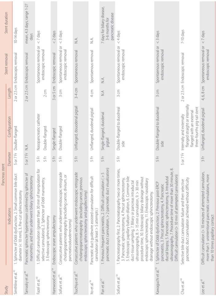
The RCTs included in the several meta-analysis, were dif- ferent from each other in terms of the pancreatic stent con- figuration, duration in placement, and the indications of stent placement were also heterogeneous, which was sum- marized in Table 1. Moreover, pancreatic stent placement are not without adverse events, which have been reported to be about 5% and continuously reported since 1993.43,49 And these include spontaneous stent migration or occlusion, bleeding, cholangitis, cholecystitis, infection, necrosis and pancreatic duct perforation.41 Therefore, several questions regarding pancreatic duct stent placement remains to be elusive. Who is the most suitable for prophylactic stent placement among the patients with risk factors for PEP?
How long does the pancreatic stent have to be in place?
Which type of pancreatic duct stent is more effective for PEP prophylaxis?
1) Who is the most suitable for prophylactic pancreatic stent placement?
Regarding the indication of prophylactic pancreatic stent placement, ESGE guideline in 2010 recommend that it should be strongly considered for high risk patients.6 In the 2014 updated version of ESGE guideline, the high risk con- ditions were also stated as follows: endoscopic ampullecto- my, known or suspected SOD, pancreatic sphincterotomy, precut biliary sphincterotomy, pancreatic guidewire-assisted biliary cannulation, endoscopic balloon sphincteroplasty, and presence of more than three of the risk factors.16 Fur- thermore, if conventional precut technique is selected as rescue technique for selective bile duct cannulation and pancreatic duct cannulation is easily accessible, a small-di- ameter (3-Fr or 5-Fr) pancreatic stent is recommended to be placed and leaved in place for a minimum of 12-24 hours.16 Kerdsirichairat et al.50 reported that pancreatic stent insertion as a salvage measures at very early phase of the
PEP within 2-48 hours also might effective for the PEP treatment.
2) Which type of pancreatic duct stent is more effective for PEP prophylaxis?
In a network meta-analysis of 6 RCTs involving 561 pa- tients (three RCTs, 5Fr straight, flanged stent; two RCTs, 5-Fr single-pigtail, unflanged stent; three RCTs, 3-Fr single- pigtail, unflanged stents), the 5-Fr pancreatic duct stent was superior to the 3-Fr pancreatic duct stent for the PEP pre- vention in high-risk patients, irrespective of the configura- tion.51 The probability of being the best was reported to be 50.3% for 5-Fr single-pigtail, unflanged stent, 46.5% for 5-Fr straight, flanged stents, and 3.1% for 3-Fr single-pigtail, unflanged stents.51 A RCT at a single center showed that 5-Fr placement was easier and faster than 3-Fr stent place- ment, while spontaneous distal migration between the two stents was not different (5-Fr stent, 68.4%; 3-Fr stent, 75.0%; p = 0.617).52 On the 2014 updated version of ESGE guideline, 5-Fr pancreatic stent was more specifically rec- ommended.16 Furthermore, Fujisawa et al.53 conducted sin- gle-center RCT with 240 patients to evaluate the prophylac- tic efficacy between short (5-Fr 3 cm) and long (5-Fr 5 cm) pancreatic stent and concluded that 5-Fr 3 cm stent was su- perior to 5-Fr 5 cm stent because the PEP rate was signifi- cantly lower in the short stent (5-Fr 3 cm, 2.0% vs. 5-Fr 5 cm, 8.8%, p = 0.035).
CONCLUSIONS
Several prophylactic techniques have been reviewed in this article. However, the most important thing for PEP prophy- laxis is the appropriate indication for ERCP. In unnecessary or low yield cases, ERCP could be replaced with MRCP or EUS. And the identification of high risk patients for PEP is also important because preventive intervention could be conducted in advance, such as pancreatic duct stenting or guidewire-assisted cannulation. In addition, the attempts of selective cannulation should be as low as possible and in cases of difficult cannulation early precut technique may be
Table 1. Discrepancies in indication, type, configuration, and length of pancreatic stent StudyPancreas stent Stent removal Stent duration IndicationsDiameterConfigurationLength Simthline et al.43 1. Sphincter of Oddi dysfunction, 2. Small common bile duct diameter (< 10 mm), 3. Pre-cut sphincterotomy5 or 7 FrDoulbe-flanged2 or 2.5 cmEndoscopic removal 10-14 days Tarnasky et al.49 Pancreatic sphincter hypertension confirmed by sphincter of manometry, and then underwent biliary sphincterotomy 5 or 7 FrN.A.2 or 2.5 cmEndoscopic removal mean, 4.3 days; range 1-27 days Fazel et al.40 1. Difficult cannulation (greater than 30 min of manipulation for selective cannulation), 2. Sphincter of Oddi manometry, 3. Endoscopic sphincterotomy
5 FrNasopancreatic catheterSpontaneous removal or endoscopic removal < 7 days 5 FrDouble-flanged2 cm Harewood et al.54 Endoscopic snare ampullectomy5 FrSingle-flanged3 or 5 cm Endoscopic removal < 2 days Sofuni et al.45 Consecutive patients who underwent endoscopic retrograde cholangiopancreatography (excluding cancer, divisum, or pancreaticoduodenectomy)
5 FrDouble-flanged 3 cmSpontaneous removal or endoscopic removal < 3 days Tsuchiya et al.55 Consecutive patients who underwent endoscopic retrograde cholangiopancreatography (excluding cancer, previous endoscopic sphincterotomy and papillectomy)
5 FrUnflanged, douodenal pigtail3-4 cmSpontaneous removal N.A. Ito et al.56 Pancreatic duct guidewire assisted cannulation for difficult cannulation (at least > 5 attempts )5 FrUnflanged, duodenal pigtail4 cmSpontaenous removalN.A. Pan et al.57 1. Previous frequent pancreatitis, 2. Difficult cannulation ( > 5 pancreatic duct cannulation, > 2 pancreatic duct visualization) 5 FrSingle-flanged, duodenal pigtail N.AN.A.7 days for biliary disease, 3-6 months for pancreatic disease Sofuni et al.58 1. Pancreatography first, 2. Pancreatography two or more times, 3. Pancreatic sphincterotomy, 4. Precut sphincterotomy, 5. Endoscopic papillary balloon dilation, 6. Common bile duct-tissue sampling, 7. Biliary or pancreas intraductal ultrasonography, 8. > 15 min cannulation, 9. > 30 min procedure time, 10. Endoscopic biliary drainage without endoscopic sphincterotomy, 11. Endoscopic nasobiliary drainage without endoscopic sphincterotomy.
5 FrDouble-flanged to duodneal side3 cmSpontaneous removal or endoscopic removal < 4 days Kawaguchi et al.59 1. Sphincter of Oddi dysfunction, 2. Previous post-ERCP pancreatitis, 3. Precut sphincterotomy, 4. Pancreatic ductal biopsy, 5. Pancreatic sphincterotomy, 6. Intradutal ultrasonography, 7. Procedure time of more than 30 minute, 8. Difficult cannulation (> 10 min of attempted cannulation)
5 FrDouble-flanged to duodenal side3 cmSpontaenous removal or endoscopic removal < 3 days Cha et al.60 Selective bile duct cannulation failure and then selective pancreatic duct cannulation achieved without difficulty5 or 7 FrInternally and externally flanged straight or internally flanged with an external three-fourths pig-tail stent 2 or 2.5 cmEndoscopic removal 7-10 days Lee et al.61 Difficult cannulation (> 10 minutes of attempted cannulation, more than 5 unintentional pancreatic cannulations, more than 10 times papillary contact
3 FrUnflanged, duodenal pigtail 4, 6, 8 cmSpontaneous removal or endoscopic removal < 7 days NA, not available; ERCP, endoscopic retrograde cholangiopancreatography.
considered as needle-knife fistulotomy is preferred in up- dated 2014 ESGE guidelines. Prophylactic placement of pancreatic stent with small diameter may also be considered if conventional precut is selected as rescue technique in dif- ficult cannulation cases.
국문 색인: 내시경 역행성 담췌관조영술, 췌장염, 예방
Conflicts of Interest
The author has no conflicts to disclose.
REFERENCES
1. Mallery JS, Baron TH, Dominitz JA, et al. Complications of ERCP. Gas- trointest Endosc 2003;57:633-638.
2. Cotton PB. Analysis of 59 ERCP lawsuits; mainly about indications.
Gastrointest Endosc 2006;63:378-382; quiz 464.
3. Elmunzer BJ, Scheiman JM, Lehman GA, et al. A randomized trial of rectal indomethacin to prevent post-ERCP pancreatitis. N Engl J Med 2012;366:1414-1422.
4. Dai HF, Wang XW, Zhao K. Role of nonsteroidal anti-inflammatory drugs in the prevention of post-ERCP pancreatitis: a meta-analysis.
Hepatobiliary Pancreat Dis Int 2009;8:11-16.
5. Cotton PB, Lehman G, Vennes J, et al. Endoscopic sphincterotomy complications and their management: an attempt at consensus. Gas- trointest Endosc 1991;37:383-393.
6. Dumonceau JM, Andriulli A, Deviere J, et al. European Society of Gas- trointestinal Endoscopy (ESGE) Guideline: prophylaxis of post-ERCP pancreatitis. Endoscopy 2010;42:503-515.
7. Masci E, Toti G, Mariani A, et al. Complications of diagnostic and therapeutic ERCP: a prospective multicenter study. Am J Gastroenterol 2001;96:417-423.
8. Loperfido S, Angelini G, Benedetti G, et al. Major early complications from diagnostic and therapeutic ERCP: a prospective multicenter study. Gastrointest Endosc 1998;48:1-10.
9. Kochar B, Akshintala VS, Afghani E, et al. Incidence, severity, and mortality of post-ERCP pancreatitis: a systematic review by using ran- domized, controlled trials. Gastrointest Endosc 2015;81:143-149 e9.
10. Gottlieb K, Sherman S. ERCP and biliary endoscopic sphincterotomy- induced pancreatitis. Gastrointest Endosc Clin N Am 1998;8:87-114.
11. Cheng CL, Sherman S, Watkins JL, et al. Risk factors for post-ERCP pancreatitis: a prospective multicenter study. Am J Gastroenterol 2006;101:139-147.
12. Freeman ML, Guda NM. Prevention of post-ERCP pancreatitis: a com- prehensive review. Gastrointest Endosc 2004;59:845-864.
13. Wang P, Li ZS, Liu F, et al. Risk factors for ERCP-related complications:
a prospective multicenter study. Am J Gastroenterol 2009;104:31-40.
14. Cotton PB, Garrow DA, Gallagher J, Romagnuolo J. Risk factors for complications after ERCP: a multivariate analysis of 11,497 procedures over 12 years. Gastrointest Endosc 2009;70:80-88.
15. Moffatt DC, Cote GA, Avula H, et al. Risk factors for ERCP-related complications in patients with pancreas divisum: a retrospective study. Gastrointest Endosc 2011;73:963-970.
16. Dumonceau JM, Andriulli A, Elmunzer BJ, et al. Prophylaxis of post- ERCP pancreatitis: European Society of Gastrointestinal Endoscopy (ESGE) Guideline - updated June 2014. Endoscopy 2014;46:799-815.
17. Freeman ML, DiSario JA, Nelson DB, et al. Risk factors for post-ERCP pancreatitis: a prospective, multicenter study. Gastrointest Endosc 2001;54:425-434.
18. Mariani A, Giussani A, Di Leo M, Testoni S, Testoni PA. Guidewire biliary cannulation does not reduce post-ERCP pancreatitis compared with the contrast injection technique in low-risk and high-risk pa- tients. Gastrointest Endosc 2012;75:339-346.
19. Kouklakis G, Gatopoulou A, Lirantzopoulos N, Efraimidou E, Manolas K. Evaluation of guide wire cannulation technique in elderly patients with choledocholithiasis. J Gastrointestin Liver Dis 2009;18:185-188.
20. Adler DG, Verma D, Hilden K, Chadha R, Thomas K. Dye-free wire- guided cannulation of the biliary tree during ERCP is associated with high success and low complication rates: outcomes in a single opera- tor experience of 822 cases. J Clin Gastroenterol 2010;44:e57-e62.
21. Cennamo V, Fuccio L, Zagari RM, et al. Can a wire-guided cannula- tion technique increase bile duct cannulation rate and prevent post- ERCP pancreatitis?: A meta-analysis of randomized controlled trials.
Am J Gastroenterol 2009;104:2343-2350.
22. Cheung J, Tsoi KK, Quan WL, Lau JY, Sung JJ. Guidewire versus conventional contrast cannulation of the common bile duct for the prevention of post-ERCP pancreatitis: a systematic review and meta- analysis. Gastrointest Endosc 2009;70:1211-1219.
23. Tse F, Yuan Y, Moayyedi P, Leontiadis GI. Guide wire-assisted cannula- tion for the prevention of post-ERCP pancreatitis: a systematic review and meta-analysis. Endoscopy 2013;45:605-618.
24. Nakai Y, Isayama H, Sasahira N, et al. Risk factors for post-ERCP pancreatitis in wire-guided cannulation for therapeutic biliary ERCP.
Gastrointest Endosc 2015;81:119-126.
25. Angsuwatcharakon P, Rerknimitr R, Ridtitid W, Ponauthai Y, Kulla- vanijaya P. Success rate and cannulation time between precut sphinc- terotomy and double-guidewire technique in truly difficult biliary cannulation. J Gastroenterol Hepatol 2012;27:356-361.
26. Yoo YW, Cha SW, Lee WC, Kim SH, Kim A, Cho YD. Double guidewire technique vs transpancreatic precut sphincterotomy in difficult biliary cannulation. World J Gastroenterol 2013;19:108-114.
27. Bailey AA, Bourke MJ, Williams SJ, et al. A prospective randomized trial of cannulation technique in ERCP: effects on technical success and post-ERCP pancreatitis. Endoscopy 2008;40:296-301.
28. Fuccio L, Cennamo V. Avoidance of early needle knife sphincterotomy to achieve deep biliary cannulation is no longer justified. Clin Gastro- enterol Hepatol 2013;11:1523.
29. Baillie J. Needle-knife papillotomy revisited. Gastrointest Endosc 1997;46:282-284.
30. Masci E, Mariani A, Curioni S, Testoni PA. Risk factors for pancreatitis following endoscopic retrograde cholangiopancreatography: a meta- analysis. Endoscopy 2003;35:830-834.
31. Bourke M. Biliary endoscopic retrograde cholangiopancreatography.
Endoscopy 2011;43:42-46.
32. Swan MP, Alexander S, Moss A, et al. Needle knife sphincterotomy does not increase the risk of pancreatitis in patients with difficult bili- ary cannulation. Clin Gastroenterol Hepatol 2013;11:430-436 e1.
33. de Weerth A, Seitz U, Zhong Y, et al. Primary precutting versus con- ventional over-the-wire sphincterotomy for bile duct access: a pro- spective randomized study. Endoscopy 2006;38:1235-1240.
34. Tang SJ, Haber GB, Kortan P, et al. Precut papillotomy versus persis- tence in difficult biliary cannulation: a prospective randomized trial.
Endoscopy 2005;37:58-65.
35. Cennamo V, Fuccio L, Repici A, et al. Timing of precut procedure does not influence success rate and complications of ERCP procedure:
a prospective randomized comparative study. Gastrointest Endosc 2009;69:473-479.
36. Manes G, Di Giorgio P, Repici A, Macarri G, Ardizzone S, Porro GB.
An analysis of the factors associated with the development of com- plications in patients undergoing precut sphincterotomy: a prospec- tive, controlled, randomized, multicenter study. Am J Gastroenterol 2009;104:2412-2417.
37. Sundaralingam P, Masson P, Bourke M. Early precut sphincterotomy does not increase risk during endoscopic retrograde cholangio- pancreatography in patients with difficult biliary access: a meta- nalysis of randomized controlled trials. Clin Gastroenterol Hepatol 2015;13:1722-1729 e2.
38. Mazaki T, Masuda H, Takayama T. Prophylactic pancreatic stent placement and post-ERCP pancreatitis: a systematic review and meta- analysis. Endoscopy 2010;42:842-853.
39. Fan JH, Qian JB, Wang YM, Shi RH, Zhao CJ. Updated meta-analysis of pancreatic stent placement in preventing post-endoscopic retro- grade cholangiopancreatography pancreatitis. World J Gastroenterol 2015;21:7577-7583.
40. Fazel A, Quadri A, Catalano MF, Meyerson SM, Geenen JE. Does a pancreatic duct stent prevent post-ERCP pancreatitis? A prospective randomized study. Gastrointest Endosc 2003;57:291-294.
41. Andriulli A, Forlano R, Napolitano G, et al. Pancreatic duct stents in the prophylaxis of pancreatic damage after endoscopic retrograde cholangiopancreatography: a systematic analysis of benefits and as- sociated risks. Digestion 2007;75:156-163.
42. Singh P, Das A, Isenberg G, et al. Does prophylactic pancreatic stent placement reduce the risk of post-ERCP acute pancreatitis? A meta-
analysis of controlled trials. Gastrointest Endosc 2004;60:544-550.
43. Smithline A, Silverman W, Rogers D, et al. Effect of prophylactic main pancreatic duct stenting on the incidence of biliary endoscopic sphincterotomy-induced pancreatitis in high-risk patients. Gastroin- test Endosc 1993;39:652-657.
44. Aizawa T, Ueno N. Stent placement in the pancreatic duct prevents pancreatitis after endoscopic sphincter dilation for removal of bile duct stones. Gastrointest Endosc 2001;54:209-213.
45. Sofuni A, Maguchi H, Itoi T, et al. Prophylaxis of post-endoscopic retrograde cholangiopancreatography pancreatitis by an endoscopic pancreatic spontaneous dislodgement stent. Clin Gastroenterol Hepa- tol 2007;5:1339-1346.
46. Choudhary A, Bechtold ML, Arif M, et al. Pancreatic stents for pro- phylaxis against post-ERCP pancreatitis: a meta-analysis and system- atic review. Gastrointest Endosc 2011;73:275-282.
47. Mazaki T, Mado K, Masuda H, Shiono M. Prophylactic pancreatic stent placement and post-ERCP pancreatitis: an updated meta-analy- sis. J Gastroenterol 2014;49:343-355.
48. Donnellan F, Byrne MF. Prevention of post-ERCP pancreatitis. Gastro- enterol Res Pract 2012;2012:796751.
49. Tarnasky PR, Palesch YY, Cunningham JT, Mauldin PD, Cotton PB, Hawes RH. Pancreatic stenting prevents pancreatitis after biliary sphincterotomy in patients with sphincter of Oddi dysfunction. Gas- troenterology 1998;115:1518-1524.
50. Kerdsirichairat T, Attam R, Arain M, Bakman Y, Radosevich D, Free- man M. Urgent ERCP with pancreatic stent placement or replacement for salvage of post-ERCP pancreatitis. Endoscopy 2014;46:1085-1094.
51. Afghani E, Akshintala VS, Khashab MA, et al. 5-Fr vs. 3-Fr pancre- atic stents for the prevention of post-ERCP pancreatitis in high-risk patients: a systematic review and network meta-analysis. Endoscopy 2014;46:573-580.
52. Zolotarevsky E, Fehmi SM, Anderson MA, et al. Prophylactic 5-Fr pancreatic duct stents are superior to 3-Fr stents: a randomized con- trolled trial. Endoscopy 2011;43:325-330.
53. Fujisawa T, Kagawa K, Ochiai K, et al. Prophylactic efficacy of 3 or 5-cm pancreatic stents for preventing post-ERCP pancreatitis: A pro- spective, randomized trial. J Clin Gastroenterol 2015 Aug 12. [epub ahead of print].
54. Harewood GC, Pochron NL, Gostout CJ. Prospective, randomized, controlled trial of prophylactic pancreatic stent placement for endo- scopic snare excision of the duodenal ampulla. Gastrointest Endosc 2005;62:367-370.
55. Tsuchiya T, Itoi T, Sofuni A, et al. Temporary pancreatic stent to prevent post endoscopic retrograde cholangiopancreatography pan- creatitis: a preliminary, single-center, randomized controlled trial. J Hepatobiliary Pancreat Surg 2007;14:302-307.
56. Ito K, Fujita N, Noda Y, et al. Can pancreatic duct stenting prevent post-ERCP pancreatitis in patients who undergo pancreatic duct guidewire placement for achieving selective biliary cannulation? A
prospective randomized controlled trial. J Gastroenterol 2010;45:1183- 1191.
57. Pan XP, Dang T, Meng XM, Xue KC, Chang ZH, Zhang YP. Clinical study on the prevention of post-ERCP pancreatitis by pancreatic duct stenting. Cell Biochem Biophys 2011;61:473-479.
58. Sofuni A, Maguchi H, Mukai T, et al. Endoscopic pancreatic duct stents reduce the incidence of post-endoscopic retrograde cholangio- pancreatography pancreatitis in high-risk patients. Clin Gastroenterol Hepatol 2011;9:851-858; quiz e110.
59. Kawaguchi Y, Ogawa M, Omata F, Ito H, Shimosegawa T, Mine T.
Randomized controlled trial of pancreatic stenting to prevent pancre-
atitis after endoscopic retrograde cholangiopancreatography. World J Gastroenterol 2012;18:1635-1641.
60. Cha SW, Leung WD, Lehman GA, et al. Does leaving a main pancre- atic duct stent in place reduce the incidence of precut biliary sphinc- terotomy-associated pancreatitis? A randomized, prospective study.
Gastrointest Endosc 2013;77:209-216.
61. Lee TH, Moon JH, Choi HJ, et al. Prophylactic temporary 3F pancreatic duct stent to prevent post-ERCP pancreatitis in patients with a diffi- cult biliary cannulation: a multicenter, prospective, randomized study.
Gastrointest Endosc 2012;76:578-585.