R E V I E W A R T I C L E O p e n A c c e s s
ARD1/NAA10 in hepatocellular carcinoma:
pathways and clinical implications
Danbi Lee 1 , Myoung-Kuk Jang 2 , Ji Hae Seo 3 , Soo Hyung Ryu 4 , Jeong A. Kim 5 and Young-Hwa Chung 1
Abstract
Hepatocellular carcinoma (HCC), a representative example of a malignancy with a poor prognosis, is characterized by high mortality because it is typically in an advanced stage at diagnosis and leaves very little hepatic functional reserve.
Despite advances in medical and surgical techniques, there is no omnipotent tool that can diagnose HCC early and then cure it medically or surgically. Several recent studies have shown that a variety of pathways are involved in the development, growth, and even metastasis of HCC. Among a variety of cytokines or molecules, some investigators have suggested that arrest-defective 1 (ARD1), an acetyltransferase, plays a key role in the development of malignancies. Although ARD1 is thought to be centrally involved in the cell cycle, cell migration, apoptosis, differentiation, and proliferation, the role of ARD1 and its potential mechanistic involvement in HCC remain unclear.
Here, we review the present literature on ARD1. First, we provide an overview of the essential structure, functions, and molecular mechanisms or pathways of ARD1 in HCC. Next, we discuss potential clinical implications and perspectives.
We hope that, by providing new insights into ARD1, this review will help to guide the next steps in the development of markers for the early detection and prognosis of HCC.
Introduction
Arrest-defective 1 (ARD1) was originally identified as an N-terminal acetyltransferase (NAT) that catalyzes N- terminal acetylation of proteins and has lysine acetyl- transferase (KAT) activity. Various isoforms of ARD1 have been identified in mammals according to differences in amino acid number, including ARD1 235 , mARD1 225 , ARD1 198 , and ARD1 131 . Each isoform has a different biological function 1,2 .
ARD1 is involved in various cellular functions that regulate cell division, proliferation, and tumorigenesis.
Furthermore, ARD1 is involved in normal development and viability 3 – 6 . ARD1 also appears to have a role in brain development. In particular, ARD1 plays an important role in the development of neuronal dendrites and is likely
involved in neurological diseases 7 – 9 . ARD1 is also impli- cated in either susceptibility to or protection against oxidative stress 9,10 .
Several reports have linked increased ARD1 expression to various human cancers, such as breast, prostate, lung, liver, cervical, bladder, and colorectal cancers 11–14 . Fur- ther, higher ARD1 expression is associated with poorer outcomes among cancer patients, including lower survival and more aggressive tumors 11,13 .
Hepatocellular carcinoma (HCC), which is common in Korea and representative of malignancies with a poor prognosis, is characterized by high mortality because it is typically in an advanced stage at diagnosis and because it leaves a poor hepatic functional reserve. To date, the potential role and signi ficance of ARD1 in HCC carci- nogenesis is unknown. In support of a potential link, one study showed higher ARD1 expression in HCC tissue than in non-HCC tissue, and patients with higher ARD1 expression also showed more frequent microvascular invasion (MVI) than patients with lower ARD1 expres- sion 15 . We previously showed that ARD1 overexpression
© The Author(s) 2018
Open Access This article is licensed under a Creative Commons Attribution 4.0 International License, which permits use, sharing, adaptation, distribution and reproduction in any medium or format, as long as you give appropriate credit to the original author(s) and the source, provide a link to the Creative Commons license, and indicate if changes were made. The images or other third party material in this article are included in the article ’s Creative Commons license, unless indicated otherwise in a credit line to the material. If material is not included in the article’s Creative Commons license and your intended use is not permitted by statutory regulation or exceeds the permitted use, you will need to obtain permission directly from the copyright holder. To view a copy of this license, visit http://creativecommons.org/licenses/by/4.0/.
Correspondence: Young-Hwa Chung ([email protected])
1
Department of Internal Medicine, University of Ulsan College of Medicine, Asan Medical Center, Seoul, Republic of Korea
2
Department of Internal Medicine, Hallym University College of Medicine, Kangdong Sacred Heart Hospital, Seoul, Republic of Korea
Full list of author information is available at the end of the article.
These authors contributed equally: Danbi Lee, Myoung-Kuk Jang, Ji Hae Seo
1234567890():,; 1234567890():,; 1234567890():,; 1234567890():,;
was associated with MVI, poor differentiation of tumors, a high cumulative recurrence rate, and poor survival 16 . Another report has suggested that ARD1 expression plays an important role in the development, progression, and potential recurrence of HCC 17 .
The present review provides a summary of recent reports regarding ARD1 and gives an overview of the essential structure, functions, and molecular mechanism or pathways of ARD1 in HCC carcinogenesis. We also discuss potential clinical implications and perspectives, such as the potential of ARD1 as a candidate tumor marker or a future target for cancer therapy.
Basic concepts concerning ARD1 Discovery of ARD1
ARD1 was first identified in Saccharomyces cerevisiae by Whiteway and Szostak in 1985 18 . As its full name of
“arrest-defective 1” suggests, yeast ARD1 is considered to have an important role in the cell cycle and cell survival.
Mutations of yeast ARD1 are associated with defects in entering the stationary phase and sporulating under nutrient-limited conditions, indicating that ARD1 is involved in controlling the switch between mitosis and alternative cell fates 18 – 20 .
ARD1 is highly conserved across organisms, from yeast to mammals. Mammalian ARD1 is located on chromo- some Xq28 and regulates diverse cellular functions, including the cell cycle, cell migration, apoptosis, autop- hagy, differentiation, development, and diseases 7,21 – 25 . Therefore, ARD1 has garnered attention for its potential role in the development of cancer 26–28 . Indeed, ARD1 is upregulated in various human cancers such as lung, breast, prostate, and colorectal cancers, and its expression level is strongly associated with clinical outcomes among cancer patients 13,14,29 – 31 . Furthermore, several mechan- istic studies have revealed that ARD1 directly or indirectly regulates diverse cellular pathways that are known critical steps in the progression of cancer 21,25,32 – 34 . Based on this evidence, ARD1 has emerged as a promising target for cancer therapy.
ARD1 variants
In mammal, various isoforms of ARD1 have been identified, including ARD1 235 , ARD1 225 , ARD1 198 , and ARD1 131 1,2 . ARD1 variants are given their names based on their different numbers of amino acids; the isoforms ARD1 235 , ARD1 225 , ARD1 198 , and ARD1 131 contain 235, 225, 198, and 131 amino acids, respectively. Among these variants, ARD1 235 and ARD1 225 have been most exten- sively characterized in human and mouse cells.
ARD1 235 and ARD1 225 isoforms share a well-conserved N-acetyltransferase domain located between amino acids 45 and 130 (aa 45 –130), although ARD1 131 and ARD1 198 have only partial N-acetyltransferase domains. The
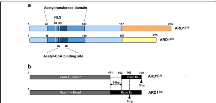
N-acetyltransferase domain is considered a core region of ARD1 because it contains an acetyl-CoA-binding site (RRLGLA) located at aa 82–87 that is critical for the acetyltransferase enzyme activity of ARD1. The N-acetyltransferase domain also contains a nuclear loca- lization signal (NLS) (KRSHRR) located at aa 78 –83, indicating a potential for dynamic translocation of ARD1 between the cytosol and the nucleus (Fig. 1a) 1,21 .
Whereas N-terminal domains are well-conserved in ARD1 variants, the variants have completely different amino acid sequences in C-terminal regions due to alternative RNA splicing. Alternative splicing at exon 8 alters the reading frame and introduces stop codons at amino acids 235 and 225, resulting in the production of two ARD1 isoforms, ARD1 235 and ARD1 225 , respectively (Fig. 1b). Although nucleotide sequences of ARD1 225 are identical to those of ARD1 235 except nucleotides 472–562 of exon 8, ARD1 235 , and ARD1 225 have different amino acid sequence in their C-terminal region, starting at amino acid 158 (aa 158).
Interestingly, mouse and human cells express different types of ARD1 variants. While human cells express two ARD1 variants (i.e., ARD1 235 and ARD 131 ), mouse cells express three (ARD1 235 , ARD1 225 , and ARD1 198 ) 1 . Thus, ARD1 225 is not expressed in humans, and mouse cells express both ARD1 225 and ARD1 235 . In contrast to mouse genomic DNA, human genomic DNA from ARD1 has no acceptor site for alternative splicing of exon 8. Thus, the ARD1 225 transcript cannot be produced from the human genome. These data suggest that the regulation of alter- native splicing of ARD1 RNA might be evolved differently between the species 2 .
Given the variation in amino acid sequence within C-
terminal domains, it is not surprising that different ARD1
variants are associated with different functional char-
acteristics. Indeed, while ARD1 225 inhibits angiogenesis
under hypoxic conditions, ARD1 235 has little relationship
with tumor angiogenesis 1,35 . Instead, ARD1 235 promotes
cell growth under normoxia, whereas ARD1 225 has no
effect on cell growth 35 . The different biological functions
of ARD1 variants might re flect their different subcellular
compartments. In mouse cells, ARD1 225 and ARD1 235
proteins are present in the cytoplasm and nucleus,
respectively 2 . In human cells, several groups have repor-
ted the subcellular localization of ARD1 235 ; however,
those results have been ambiguous and discrepant. Earlier
studies reported that the majority of human ARD1 235
expression is in the nucleus, with low levels of expression
in the cytoplasm 7,21 . By contrast, other studies suggest
that human ARD1 235 is predominantly located in the
cytoplasm rather than in the nucleus 13,36 . In light of these
discrepancies, the subcellular localization of human
ARD1 235 appears to differ based on the cell line, sug-
gesting that localization of human ARD1 235 is dependent
on the cellular context 28 . Recently, Park et al. provided one possible explanation for these complicated and often conflicting results 37 . They found that human ARD1 235 is imported to the nucleus of growing cells, especially during the S phase of the cell cycle. Furthermore, nuclear translocation of ARD1 235 is mediated by NLS, and this translocation is essential for proper cell cycle progression.
Based on these results combined, ARD1 appears to have variable properties depending on subcellular localization, speci fic isoform, and physiological conditions. Therefore, it will be critical to consider the role of these factors in modulating ARD1 activity when evaluating the potential for cancer treatment that targets ARD1.
ARD1 as an acetyltransferase N-terminal acetyltransferase activity
ARD1 was originally identified as a NAT that catalyzes N-terminal acetylation of proteins 18 . NATs transfer acetyl groups from acetyl-CoA to the first amino acid of a given protein. N-terminal acetylation is one of the major protein modifications in eukaryotes, occurring in 80–90% of proteins in humans and in 50–70% of proteins in yeast 19,38 . When proteins are synthesized and extruded from the ribosome, NATs associated with the ribosome acetylate the N-terminal α-amino group of the nascent polypeptide 39 . Thus, protein N-terminal acetylation occurs co-translationally and irreversibly.
To date, six human NAT complexes have been identi- fied: NatA, NatB, NatC, NatD, NatE, and NatF. The naming of each complex reflects the specific amino acid sequences contained in the N-terminus. ARD1 is a cata- lytic subunit of the NatA complex that acetylates Ser, Ala, Gly, Thr, and Val within the N-terminus of a protein after an initiator methionine is cleaved by aminopeptidase 27 . N-terminal acetylation is thought to play a role in protein stability by inhibiting protein degradation via blockade of N-terminal ubiquitination 40 . Indeed, ARD1 acetylates the first methionine of the tuberous sclerosis 2 (TSC2) pro- tein, which, in turn, stabilizes the TSC2 protein 28 . How- ever, other studies have reported that N-terminal acetylation does not block protein degradation. In fact, N- terminal acetylation of a protein may even create speci fic degradation signals 41 . Beyond protein stability, several studies report other potential roles of protein N-terminal acetylation, including protein–protein interactions and targeting subcellular localization 42,43 . Taken together, the existing data imply that the role of N-terminal acetylation may differ based on the substrate protein.
Recently, N-terminal acetylation activity by ARD1 235 was found to relate strongly to genetic disease in humans.
In particular, a Ser37Pro mutation of ARD1 235 was recently discovered in Ogden syndrome, resulting in the lethality of male infants 44 . Subsequent in vitro experi- ments have revealed that the Ser37Pro mutation
Fig. 1 The structure of ARD1. a Structures of ARD1 isoforms. The ARD1
235and ARD1
225isoforms share a highly conserved N-acetyltransferase domain located between amino acids 45 and 130 (aa 45 –130). The N-acetyltransferase domain contains an acetyl-CoA-binding site (RRLGLA) located at aa 82 –87 and a nuclear localization signal (NLS) (KRSHRR) located at aa 78–83. ARD1
235and ARD1
225have completely different amino acid sequences in their C-terminal region, starting at aa 158. b Alternative splicing of ARD1 mRNA. ARD1 variants are derived from alternative RNA splicing.
Alternative splicing at exon 8 alters the reading frame and introduces stop codons at amino acids 235 and 225, resulting in the production of two
ARD1 isoforms, ARD1
235and ARD1
225, respectively
significantly reduces the N-terminal acetylation activity of ARD1 235 , suggesting a potential link between the NAT function of ARD1 235 and human disease 44–46 .
Lysine acetyltransferase activity
While co-translational N-terminal acetylation is one of the most common protein modi fications, a less common but potentially more important form of protein acetyla- tion occurs post-translationally on lysine residues via KATs. The addition of an acetyl group to lysine prevents the formation of positive charges on the amino group.
This modi fication therefore has a significant impact on the electrostatic properties of the protein. Lysine acetylation is crucial for regulating various protein func- tions, including chromatin-based transcriptional control, protein–protein interactions, and protein stability 47 .
Although ARD1 was originally identified as a NAT, its subcellular localization in non-ribosomal forms and in the nucleus strongly suggests that ARD1 has other functions.
Indeed, mammalian ARD1 has both NAT and KAT activity. Mouse ARD1 225 was first proposed to possess KAT activity because it acetylated the lysine 532 residue of HIF-1 α, resulting in the degradation of HIF-1α via the ubiquitin –proteasome system 48 . Thereafter, human ARD1 235 was also shown to have KAT activity. Human ARD1 235 acetylates β-catenin and is involved in the pro- liferation of lung cancer cells, although the target site of lysine acetylation has not yet been identi fied 49,50 . ARD1 post-translationally mediates the acetylation of a micro- tubule fraction purified from porcine brain in vitro 8 . However, the specific substrate from the microtubule fraction containing α/β-tubulin has not been identified.
Furthermore, ARD1 235 acetylates myosin light chain kinase (MLCK), deactivating that enzyme 51 . In prostate cancer cells, ARD1 acetylates the androgen receptor (AR) and this acetylation is required for AR-mediated gene transcription 30 . Recent studies have uncovered substrate proteins of ARD1 and their speci fic target sites of lysine acetylation. In particular, during bone formation, ARD1 acetylates the lysine-225 residue of Runt-related tran- scription factor 2 (Runx2). Runx2 acetylation, in turn, leads to the inhibition of Runx2-mediated gene tran- scription, which results in the regulation of osteoblast differentiation 52 . Recently, Heat Shock Protein 70 (Hsp70) was identified as a new substrate of ARD1. Under stress conditions, the lysine 77 residue of Hsp70 is acetylated by ARD1, and this acetylation is essential for the enzyme activity of Hsp70 and control stress response of cells 9 . Furthermore, under glutamine deprivation, ARD1 acet- ylates the lysine-388 residue of phosphoglycerate kinase 1 (PGK1), a metabolic enzyme. PGK1 acetylation via ARD1 enhances kinase functions of PGK1 and contributes to the maintenance of cellular homeostasis in a nutrient-starved state 53 . Based on evidence regarding the KAT activity of
ARD1, it is notable that ARD1 is the first identified pro- tein with both NAT and KAT activity. However, whether KAT and NAT activity of ARD1 are regulated in similar or different ways remains unclear.
Biological function of ARD1 ARD1 in non-cancerous conditions
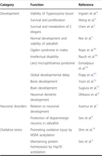
ARD1 is involved in various cellular functions (Table 1).
In particular, emerging research has shed new light on the importance of ARD1 during developmental stage.
Depletion or mutation of ARD1 leads to lethality in Drosophila, Danio rerio, Caenorhabditis elegans, and Trypanosoma brucei, highlighting an essential role for ARD1 in typical development and viability 3 – 6 . Dysfunc- tion of ARD1 is also fatal in humans, given that ARD1 mutations that cause a reduction in NAT activity can result in infant mortality, intellectual disability, develop- ment delay, and Lenz microphthalmia syndrome 44,54–56 . In mouse models, mice with depleted levels of the ARD1 225 and ARD1 235 variants display severe defects in bone development 52 . ARD1 235 is also implicated in brain
Table 1 The biological functions of ARD1
Category Function Reference
Development Viability of Trypanosoma brucei Ingram et al.
4Survival and proliferation Wang et al.
6Survival and metabolism of C
elegans
Chen et al.
3Normal development and viability of zebra fish
Ree et al.
5Ogden syndrome in males Rope et al.
44Intellectual disability Rauch et al.
56Lenz microphthalmia syndrome Esmailpour
et al.
54Global developmental delay Popp et al.
55Bone development Yoon et al.
52Brain development Sugiura et al.
57Neuronal dendritic
development
Ohkawa et al.
8Neuronal disorders Relation to neuronal development
Asamui et al.
7Protection of dopaminergic neurons in zebra fish
Seo et al.
9Oxidative stress Promoting oxidative injury by MSRA acetylation
Shin et al.
10Maintaining protein homeostasis by Hsp70 acetylation
Seo et al.
9development. Mouse models suggest that ARD1 235 is expressed in regions of cell division and migration throughout the course of brain development, and expressions of these proteins is downregulated as neurons mature and form appropriate synaptic connections 57 . Similarly, downregulation of ARD1 235 is observed during differentiation of neurons, suggesting that ARD1 235 plays an important role in development of dendrites in the brain 8 .
In addition to development, ARD1 is implicated in various neurological diseases. In particular, through direct interaction with amyloid- β protein (Aβ), ARD1 235 sup- presses the secretion of Aβ, which is the first step in the development of Alzheimer’s disease 7 . Another study using a zebrafish model of Parkinson’s disease showed that ectopic expression of dominant-negative human ARD1 235 exacerbated dopaminergic neuronal loss and locomotor deficits compared with wild-type human ARD1 235 expression 9 . These results suggest that ARD1 235 may be protective against the development of neurodegenerative disease.
Recent studies have revealed that ARD1 is also involved in cellular response to oxidative stress. ARD1 235 represses the enzymatic activity of methionine sulfoxide reductase A, an enzyme that protects cells from oxidative stress by removing reactive oxygen species. In support of a pro- oxidant role of ARD1 235 , an ARD1 235 transgenic mouse is more susceptible to oxidative stress and shows more severe tissue injury compared with wild-type mouse 10 . However, results of a recent study suggest the opposite effect-that ARD1 235 may be protective against oxidative stress. In this study, ARD1 235 prevented protein aggre- gation under oxidative stress by regulating chaperone function of Hsp70 required for the maintenance of pro- tein homeostasis. The authors therefore concluded that ARD1 235 protects cells against oxidative stress and func- tions as an anti-oxidant protein 9 . Although it is presently unclear whether ARD1 has a protective or apoptotic role under oxidative stress, existing data suggest that ARD1 is a potential target for ameliorating oxidative stress in diverse human health conditions, such as cancer, neuro- logical disease, in flammation, and metabolic disease.
ARD1 in cancerous conditions
Role of ARD1 as a tumor suppressor Mammalian ARD1 was first reported to act as a tumor suppressor, given that mouse ARD1 225 inhibits HIF-1α stabilization and angiogenesis by acetylating the lysine-532 residue 48 . In contrast to ARD1 225 , ARD1 235 has no effect on HIF-1α acetylation or stability. However, a crucial role of ARD1 225 in HIF-1 α stability was confirmed using a mouse tumor model 11 . In that study, the authors generated Apc Min/ + /ARD1 225 transgenic mice and demonstrated the anti- tumor effect of ARD1 225 in mice. In particular, they found
a significant decrease in the quantity of intestinal polyps among Apc Min/+ /ARD1 225 transgenic mice compared with Apc Min/+ transgenic mice. Within the intestinal polyps of Apc Min/+ /ARD1 225 transgenic mice, they found that VEGF expression was also reduced, which, in turn, suppressed tumor angiogenesis and growth.
In addition to ARD1 225 , human ARD1 235 is also reported to have tumor suppressor properties. In breast cancer, higher ARD1 235 expression levels are correlated with better clinical outcome from the patients patients, including higher survival, smaller tumor volume, and lower rates of lymph node metastasis 34,58 . In lung and gastric cancers, ARD1 235 expression is significantly down- regulated in malignant tumors with lymph node metas- tasis compared with primary tumors without metastasis, suggesting that downregulation of ARD1 plays a causal role in tumor development 29 .
Investigations into the underlying molecular mechan- isms have led to new insights into mechanisms through which ARD1 reduces proliferation and metastasis. In breast cancer cells, inhibition of ARD1 235 induces autophagy by inhibiting mTOR signaling and subse- quently reduces cancer cell growth 58 . Yi et al. reported that ARD1 235 is required for caspase activation during apoptosis and that depletion of ARD1 235 may thus provide robust protection against DNA damage-induced cell death 25,59 . These data suggest a pro-apoptotic role of ARD1 235 . Regarding cell motility, ARD1 235 directly acetylates the lysine 608 residue of MLCK, leading to the inhibition of MLCK activity required for cell contraction and migration 51 . In lung cancer cells, ARD1 235 binds to p21-activated kinase-interacting exchange factor to block its downstream Rac1/Cdc42 pathway. Blockade of the Rac1/Cdc42 pathway results in the inhibition of cancer cell metastasis, although this effect appears to be independent of the acetyltransferase activity of ARD1 235 29 . In breast cancer cells, ARD1 235 inhibits cancer cell migration by targeting signal transdu- cer and activator of transcription 5a (STAT5a) indepen- dently of acetyltransferase activity-independent manner 34 . Taken together, although ARD1 is an acetyltransferase enzyme, these results suggest that ARD1 235 can exert oncogenic functions, independent of its catalytic activity.
Role of ARD1 as an oncogene Although several studies
have demonstrated that ARD1 is an important molecule
involved in cancer progression, it is still under debate
whether human ARD1 235 serves as an oncogene or a
tumor suppressor. In fact, a growing number of studies
have highlighted potential oncogenic properties of ARD1
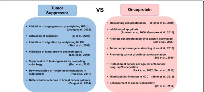
in cancer progression (Fig. 2). At present, it is widely
accepted that ARD1 235 relates positively to cellular
growth. Fisher et al. first suggested that ARD1 235 is
required to maintain division and proliferation of liver
cancer cells under hypoxia 60 . Furthermore, a fluorescence-activated cell sorting analysis revealed that ARD1 235 promotes cell proliferation by regulating the G1/
S and G2/M checkpoints of the cell cycle in breast and lung cancer cells, indicating that ARD1 235 is relevant for cell proliferation 14,32,50 . Regarding the molecular mechan- isms, ARD1 235 promotes tumor growth by enhancing cyclin D1 expression, and these effects are mediated by the activation of β-catenin and activator protein 1 32,50 . Recently, SAM domain and HD domain-containing protein 1—the latter of which is involved in the regulation of intracellular dNTP pools required for DNA replication
—were found to be involved in ARD1 235 -mediated cancer cell growth 61 .
Indeed, a number of links have been found between increased expression of ARD1 235 and human cancers, such as breast, prostate, lung, liver, cervical, bladder, and colorectal cancers 12 – 14,62 . In addition, high levels of ARD1 235 expression correspond with poorer outcomes among cancer patients, such as low survival rate and more aggressive tumors 13,62 . These point to the potential role of ARD1 235 as an oncoprotein.
In addition, ARD1 235 seems to promote the invasive properties of cancer cells. In lung cancer cells, ARD1 downregulates E-cadherin by regulation of DNA methyl- transferase 1 62 . In breast cancer cells, ARD1 235 enhances cell motility by activation of Aurora A kinase function 33 . Based on these results, ARD1 235 expression is closely related to MVI in HCC patients 15 .
Other studies demonstrate that ARD1 235 has a protec- tive effect against cell death. Depletion of ARD1 235 induces the transcription of p53-dependent pro-
apoptotic genes and activation of the caspase- dependent apoptosis pathway 22,63 . Depletion of ARD1 235 also revokes doxorubicin —but not tumor necrosis factor α (TNFα)-induced activation of the nuclear transcription factor-κB (NF-κB) 64 . Neuroblas- toma cells expressing dominant-negative ARD1 235 are more sensitive to etoposide compared with wild-type ARD1-expressing cells 9 . These results suggest that ARD1 235 is involved in drug-resistant mechanisms of cancer cells. Thus, ARD1 235 could be a potential marker for the prediction of chemoresistance in cancer patients.
In-depth studies are needed to address the association between ARD1 235 expression and drug sensitivity or resistance of cancer patients and to elucidate the detailed molecular mechanism by which ARD1 235 protects cancer cells against anti-tumor drugs.
General consideration of ARD1 in clinical respects HCC is the malignancy with the poorest prognosis among the most common types of cancers 65 . In line with this poor prognosis, the majority of HCC patients are hardly candidates for the best curative option, curative surgical resection, because of frequent vascular invasion and/or intrahepatic or extrahepatic metastases at the time of diagnosis 66 . Poorly reserved underlying liver function and frequent postoperative recurrence of HCC represent additional hindrances to successful disease manage- ment 67 . To expand the understanding of potential clinical applications of ARD1, now we are going to enter into the clinical implications of ARD1, which will be focused in a field of HCC.
Fig. 2 Controversial roles of ARD1 in cancer development and progression. Many studies have reported an important role of ARD1 in cancer
progression. However, it is still debated whether human ARD1 serves as an oncogene or a tumor suppressor
Basic clinical concepts of ARD1 in hepatocarcinogenesis HCC is typically characterized by hypervascularity.
Hypervascularity may be due to the fact that HCC typically develops in hypoxic environments caused by underlying chronic liver diseases, such as liver cirrhosis. In most HCCs, underlying chronic liver diseases necessarily pro- voke an alteration (i.e., decrement) in intrahepatic blood flow, which results in poor oxygen supply (i.e., hypoxia) to the liver. This hypoxia accelerates the production of angiogenic factors, including VEGF, basic fibroblast growth factor, and insulin-like growth factor-II (IGF-II). Over- expression of angiogenic factors subsequently promotes neovascularization, which further contributes to the development of HCC and leads to increased vascular density in tumors (i.e., hypervascular tumors). At the same time, overexpression or hyperactivation of angiogenic fac- tors can enhance the growth of HCC and also promote intrahepatic or extrahepatic metastases 68 .
In previous studies, we sought to identify markers for the early detection and prediction of post-treatment prognosis among HCC patients using serological or his- tological features. Although we demonstrated their clin- ical signi ficance and found their usefulness in clinical practice, the many ways to the destination mean that there is no single, perfectly effective way to cover all of them. Thus, it is essentially impossible to understand all forms of hepatocarcinogenesis and to cure HCC com- pletely by conquering a sole pathway because it is too complex 68,69 . Indeed, we found that several factors are involved in hepatocarcinogenesis, including IGF-II, TNF- α, transforming growth factor beta 1 (TGF-β), osteo- pontin, metastatic tumor antigen (MTA) 1, and MTA2.
These markers can be used not only for diagnosis/prog- nosis but also as therapeutic targets for postoperative adjuvant therapies 70 – 80 .
A separate line of work suggests that hypoxia is asso- ciated with tumorigenesis. In particular, HIF-1 α has been reported to play a critical role in hepatocarcinogenesis 81 . In HCC, intratumoral HIF-1 α and VEGF expression sig- ni ficantly increase to promote new vessel formation by stimulating transcription of cytokines, which are involved in angiogenesis 82 . Relying on various conditions, HIF-1 α reacts differently according to the oxygen concentration in the tissues 83 . Under normoxia, HIF-1α typically has a short half-life of 5 min; thus, it is immediately degraded into biologically inactive products. That is, under nor- moxia, the proline residues at P402 and P564 of the ODD domain of HIF-1α undergo hydroxylation, then ubiquiti- nation by the von Hippel-Lindau (pVHL) protein, fol- lowed by degradation via a proteasome. At this step, the ODD domain of HIF-1 α plays an important role in degradation as well as the prevention of its over- expression 84 – 86 . In contrast to normoxia, hypoxia stabi- lizes HIF-1 α to accelerate the development, growth, and
subsequent metastasis of cancers via stimulating the transcription of angiogenic factors (e.g., VEGF) 87 . These results provide a theoretical basis for notion that the HIF- 1α pathway is a promising target for cancer therapies 88 .
As a first step, it may be worth briefly mentioning the association between HIF-1 α and MTA1 in hepatocarci- nogenesis. For more in-depth discussion, we refer the reader to previous papers 80,89,90 . Based on our prior stu- dies, we found that MTA1 is more highly overexpressed in hepatitis B virus (HBV)-associated HCC than in non- HBV-associated HCC 76 . This may be due to the HBx protein of HBV that can induce MTA1 expression and result in the enhancement of hypoxia signaling via HIF-1α stabilization. Consequently, stabilization of HIF-1α accelerates angiogenesis under hypoxia by reducing the binding of pVHL protein to HIF-1α and preventing the ubiquitin-dependent degradation of HIF-1α. Further- more, MTA1 itself may increase the transcription and stabilization of HIF-1α 80 . MTA1 activation by TGF-β1 is also involved in tumor progression through the promo- tion of epithelial –mesenchymal transition (EMT) 91,92 .
On the other hand, it seems that the association between HIF-1 α and ARD1 in human HCC is not yet clear. In the acetylation process, ARD1 which exists in all mammalian cells including brain, heart, bone marrow, and liver, acts as a key molecule. ARD1 was named because its deletion impairs cell mitosis and arrests cell division. In particular, ARD1 is detected in high con- centrations in tumor cells of colorectal and breast cancers in some patients 1,21 . Furthermore, concentrations of ARD1 are high in organs that are highly vascular (e.g., heart, liver), and thus, ARD1 may play an important role in determining the destiny of cells during cell cycles. It, therefore, is thought of ARD1 that it should be deeply involved in the progression of malignancies as the cell division increases.
The existing data, in vitro, suggest that mouse ARD1 plays a pivotal role as a negative controller of HIF-1 α stability under normoxia 48,93 . It is assumed that mouse ARD1 acetylates HIF-1 α protein by direct reaction with Lys 532 of the ODD domain. This deepens the instability of HIF-1 α and then suppresses the expression of angiogenic factors such as VEGF, which lead to be able to prevent development and/or progression of malignancies by inhibiting neovascularization. On the other hand, recent reports suggest that human ARD1 may have no impact on the stability of HIF-1α 36,94 . In addition, there is a view that the ARD1 is not regulated by hypoxia in several human tumor cell lines including cervical adenocarcinoma, fibrosarcoma, and breast cancer cells.
Notably, as mentioned before, HCC is a typical hyper-
vascular tumor and also it has been developed generally in
hypoxic condition such as liver cirrhosis. Additionally, the
HIF-1 α signaling pathway is the representative crucial
mechanism in hepatocarcinogenesis and angiogenesis.
Although, to date, the biologic functions of ARD1 have not been clearly defined in malignancies, especially HCC, and the direct association of ARD1 with HIF-1α in HCC of human is not also clarified, we still wonder whether ARD1 will have a direct or indirect relationship with HIF- 1 α in human hepatocarcinogenesis.
On the basis of the facts described above, it is possible that ARD1 is also implicated in the development of HCC and its progression, including tumor growth and metas- tasis. Therefore, it is very valuable to investigate ARD1 expression and to ascertain its potential association with clinical characteristics for clarifying hepatocarcinogenesis and setting up the preventive strategies in the future. At the same time, it will be useful to amend current treat- ment modalities and develop new effective therapies.
ARD1 expression and its clinical signi ficance in non-HCC neoplasia
Dose ARD1 act as an oncoprotein or a tumor sup- pressor in human cancers? This point remains to be
clarified, but a number of in vitro studies on the molecular mechanisms of carcinogenesis have been published in favor of the oncoprotein role 27,80,89,90
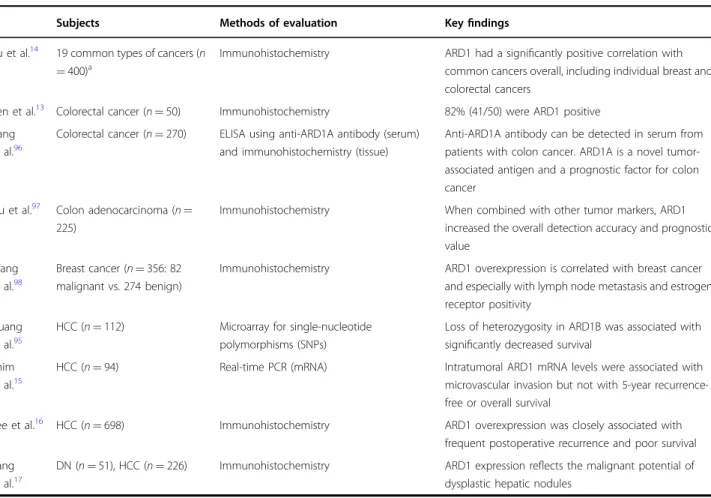
. Research on ARD1 in clinical settings, however, remains in its very early stages. In fact, there are very few reports on ARD1 using human neoplastic tissues. An even smaller number of studies examined the potential association between ARD1 expression and recurrence or survival in patients with cancers 13 – 17,95 – 98 . These studies are summarized in Table 2.
Yu et al. investigated human ARD1 expression in cancer tissues and compared this expression to that in non- neoplastic tissues 14 . For this study, the investigators enrolled a relatively large number of total subjects (400 with common cancers vs. 133 with matching non- cancerous tissues). Across the sample, the rates of ARD1-positive tissue were 52.3% (209/400) of cancer tissues and 31.5% (42 of 133) of non-cancerous tissues.
Although the overall difference in ARD1-positive rates between cancer and non-cancerous tissues reached sta- tistical signi ficance (p < 0.005), ARD1 was still detected in
Table 2 Clinical studies of ARD1 overexpression in malignancies including hepatocellular carcinoma in humans
Subjects Methods of evaluation Key findings
Yu et al.
1419 common types of cancers (n
= 400)
aImmunohistochemistry ARD1 had a signi ficantly positive correlation with common cancers overall, including individual breast and colorectal cancers
Ren et al.
13Colorectal cancer (n = 50) Immunohistochemistry 82% (41/50) were ARD1 positive Jiang
et al.
96Colorectal cancer (n = 270) ELISA using anti-ARD1A antibody (serum) and immunohistochemistry (tissue)
Anti-ARD1A antibody can be detected in serum from patients with colon cancer. ARD1A is a novel tumor- associated antigen and a prognostic factor for colon cancer
Liu et al.
97Colon adenocarcinoma (n = 225)
Immunohistochemistry When combined with other tumor markers, ARD1
increased the overall detection accuracy and prognostic value
Wang et al.
98Breast cancer (n = 356: 82 malignant vs. 274 benign)
Immunohistochemistry ARD1 overexpression is correlated with breast cancer and especially with lymph node metastasis and estrogen receptor positivity
Huang et al.
95HCC (n = 112) Microarray for single-nucleotide
polymorphisms (SNPs)
Loss of heterozygosity in ARD1B was associated with signi ficantly decreased survival
Shim et al.
15HCC (n = 94) Real-time PCR (mRNA) Intratumoral ARD1 mRNA levels were associated with
microvascular invasion but not with 5-year recurrence- free or overall survival
Lee et al.
16HCC (n = 698) Immunohistochemistry ARD1 overexpression was closely associated with
frequent postoperative recurrence and poor survival Kang
et al.
17DN (n = 51), HCC (n = 226) Immunohistochemistry ARD1 expression re flects the malignant potential of dysplastic hepatic nodules
a
Total sample (N = 400; p < 0.005); urinary bladder, n = 21; breast, n = 48 (p < 0.05); cervix, n = 14; hypothyroid, n = 12; stomach, n = 56; lung, n = 29; colorectum, n =
125 (p < 0.01); prostate, n = 12; nasopharynx, n = 11 (p < 0.1); liver, n = 9; esophagus, n = 19; skin, n = 14; kidney, n = 10 (p < 0.1); gallbladder, n = 4; eye, n = 3; vocal
cords, n = 5 (p < 0.1); pancreas, n = 2; ovary, n = 3; brain, n = 3
a substantial number of non-cancerous tissues (i.e., 31.5%), and this rate was not significantly different from specific cancer types (refers to footnote of Table 2). This is because only small number of cancer cases of each type were merged into the same overall group and then ana- lyzed as a whole. Across the cancer types, there was a signi ficant association between ARD1 overexpression and colorectal (n = 125, 48.0% vs. 18.2%; p < 0.01) and breast cancers (n = 48, 62.5% vs. 38.1%; p < 0.05). At any rate, the study con firmed that ARD1 expression is widespread in various cancers and, at the very least, showed its close association with colorectal and breast cancers. A link between ARD1 expression and colorectal cancers is con- sistent with a previous study by Ren et al 13 . In that study, the authors used immunohistochemistry and compared ARD1 expression in colorectal cancer and colitis tissues.
They found that ARD1 was positive in 41 out of 50 (82%) tested colorectal cancer tissues, but weakly positive in only 12 of 50 (24%) matched normal tissues (p < 0.001) and negative in all of 20 colitis tissues (p < 0.001).
Importantly, they showed the possibility of ARD1 as a potential biomarker in patients with colorectal cancer or even other malignancies. In fact, in the next study by the same investigators, they acquired the result with ARD1 overexpression in 84.1% (227 out of 270) of tested colon cancer tissues and, strikingly, successfully isolated the autoantibody against ARD1 in serum obtained from patients 96 . They suggested for the first time that ARD1 is a novel tumor antigen in colon cancers and also has a potential to predict prognosis in the patients with colon cancer. In a subsequent study, the investigators used four autoantibodies (synuclein-gamma (SNCG), a human homolog of piwi (Hiwi), phosphatase of regenerating liver-3 (PRL-3), and ARD1) from 225 colon cancer patients, which were all isolated from serum, to evaluate whether the combination of them improve the values as tumor markers in terms of early detection and prognostic prediction of cancer 97 . Results showed that the combi- nation of autoantibodies was associated with an elevated positive tumor marker rate (76.9% or 173/225) and an increased detection accuracy of cancer, with 61.9% (252/
407) for SNCG and 82.6% (336/407) for the combination, with a hazard ratio 3.2 (p < 0.001) for poor prognosis. It is somewhat conclusive that ARD1 is clearly overexpressed and can be considered a tumor-specific antigen in colon cancer.
In terms of other malignancies, fewer studies have been reported, for which reason it is difficult to conclude whether ARD1 has a potential clinical role. Consistent in part with the present conditions, ARD1 is overexpressed in malignant breast neoplasia compared with benign conditions, e.g., fibroadenomas, hyperplasia, inflammatory breast diseases, and cysts. According to a study by Yu et al., the ARD1-positive rate was signi ficantly higher in
breast cancer tissues than in non-cancerous ones (62.5%
(30/48) vs. 38.1% (8/21); p < 0.05) 14 . Their findings are comparable to the ARD1-positive rate reported by Wang et al. in invasive ductal carcinomas (61.0% or 50/82) compared with benign conditions listed above (48.8% or 119/244) 98 . Although they demonstrated the implications of ARD1 in malignant breast neoplasia, there were some limitations. Above all, the staining intensity was not gra- ded, and the tissues of substantial proportion of benign diseases also stained positive for ARD1, which did not make ARD1 a suitable indicator for differentiating breast cancers from non-neoplastic diseases, i.e., a tumor- specific marker. Further, there was a report in contrast to it that ARD1 is a tumor suppressor in breast cancer by reducing cell proliferation and autophagy via inhibiting mTOR signaling pathway 58 . In thyroid cancer, as well, it remains inconclusive whether ARD1 is an oncogenic protein or a tumor suppressor 21,99 .
ARD1 expression and its clinical signi ficance in HCC So far, to our knowledge, there has been only one clinical study showing ARD1 overexpression by immu- nohistochemistry in human HCC samples 14 . However, the subjects with HCC were composed of only a very small portion of the total study population (<3%, 9/400);
therefore, positive rates of ARD1 detection could not be statistically compared between the HCC and non-HCC groups [33.3% (3/9) vs. 0% (0/1), p = not applicable]. Also, the analyses for clinical outcomes could not be done in HCC because there were no clinical data about the patients such as HCC stages or the patients’ survival.
Although another microarray study showed the associa- tion of a specific single-nucleotide polymorphism (loss of heterozygosity in ARD1B) with clinicopathological char- acteristics and survival in HCC patients, it could not directly demonstrate the signi ficance of ARD1 at the protein levels for the development, progression, and/or prognosis of HCC.
Recently, in light of the potential of ARD1 as a predictor of clinical outcomes, we published an important paper using 94 HCC patients 15 . In the study, we hypothesized that ARD1 is overexpressed in HCC but not in non-HCC and thus may predict patients ’ clinical outcomes in terms of recurrence and survival. The theoretical basis has been sufficiently described above: the main feature of HCC is hypoxia in the central lesion, resulting in hypervascularity, which is mediated by HIF1-α pathway. In the study, we quantified ARD1 mRNA levels in HCC and matching non-HCC tissues using real-time PCR. Based on these results, patients were regrouped into high (n = 38) and low levels (n = 56) of ARD1 expression. We found that the high-expression group had more frequent MVI than the low-expression group (32% vs. 14%; p < 0.05).
Regrettably, however, we could not see the findings that
we expected in 5-year recurrence-free (34% vs. 46%, p = 0.98) and overall survival rates (76% vs. 73%, p = 0.52).
Importantly, intratumoral ARD1 mRNA levels were sig- nificantly high in HCCs with MVI, suggesting novel potential links between ARD1 overexpression and the clinical courses of HCC patients.
Very importantly, in April 2017, we presented novel findings from two large-scale clinical studies on the sig- ni ficance of ARD1 in HCC at the 52nd Annual Meeting of the European Association for the Study of the Liver in Amsterdam, the Netherlands. In the first study, results clearly demonstrated ARD1 overexpression in HCC, and a good correlationship between ARD1 expression and postoperative recurrence and patient survival. We enrol- led 698 histologically confirmed HCC patients (162 with liver transplantation and 536 with curative surgical resection). By grading the intensity of ARD1 immuno- histochemical staining, we investigated the association of ARD1 with the histological and radiological characters of the tumors, as well as patient clinical outcomes. The results demonstrated that ARD1 was stainable in 87%
(605/698) of tumors, and, as expected, ARD1 over- expression was positively associated with MVI (p < 0.001) and poor differentiation of the tumors (p < 0.001). Inter- estingly, the 5-year cumulative recurrence rates of HCC increased as the intensity of ARD1 expression in HCC tissues increased (p < 0.05). Further, the survival of the patients also decreased with higher ARD1 expression (p <
0.01) 16 . In another presentation, we showed that ARD1 expression may reflect the malignant potential of dys- plastic nodules, a considerable portion of which subse- quently leads to HCC 17 . In brief, 63% (32/51) were stained positive for ARD1 in dysplastic nodules, whereas 93%
(210/226) of HCC were stained positive ARD1 and all normal livers tested were negative ARD1 (0/4). Impor- tantly, in the subgroups of patients histologically de fined as having low-grade or high-grade dysplasia, both ARD1- positive rate and its staining intensity were signi ficantly higher in high-grade dysplastic nodules than in low-grade ones. Altogether, we found that higher intensity of ARD1 staining via immunohistochemistry is associated with higher malignant potential, even in the dysplastic stages. Taken together, the evidence suggests that ARD1 in the liver may play an important role both in the early stages of HCC development and in its progression to the advanced stages.
Clinical prospective of ARD1 in HCC
It is established that ARD1 widely exists in mammalian cells and acts as an acetyltransferase by catalyzing protein acetylation, which serves to regulate cell division, pro- liferation, and even tumorigenesis 11 . All reviewed data are in accordance with clinical observations from all color- ectal cancers and selected groups with breast neoplasia as
summarized in Table 2. First, what we wish to emphasize here is that ARD1 overexpression in the liver con- tinuously changes in a stepwise pattern from low-grade dysplasia, to high-grade dysplasia to HCC, as determined histologically. Moreover, intensity of ARD1 expression is well correlated with HCC recurrence and patient long- term survival. This means that ARD1 not only takes part in early development (i.e., from dysplasia to cancer) but also plays a key role in tumor progression (i.e., poor dif- ferentiation and MVI) and even its recurrence.
A number of obstacles still lie ahead in the study of ARD1. To use the understanding of the clinical sig- nificance of ARD1 overexpression in HCC in a clinical practice, it should be applicable for early detection (as a specific tumor marker), and curative or postoperative adjuvant treatment (as a therapeutic target). These goals are promising, given recent research findings by our group and others. In addition to other molecules (e.g., MTA1), ARD1 can be considered as a potential target in hepatocarcinogenesis and conquering HCC in the future.
Conclusion
Since ARD1 was first identified in 1985, its potential roles have been discussed constantly. ARD1 has several isoforms and plays different roles in different species. In humans, we suggest that ARD1 has speci fic roles in cel- lular growth, differentiation, neuronal development, and cancer development/progression. In particular, ARD1 may play opposite roles—as a tumor suppressor or an oncoprotein—in the tumorigenesis of different cancer types. On the basis of our recent studies, however, ARD1 might be an oncogenic protein in hepatocarcinogenesis.
Furthermore, we have revealed that ARD1 is a useful diagnostic and/or prognostic index in patients with HCC. Nonetheless, the molecular signaling pathway used by ARD1 in HCC has not been completely determined. Further investigations should continue to clarify the precise molecular mechanism of ARD1 in hepatocarcinogenesis.
Acknowledgements
This work was supported by Basic Science Research Program through the National Research Foundation of Korea (NRF) funded by the Ministry of Education (NRF-2016R1A6A1A03011325, No 2016R1D1A1B03935560), and by a National Research Foundation of Korea (NRF) grant funded by the Korean Government (MSIP) (No. 2014R1A5A2010008).
Author details
1