The prognosis in cases of hepatocellular carcinoma after hepatectomy: young patients versus older patients
Ji Soo Lee, Jong Man Kim, Seunghwan Lee, Jin-Yong Choi, Wontae Cho, Gyu-Seoung Choi, Jae Berm Park, Choon Hyuck David Kwon, Sung Joo Kim, and Jae-Won Joh
Department of Surgery, Samsung Medical Center, Sungkyunkwan University School of Medicine, Seoul, Korea
Backgrounds/Aims: Hepatocellular carcinoma (HCC) is uncommon in young adults and the prognosis of these patients is still unclear. In this retrospective study, we compared the clinicopathological characteristics and outcomes of young patients with HCC with those of older patients with HCC. Methods: We retrospectively reviewed the clinicopathological characteristics of a total of 1,124 patients with HCC who underwent hepatectomy at our institution between 2006 and 2010. Patients ≤40 years of age at the time of HCC diagnosis were classified in the younger group. Results: One hundred and three patients (9.2%) were classified in the younger group. whereas, 1021 patients were classified in the older group. The incidences of hepatitis B virus infection, alpha-fetoprotein (AFP) levels, and indocyanine green retention test were all higher in younger patients than in older patients (p<0.05). Disease-free survival and overall survival were longer in older patients than in younger patients, without statistical significance. In younger patients, increased levels of protein induced by vitamin K antagonist-II (PIVKA-II) and alkaline phosphatase, portal vein tumor thrombosis, and intrahepatic metastasis were all predisposing factors for tumor recurrence after hepatectomy.
Conclusions: Although the AFP levels were higher in younger patients with HCC than in older patients with HCC, disease-free survival and overall survival after liver resection were not significantly different between the two groups.
(Korean J Hepatobiliary Pancreat Surg 2015;19:154-160)
Key Words: Hepatocellular carcinoma; Hepatectomy; Young age; Prognosis; Survival
Received: August 27, 2015; Revised: September 25, 2015; Accepted: September 30, 2015 Corresponding author: Jong Man Kim
Department of Surgery, Samsung Medical Center, Sungkyunkwan University School of Medicine, 81 Irwon-ro, Gangnam-gu, Seoul 06351, Korea Tel: +82-2-3410-1719, Fax: +82-2-3410-0040, E-mail: [email protected]
Copyright Ⓒ 2015 by The Korean Association of Hepato-Biliary-Pancreatic Surgery
This is an Open Access article distributed under the terms of the Creative Commons Attribution Non-Commercial License (http://creativecommons.org/
licenses/by-nc/4.0) which permits unrestricted non-commercial use, distribution, and reproduction in any medium, provided the original work is properly cited.
Korean Journal of Hepato-Biliary-Pancreatic Surgery ∙ pISSN: 1738-6349ㆍeISSN: 2288-9213
INTRODUCTION
Hepatocellular carcinoma (HCC) is a common malig- nancy worldwide. Although HCC is generally diagnosed in middle-aged and elderly individuals, the age of peak incidence differs substantially according to etiology and geographical region. In low-risk areas of western coun- tries, HCC is rarely diagnosed in patients younger than 40 years of age. However, in high-risk areas such as East Asia, HCC is often diagnosed in individuals aged between 20 to 40 years.1 Therefore, HCC is a serious threat to pub- lic health in East Asia.
Patient age at the time of diagnosis has important prog- nostic value for certain cancers. While the prognosis for thyroid cancer is favorable if the patient is diagnosed at a young age,2 this is an unfavorable prognostic factor for breast cancer.3 The impact of diagnosis at a young age
on the outcome of HCC remains controversial. This con- troversy could be due to geographic variability and differ- ences in risk profiles between populations.4-8
Several reports described the factors associated with poor prognosis for young patients with HCC. Most of these studies included patients with advanced disease at presentation and high serum alpha-fetoprotein (AFP) levels.4,6,8 However, the difference in prognosis between young and old patients with HCC is not definitively established. There are several reports of worse outcomes for younger patients with HCC, as compared with older patients with HCC;5,7 whereas, others have reported no differences in survival between these two groups.4,6,8
In this study, we compared the clinicopathological char- acteristics and outcomes of younger patients with HCC with those of older patients with HCC treated at our institution.
MATERIALS AND METHODS
Patients
The study sample included 1,124 patients who under- went surgical resection of HCC between January 2005 and December 2010 at our institution. Of these patients, 103 (9.2%) were 40 years of age or younger (defined as the younger patient group), while 1021 patients (90.8%) were over 40 years of age (defined as the older patient group).
The study was approved by the Samsung Medical Center Institutional Review Board and performed according to the guidelines of the Helsinki Declaration. The exclusion cri- teria were as follows: mixed HCC and cholangiocarcinoma on pathology; age <18 years; previous locoregional thera- pies such as hepatectomy, radiation, transarterial chemo- embolization (TACE), radiofrequency ablation (RFA), and percutaneous ethanol injection (PEI); intraoperative RFA;
fibrolamellar HCC, and loss to follow-up after hepatectomy.
Demographic, preoperative laboratory, and pathologic data collected from the patients’ electronic medical records were retrospectively reviewed.
None of the patients in either group received post- operative adjuvant therapy before recurrence. Patients with intrahepatic recurrences were treated with RFA, TACE, or radiation according to their functional liver reserve and the pattern of recurrence.
Surgery and pathology
Liver function was evaluated using the Child-Pugh classification system. Patients were required to have Child-Pugh class A liver function. Patients were not con- sidered for resection if they had a serum total bilirubin level ≥1.5 mg/dl, an indocyanine green retention test at 15 minutes (ICG-R15) ≥20%, or ascites. Patients with gross vascular invasion on imaging were only considered for resection if the main portal vein and the portal branch to the remaining liver lobe were patent. The platelet count and international normalization ratio (INR) were also re- corded, in addition to the levels of serum albumin, total bilirubin, aspartate transaminase (AST), alanine trans- aminase (ALT), alkaline phosphatase (ALP), creatinine, alpha-fetoprotein (AFP), and protein induced by vitamin K antagonist-II (PIVKA-II).
Standard operative techniques for hepatectomy are pre- viously described.9,10 Major hepatectomy was defined as
the resection of ≥3Couinaud segments and minor hep- atectomy was defined as the resection of <3 segments.
Anatomic resections involved resection of the tumor with the related portal vein branches and the corresponding hepatic territory. Both peripheral tumors and central tu- mors were treated by non-anatomic resection. Peripheral tumors and tumors with extrahepatic growths were treated by partial hepatectomy, because this method reportedly yields adequate surgical outcomes. Central tumors near the hepatic hilum and major vessels were treated by enucleation. In such cases, removing a large liver tissue only to obtain adequate margins was considered to be too difficult or risky.10
Postoperative histological parameters assessed included tumor diameter, number of tumors, encapsulation, portal vein tumor thrombosis, microvascular invasion, intra- hepatic metastasis, multicentric occurrence, serosal in- volvement, and fibrosis grade. Intrahepatic metastasis and multicentric occurrence were defined based on guidelines from the Liver Cancer Study Group of Japan.11 The Edmonson-Steiner scale was used to grade HCC as well differentiated (grade I), moderately differentiated (grade II), or poorly differentiated (grade III, IV).12 Modified UICC staging was used to stage HCC.13 The Ludwig-Batts scoring system was used to assess hepatic fibrosis (stage) on the following scale of 0-4: F0=absent, F1=portal fib- rosis, F2=periportal fibrosis, F3=bridging fibrosis; and F4=cirrhosis. Tumor recurrence and survival data were al- so recorded.
Surveillance after surgical resection
Patients were followed postoperatively every 2 to 3 months after surgery. Follow-up evaluations included phys- ical examinations, AFP measurements, PIVKA-II measure- ments, liver function tests, and chest X-rays. Abdominal computed tomography was performed every 3 months or when recurrence was suspected. If no definitive evidence of recurrence was visible by CT, magnetic resonance imag- ing and/or positron emission tomography scanning were performed. Detailed information was recorded for each pa- tient with recurrence. Patients with intrahepatic recurrences were treated with RFA, TACE, or sorafenib according to their functional liver reserve and recurrence pattern.
Follow-up time was defined as the time from surgery to the time of the last follow-up or death.9,10
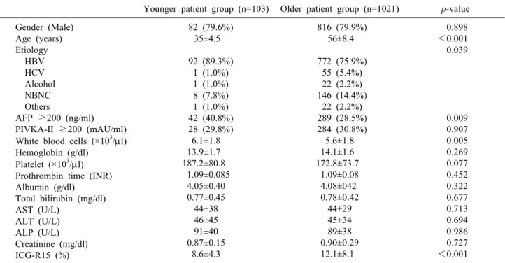
Table 1. Demographic characteristics
Younger patient group (n=103) Older patient group (n=1021) p-value Gender (Male)
Age (years) Etiology HBV HCV Alcohol NBNC Others
AFP ≥200 (ng/ml) PIVKA-II ≥200 (mAU/ml) White blood cells (×103/l) Hemoglobin (g/dl)
Platelet (×103/l) Prothrombin time (INR) Albumin (g/dl)
Total bilirubin (mg/dl) AST (U/L)
ALT (U/L) ALP (U/L) Creatinine (mg/dl) ICG-R15 (%)
82 (79.6%) 35±4.5 92 (89.3%)
1 (1.0%) 1 (1.0%) 8 (7.8%) 1 (1.0%) 42 (40.8%) 28 (29.8%)
6.1±1.8 13.9±1.7 187.2±80.8 1.09±0.085
4.05±0.40 0.77±0.45 44±38 46±45 91±40 0.87±0.15
8.6±4.3
816 (79.9%) 56±8.4 772 (75.9%)
55 (5.4%) 22 (2.2%) 146 (14.4%)
22 (2.2%) 289 (28.5%) 284 (30.8%)
5.6±1.8 14.1±1.6 172.8±73.7
1.09±0.08 4.08±042 0.78±0.42
44±29 45±34 89±38 0.90±0.29 12.1±8.1
0.898
<0.001 0.039
0.009 0.907 0.005 0.269 0.077 0.452 0.322 0.677 0.713 0.694 0.986 0.727
<0.001
*HBV, hepatitis B virus; HCV, hepatitis C virus; NBNC, non-B non-C; AFP, alpha-fetoprotein; PIVKA-II, protein induced by vitamin K antagonist-II; INR, internal normalization ratio; AST, aspartate transaminase; ALT, alanine transaminase; ALP, alkaline phosphatase; ICG-15, indocyanine green retention rate at 15 minutes
Statistical analysis
Categorical variables were expressed as percentages and compared using the 2 test or Fisher’s exact test.
Continuous variables were expressed as means and stand- ard deviations and compared using the Mann-Whitney U test. Patient survival and recurrence were calculated using the Kaplan-Meier method and compared using the log-rank test. The most suitable cut-off value for each continuous variable was determined by the receiver oper- ating characteristics (ROC) curve. Clinical and pathologic variables with prognostic significance in univariate analy- sis were entered into a Cox multivariate proportional haz- ards model to determine which factors were independently predictive of HCC recurrence. p<0.05 was considered to indicate significance. Analyses were carried out using SPSS 21.0 (SPSS, Chicago, IL, USA).
RESULTS
Preoperative patient characteristics
The demographic and preoperative characteristics of the patients were shown in Table 1. A total of 10.2% of the patients with HCC were classified in the young group.
The ratio of males to females in the younger group was not significantly different from that of the older group.
The younger group showed a higher incidence of HBV infection than the older group (89.3% vs. 79.9%); how- ever, the incidences of non-B, non-C in the younger group were lower than in the older group (7.8% vs. 14.4%).
The mean AFP level in the younger group was higher than in the older group (5885.3±19750.5 ng/ml vs.
5644.2±69285.3 ng/ml; p<0.001); however, the mean PIVKA-II levels of the 2 groups were not significantly different (232.3±359.2 mAU/ml vs. 214.2±308.5 mAU/ml;
p=0.496). Although the serum albumin and total bilirubin levels were not significantly different between the 2 groups, the younger group showed a lower mean ICG-R15 level than the older group (8.6% vs. 12.1%; p<0.001).
Perioperative and pathological characteristics The rates of anatomical resection and major resections in the younger group were not significantly different from those of the older group. The mean tumor sizes in the younger and older groups were 4.8±4.0 cm and 4.4±3.1 cm, respectively. No significant differences in tumor size, tumor number, grade, encapsulation, microvascular in-
Fig. 1. (A) Disease-free survival and (B) overall survival in patients with hepatocellular carcinoma who underwent hepatic resection.
Table 2. Perioperative and pathologic characteristics
Younger patient group (n=103) Older patient group (n=1021) p-value Anatomical resection
Major resection Tumor size ≤3 cm 3-5 cm ≥5 cm
Tumor number (Multiple)
Tumor differentiation grade 3 and 4 Encapsulation
Microvascular invasion Portal vein tumor thrombosis Serosal involvement
Intrahepatic metastasis Multicentric occurrence Fibrosis
None Portal Periportal Septal Cirrhosis
Free resection margin (mm) Hospitalization (days) Postoperative mortality
55 (53.4%) 31 (30.1%) 47 (45.6%) 26 (25.2%) 30 (29.1%) 2 (1.9%) 13 (12.9%) 92 (91.1%) 58 (56.3%) 10 (9.9%)
2 (2.0%) 18 (17.8%) 19 (18.8%) 8 (7.9%) 1 (1.0%) 4 (4.0%) 44 (43.6%) 44 (43.6%)
11±12 11.3±5.8
0 (0%)
541 (53.0%) 285 (27.9%) 436 (42.7%) 310 (30.4%) 275 (26.9%) 55 (5.4%) 81 (8.1%) 917 (91.4%) 527 (51.6%) 67 (6.7%) 11 (1.1%) 128 (12.7%) 176 (17.5%) 76 (7.7%) 13 (1.3%) 36 (3.6%) 408 (41.1%) 460 (46.3%)
10±10 11.2±7.4
4 (0.4%)
0.937 0.646 0.931
0.159 0.131 0.853 0.408 0.219 0.336 0.164 0.784 0.777
0.067 0.793 0.953 vasion, portal vein tumor thrombosis, serosal involvement,
intrahepatic metastasis, multicentric occurrence, fibrosis, or hospitalization were observed between the 2 groups.
There was no mortality after treatment among patients in the younger group (Table 2).
Disease-free and overall survival
The mean follow-up duration in the younger and older
groups were 35±18 months and 32.3±16.7 months, re- spectively (p=0.156). The 1-year, 2-year and 3-year dis- ease-free survival rates were 65.5%, 53.9%, and 44.6% in the younger group, and 72.5%, 62.1%, and 57.3% in the older group, respectively. Disease-free survival in the younger group was lower than in the older group, without statistical significance (p=0.071) (Fig. 1). The 1-year, 2-year, and 3-year overall survival rates were 93.1%,
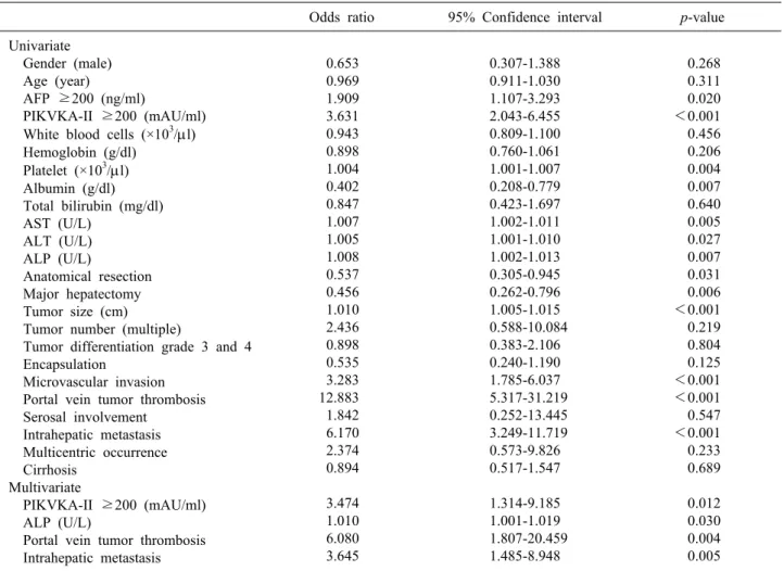
Table 3. Risk factors related to tumor recurrence in patients with large HCC after hepatic resection
Odds ratio 95% Confidence interval p-value Univariate
Gender (male) Age (year)
AFP ≥200 (ng/ml) PIKVKA-II ≥200 (mAU/ml) White blood cells (×103/l) Hemoglobin (g/dl)
Platelet (×103/l) Albumin (g/dl) Total bilirubin (mg/dl) AST (U/L)
ALT (U/L) ALP (U/L)
Anatomical resection Major hepatectomy Tumor size (cm) Tumor number (multiple)
Tumor differentiation grade 3 and 4 Encapsulation
Microvascular invasion Portal vein tumor thrombosis Serosal involvement
Intrahepatic metastasis Multicentric occurrence Cirrhosis
Multivariate
PIKVKA-II ≥200 (mAU/ml) ALP (U/L)
Portal vein tumor thrombosis Intrahepatic metastasis
0.653 0.969 1.909 3.631 0.943 0.898 1.004 0.402 0.847 1.007 1.005 1.008 0.537 0.456 1.010 2.436 0.898 0.535 3.283 12.883 1.842 6.170 2.374 0.894 3.474 1.010 6.080 3.645
0.307-1.388 0.911-1.030 1.107-3.293 2.043-6.455 0.809-1.100 0.760-1.061 1.001-1.007 0.208-0.779 0.423-1.697 1.002-1.011 1.001-1.010 1.002-1.013 0.305-0.945 0.262-0.796 1.005-1.015 0.588-10.084
0.383-2.106 0.240-1.190 1.785-6.037 5.317-31.219 0.252-13.445 3.249-11.719 0.573-9.826 0.517-1.547 1.314-9.185 1.001-1.019 1.807-20.459
1.485-8.948
0.268 0.311 0.020
<0.001 0.456 0.206 0.004 0.007 0.640 0.005 0.027 0.007 0.031 0.006
<0.001 0.219 0.804 0.125
<0.001
<0.001 0.547
<0.001 0.233 0.689 0.012 0.030 0.004 0.005
*AFP, alpha-fetoprotein; PIVKA-II, protein induced by vitamin K antagonist-II; AST, aspartate transaminase; ALT, alanine trans- aminase; ALP, alkaline phosphatase; ICG-15, indocyanine green retention rate at 15 minutes
82.5%, and 79.7% in the younger group, and 93.6%, 87.8%, and 83.1% in the older group, respectively (p=0.267).
Tumor recurrence
Tumor recurrence was observed in 52 (50.5%) younger patients and 402 (39.7%) older patients. Intrahepatic re- currence occurred in 31 (59.6%) younger patients and 239 (59.5%) older patients. The differences in systemic re- currence (n=5, 9.6% vs. n=49, 12.2%) and concurrent in- trahepatic and systemic recurrence (n=16, 30.8% vs.
n=114, 28.4%) between the 2 groups were not significant.
The risk factors for HCC recurrence after hepatectomy in the younger group were summarized in Table 3.
Multivariate analysis revealed that increased PIVKA-II levels, increased ALP levels, portal vein tumor thrombo- sis, and intrahepatic metastasis were independent predis- posing factors for tumor recurrence.
DISCUSSION
The prognosis of young adults with HCC is controversial.
In the present study, younger patients with HCC who un- derwent hepatectomy were more likely than older patients to be HBV-positive, to have high AFP levels at diagnosis, and better liver function status. However, tumor size, tumor number, serum PIVKA-II levels at presentation and extent of liver resection were not significantly different in younger patients, as compared to older patients. Therefore, the sur- vival outcomes of young adults with HCC are not measur- ably different from those of older patients after liver resection.
The outcome of young patients with HCC is highly debated. Among the tumor factors, AFP levels are re- portedly higher in younger patients than older patients.
Specifically, more than 50% of all patients with HCC are
shown to have an AFP level equal to or exceeding 400 ng/ml.5,6,14 In addition, younger patients tend to have larger tumors than older patients, with the maximum tumor di- ameter in younger patients exceeding 6 cm.6,8,14,15 Thus, younger patients with HCC should be offered aggressive hepatectomy to preserve liver function. In the present study, the younger group had higher AFP levels and showed better liver function than the older group; these findings are consistent with previous reports.6,14,15 However, tumor size did not differ significantly between the 2 groups. In addition, the presence of initial metastases was similar between the 2 groups. Specifically, younger patients with HCC had worse disease-free and overall sur- vival rates, presumably because they presented with higher AFP levels. However, the survival rates of younger and older patients were not significantly different. Therefore, the natural histories of younger patients with HCC may be similar to those of older patients with HCC after hepatectomy. Thus, early detection may be the best way to improve the prognosis of young patients.
Previous studies have reported that young patients with HCC (aged <40 years) tend to have poor prognoses, as compared to older patients. This is most likely because young patients are often diagnosed at more advanced stages of disease, as indicated by their high serum AFP levels and advanced TNM stages.4,6,8 The results of our study are consistent with previous reports on the im- portance of higher serum AFP levels; however, we did not find any significant differences in disease-free or overall survival between younger and older patients with HCC.
In addition, high serum AFP levels were not associated with tumor recurrence in younger patients.
We defined younger patients with HCC as patients less than 40 years of age at the time of HCC diagnosis. In the present study, 10.2% of all patients with HCC were classified as younger patients. The rates of HCC in this age category (40 years and younger) are reported to range from 0.6 to 10.9% in Asia.7,8,14,15 Although most studies were carried out in Asia, the frequency of HCC is shown to vary according to region. Moreover, many young Asian patients with HCC are also HBV-positive. HBV is an un- derlying cause of HCC in young patients, and many car- riers of HBV live in Asia.16 Moreover, HCC can also de- velop in non-cirrhotic livers. According to our data, about 57% of all patients with HCC develop cancer in the ab-
sence of cirrhosis.
The American Association for the Study of Liver Diseases (AASLD) recommends HCC screening for all pa- tients with cirrhotic HBV infection, Asian male patients older than 40 years with HBV infection, and Asian female patients older than 50 years with HBV infection.17 Based on a strict application of these guidelines, young patients (under the age of 40 years) should be excluded from can- cer screening programs. However, our study results sug- gest that patients less than 40 years of age with HBV in- fection should receive cancer screening, because early di- agnosis of HCC in young patients may improve survival.
The present study had some limitations. First, the signs and symptoms of the patients with HCC were not included in the clinical data because it was impossible to identify these warning signs or symptoms for early detection in young patients. Second, the detailed treatment records tak- en at the time when young patients experienced HCC re- currence did not include sufficient data for analysis. It was difficult to collect treatment data for patients with re- current HCC because many of these patients were treated at other centers. Third, since the observational data were analyzed at a single institution, there was possible se- lection bias; specifically, younger patients may have been more likely to be referred to our institution for evaluation and treatment, while older patients may have received less aggressive treatments and had lower referral rates. In sup- port of this idea, older patients are often managed at com- munity centers. Fourth, patients with unresectable HCC were not included in our analysis. Our study focused on patients who underwent hepatectomy, and its results may therefore have been biased.
In conclusion, younger patients with HCC have differ- ent clinical characteristics than older patients with HCC.
Specifically, younger patients with HCC are more likely to have hepatitis B infection, high AFP levels, and better liver function. However, younger and older patients with HCC after surgical resection did not exhibit any sig- nificant differences regarding disease-free survival and overall survival.
REFERENCES
1. Bosch FX, Ribes J, Díaz M, Cléries R. Primary liver cancer:
worldwide incidence and trends. Gastroenterology 2004;127(5
Suppl 1):S5-S16.
2. Gilliland FD, Hunt WC, Morris DM, Key CR. Prognostic factors for thyroid carcinoma. A population-based study of 15,698 cases from the Surveillance, Epidemiology and End Results (SEER) program 1973-1991. Cancer 1997;79:564-573.
3. Peres J. Advanced breast cancer in young women. J Natl Cancer Inst 2013;105:1257-1258.
4. Yamazaki Y, Kakizaki S, Sohara N, Sato K, Takagi H, Arai H, et al. Hepatocellular carcinoma in young adults: the clinical char- acteristics, prognosis, and findings of a patient survival analysis.
Dig Dis Sci 2007;52:1103-1107.
5. Cho SJ, Yoon JH, Hwang SS, Lee HS. Do young hepatocellular carcinoma patients with relatively good liver function have poor- er outcomes than elderly patients? J Gastroenterol Hepatol 2007;
22:1226-1231.
6. Kim JH, Choi MS, Lee H, Kim do Y, Lee JH, Koh KC, et al.
Clinical features and prognosis of hepatocellular carcinoma in young patients from a hepatitis B-endemic area. J Gastroenterol Hepatol 2006;21:588-594.
7. Chen CH, Chang TT, Cheng KS, Su WW, Yang SS, Lin HH, et al. Do young hepatocellular carcinoma patients have worse prognosis? The paradox of age as a prognostic factor in the sur- vival of hepatocellular carcinoma patients. Liver Int 2006;26:
766-773.
8. Lam CM, Chan AO, Ho P, Ng IO, Lo CM, Liu CL, et al.
Different presentation of hepatitis B-related hepatocellular carci- noma in a cohort of 1863 young and old patients - implications for screening. Aliment Pharmacol Ther 2004;19:771-777.
9. Kim JM, Kwon CH, Joh JW, Park JB, Lee JH, Kim SJ, et al.
Outcomes after curative hepatectomy in patients with non-B
non-C hepatocellular carcinoma and hepatitis B virus hep- atocellular carcinoma from non-cirrhotic liver. J Surg Oncol 2014;110:976-981.
10. Kim JM, Kwon CH, Joh JW, Park JB, Lee JH, Kim SJ, et al.
Differences between hepatocellular carcinoma and hepatitis B vi- rus infection in patients with and without cirrhosis. Ann Surg Oncol 2014;21:458-465.
11. Liver Cancer Study Group of Japan. General rules for the clin- ical and pathological study of primary liver cancer. 2nd ed.
Tokyo: Kaneharap, 2003.
12. Edmondson HA, Steiner PE. Primary carcinoma of the liver: a study of 100 cases among 48,900 necropsies. Cancer 1954;7:
462-503.
13. Korean Liver Cancer Study Group and National Cancer Center, Korea. Practice guidelines for management of hepatocellular car- cinoma 2009. Korean J Hepatol 2009;15:391-423.
14. Chang PE, Ong WC, Lui HF, Tan CK. Is the prognosis of young patients with hepatocellular carcinoma poorer than the prognosis of older patients? A comparative analysis of clinical character- istics, prognostic features, and survival outcome. J Gastroenterol 2008;43:881-888.
15. Takeishi K, Shirabe K, Muto J, Toshima T, Taketomi A, Maehara Y. Clinicopathological features and outcomes of young patients with hepatocellular carcinoma after hepatectomy. World J Surg 2011;35:1063-1071.
16. Dan YY, Aung MO, Lim SG. The economics of treating chronic hepatitis B in Asia. Hepatol Int 2008;2:284-295.
17. Bruix J, Sherman M; Practice Guidelines Committee, American Association for the Study of Liver Diseases. Management of hepatocellular carcinoma. Hepatology 2005;42:1208-1236.