148
서 론
암세포의 침윤과 전이에 세포부착인자(adhesion molecule) 가 중요한 역할을 하는 것으로 알려지면서 암의 진행과정 예측에 있어 이들에 대한 관심이 고조되고 있다. 다양한 세포부착분자들에 의한 세포와 세포의 상호작용은 세포 의 기능과 분화에 아주 중요한 역할을 한다. 현재 세포 부착분자는 100가지 이상이 밝혀졌고 그 유전자도 복제 (clon)되어 cadherin, integrin, immunoglobulin superfamily, selectin, CD44 등 5군으로 나누어져 있으며, 이들 각 군의 분자는 생화학적, 유전학적 특징은 다르나, 기능은 서로 연관되어 있다.(1) 세포부착분자 중 cadherin은 가장 중요 한 칼슘-의존성(calcium-dependent) 부착인자로 동종성 상 호작용(homophilic interaction)에 의해 같은 종류의 세포끼 리 부착을 매개하는 단백질이며, 칼슘을 제거하면 트립신 (trypsin)에 의해 분해되는 칼슘-의존성 막 당단백질로, 면 역학적 성질과 분포하고 있는 조직에 따라서 현재 11종류 가 알려져 있다. 그중 E-cadherin은 모든 점막 상피의 세포 경계연에 국한하여 발현하여 상피의 구조를 유지하는 데 중요한 성분으로 이 분자는 세포 사이의 접착제 구실만 하는 것이 아니라, 세포 상호작용을 매개하여 조직의 구 조 형성과 유지, 그리고 태아 형태 발달에 중요한 역할을 한다.(2) 또한 하향조절(down-regulation)에 의해 활성도가 저하되면 상피세포가 서로 분리되어 침윤할 가능성이 높 아진다.(3) E-cadherin 발현의 감소 및 소실이 위암, 유방 암, 방광암, 전립선암, 대장암, 여성 생식기암 등 사람의 여러 암종에서 발견되었다. 이 중 유방암에서 E-cadherin 은 보고자들에 따라 약간의 차이는 있으나 50∼60%에서 발현 감소 및 소실을 보이고 있으며, 유방암에서 E-cadherin 발현의 소실 및 감소와 유형, 분화도, 림프절 전이여부의 관련성에 대한 많은 연구가 있으나 아직 논쟁의 대상이
유방암 환자에서 E-cadherin의 발현 및 임상적 의의
순천향대학교 천안병원 외과학교실, 1병리학교실
김정만ㆍ김태윤ㆍ송 단ㆍ김성용․백무준․이문수․김창호ㆍ양승하1ㆍ이민혁ㆍ조무식
Expression of E-cadherin and Clinical Correlation in Patients with Breast Carcinoma
Jung Man Kim, Tae Yoon Kim, Dan Song, Sung Yong Kim, Moo Jun Baek, Moon Soo Lee, Chang Ho Kim, Seung Ha Yang, Min Hyuk Lee and Moo Sik Cho
Departments of Surgery and 1Pathology, College of Medicine, Soonchunhyang University, Cheonan, Korea
Purpose: E-cadherin is a Ca (2+)-dependent cell adhesion
molecule that plays an important role in normal growth and development via the mediation of homotypic, homophilic cell-cell interactions. Recent studies suggest that E-cadherin may also be important in neoplastic progression, particularly as a suppressor of invasion.Methods: To evaluate the expression of E-cadherin, ac-
cording to the histopathological features and the relation between E-cadherin and prognosis in breast carcinoma, immunohistochemical staining, using mouse HECD-1 was performed for 66 cases of breast carcinoma.Results: The breast carcinomas showed loss or marked
reduction of E-cadherin expression in 42 (63.6%) of the 66 cases. In a multivariate analysis, E-cadherin expression had no independent prognostic value, while the axillary lymph node status, tumor diameter, and TNM stage were in- dependent prognostic factors. The results indicate that E-cadherin expression was related to several histological features in breast cancer, but had no independent prognostic value over standard prognostic factors. No significant corre- lation was found between E-cadherin expression and tumor size, stage, nuclear grade, lymph node status, histologic type, or hormonal status.Conclusion: The loss or marked reduction of E-cadherin
expression in breast carcinomas was not related to the prognosis of the patients. (Journal of Korean Breast Cancer
Society 2004;7:148-153)
ꠏꠏꠏꠏꠏꠏꠏꠏꠏꠏꠏꠏꠏꠏꠏꠏꠏꠏꠏꠏꠏꠏꠏꠏꠏꠏꠏꠏꠏꠏꠏꠏꠏꠏꠏꠏꠏꠏꠏꠏꠏꠏꠏꠏꠏ
Key Words: Breast cancer, E-cadherin
중심 단어: 유방암, E-cadherin
책임저자:김성용, 충남 천안시 봉명동 23-20
ꂕ 330-721, 순천향대학교 천안병원 외과학교실 Tel: 041-570-2145, Fax: 041-571-0129
E-mail: [email protected]
접수일:2004년 4월 7일, 게재승인일:2004년 8월 13일
되고 있다. 저자들은 유방암 환자를 대상으로 이 항원의 일차항체를 이용한 면역조직 화학적 염색을 실시하여 E-cadherin 발현의 소실 및 감소가 유방암의 조직학적 분 류(histologic type)나 핵 등급(nuclear grade), 액와 림프절 (axillary lymph node) 전이여부와 연관성 및 생존율과 관련 된 예후인자로서의 의의가 있는지를 알아보고자 하였다.
방 법
1) 실험대상
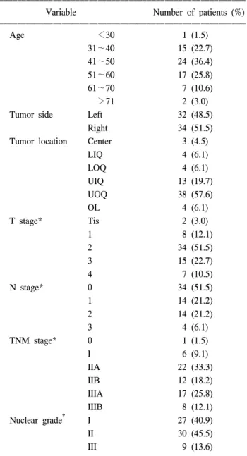
1994년부터 1998년까지 순천향대학교 천안병원에서 외 과학교실에서 조직학적으로 진단받고 수술을 시행한 환 자 중 외래추적관찰이 가능했던 66명의 유방암 환자를 대 상으로 하였다. 대상연령은 40대가 가장 많았고, 상외측이 호발부위였으며, AJCC 종양 병기(Tumor stage)상 2기(stage II)가 가장 많았다. 유방암을 Nottingham modification of the Bloom-Richardson system에 의한 grade에 따라 분류하면 grade I이 27예, grade II가 30예 그리고 grade III가 9예였다 (Table 1).
2) 실험방법
파라핀절편을 탈파라핀과 grade alcohol을 거쳐 함수시 킨 후, 항원부활을 위해 microwave oven에서 10분간 가열 처리하였다. 조직내의 내인성 peroxidase를 차단하기 위해 3% 과산화수소-메타놀용액에 30분간 작용시킨 후 증류수 로 수세하였고, avidin과 biotin의 blocking은 blocking kit (Vector, USA)를 사용하였으며, 실온에서 각각 30분씩 작 용시킨 후 PBS로 3분씩 3회 수세하였다. E-cadherin에 대 한 단일항체인 HECD-1 (Alexis, USA)을 작용시킨 후 2시 간 부란시켰다. 그 후 PBS로 3분씩 3회 수세하고 LAB kit (DAKO, USA)를 이용하여 이차항체와 peroxidase 표식 streptoavidin을 각각 30분씩 적용시켰다. PBS로 3분씩 3회 수세한 후 DAB kit (Vector, USA)로 발색시키고 수세하였 으며 hematoxylin으로 핵염색을 한 후 탈수 및 투명과정을 거쳐 Balsam 봉입 후 광학현미경으로 관찰하였다.
3) 판정 및 통계학적분석
E-cadherin은 종양세포의 세포막에 염색된 것을 발현된 것으로 하였으며, 이 항원의 발현이 종양조직 중 30% 이 하는 1+, 30∼50%은 ++, 50% 이상을 +++로 하였다.
유방암의 유형 및 조직병리학적 소견에 따른 발현율을 비 교검토하기 위하여 Chi-square 방법에 의한 P-value를 측정 하였고 생존율은 Kaplan-Meier 방법을 이용하여 산출하였 으며 생존율에 영향을 미치는 혼란변수를 통제하기 위한 다변량 분석은 Cox proportional hazard model을 이용하여 독립적인 설명변수를 알아보고자 하였다. 모든 통계적 분 석은 양측 검정을 이용하였으며 유의성은 P 값이 0.05이
하인 경우 통계적인 유의성이 있는 것으로 판정하였다.
결 과
유방암에서 E-cadherin은 종양조직의 국소 및 전체부위 에서 발현되었으며, 종양세포의 세포질과 세포막에서 발 현되었다. 유방암에서 E-cadherin 발현이 주변 정상조직에 비해 소실 및 심한 감소(이하 -/+로 표기)를 보인 예는 66예 중 42예(63.6%), 약간의 감소 및 같은(이하 ++/+++
Table 1. General characteristics of 66 study subject ꠚꠚꠚꠚꠚꠚꠚꠚꠚꠚꠚꠚꠚꠚꠚꠚꠚꠚꠚꠚꠚꠚꠚꠚꠚꠚꠚꠚꠚꠚꠚꠚꠚꠚꠚꠚꠚꠚꠚꠚꠚꠚꠚꠚꠚꠚꠚꠚꠚꠚꠚꠚꠚꠚꠚ
Variable Number of patients (%)
ꠏꠏꠏꠏꠏꠏꠏꠏꠏꠏꠏꠏꠏꠏꠏꠏꠏꠏꠏꠏꠏꠏꠏꠏꠏꠏꠏꠏꠏꠏꠏꠏꠏꠏꠏꠏꠏꠏꠏꠏꠏꠏꠏꠏꠏꠏꠏꠏꠏꠏꠏꠏꠏꠏꠏ
Age <30 1 (1.5)
31∼40 15 (22.7)
41∼50 24 (36.4)
51∼60 17 (25.8)
61∼70 7 (10.6)
>71 2 (3.0)
Tumor side Left 32 (48.5)
Right 34 (51.5)
Tumor location Center 3 (4.5)
LIQ 4 (6.1)
LOQ 4 (6.1)
UIQ 13 (19.7)
UOQ 38 (57.6)
OL 4 (6.1)
T stage* Tis 2 (3.0)
1 8 (12.1)
2 34 (51.5)
3 15 (22.7)
4 7 (10.5)
N stage* 0 34 (51.5)
1 14 (21.2)
2 14 (21.2)
3 4 (6.1)
TNM stage* 0 1 (1.5)
I 6 (9.1)
IIA 22 (33.3)
IIB 12 (18.2)
IIIA 17 (25.8)
IIIB 8 (12.1)
Nuclear grade† I 27 (40.9)
II 30 (45.5)
III 9 (13.6)
ꠏꠏꠏꠏꠏꠏꠏꠏꠏꠏꠏꠏꠏꠏꠏꠏꠏꠏꠏꠏꠏꠏꠏꠏꠏꠏꠏꠏꠏꠏꠏꠏꠏꠏꠏꠏꠏꠏꠏꠏꠏꠏꠏꠏꠏꠏꠏꠏꠏꠏꠏꠏꠏꠏꠏ
* = AJCC cancer staging manual; † = Nottingham modification of Bloom-Richardson System. LIQ = lower inner quadrant; LOQ
= lower outer quadrant; UIQ = upper inner quadrant; UOQ = upper outer quadrant; OL = overlapping lesion.
로 표기)예는 24예(36.4%)였다(Fig. 1). 병리학적 특성과 종 양 유전자 표출에 따른 생존율을 보면 종양병기, 종양의 크기, 액와 림프절 전이 유무 및 여성 호르몬 수용체 여부 (estrogen receptor status)는 통계학적인 의의가 있었으나 E-cadherin 발현 및 핵 등급은 통계학적인 의의는 없었다.
하지만 E-cadherin 발현의 소실이 있는 경우 및 핵 등급이 안 좋을수록 예후가 나쁜 경향을 발견할 수 있었다(Fig. 2).
여성호르몬 수용체 발현여부에 따라서는 사망율(mortality) 과 연관성이 있었으며, 종양의 크기, 액와 림프절 전이 및 종양병기에 따라서는 생존 기간(survival time) 및 사망율
Table 2. Survival by pathologic parameter and tumor marker expression
ꠚꠚꠚꠚꠚꠚꠚꠚꠚꠚꠚꠚꠚꠚꠚꠚꠚꠚꠚꠚꠚꠚꠚꠚꠚꠚꠚꠚꠚꠚꠚꠚꠚꠚꠚꠚꠚꠚꠚꠚꠚꠚꠚꠚꠚꠚꠚꠚꠚꠚꠚꠚꠚꠚꠚꠚꠚꠚꠚꠚꠚꠚꠚꠚꠚꠚꠚꠚꠚꠚꠚꠚꠚꠚꠚꠚꠚꠚꠚꠚꠚꠚꠚꠚꠚꠚꠚꠚꠚꠚꠚꠚꠚꠚꠚꠚꠚꠚꠚꠚꠚꠚꠚꠚꠚꠚꠚꠚꠚꠚꠚꠚꠚꠚꠚ
Variable Survival time mean (SD) P-value Mortality, n/N (%) P-value‡
ꠏꠏꠏꠏꠏꠏꠏꠏꠏꠏꠏꠏꠏꠏꠏꠏꠏꠏꠏꠏꠏꠏꠏꠏꠏꠏꠏꠏꠏꠏꠏꠏꠏꠏꠏꠏꠏꠏꠏꠏꠏꠏꠏꠏꠏꠏꠏꠏꠏꠏꠏꠏꠏꠏꠏꠏꠏꠏꠏꠏꠏꠏꠏꠏꠏꠏꠏꠏꠏꠏꠏꠏꠏꠏꠏꠏꠏꠏꠏꠏꠏꠏꠏꠏꠏꠏꠏꠏꠏꠏꠏꠏꠏꠏꠏꠏꠏꠏꠏꠏꠏꠏꠏꠏꠏꠏꠏꠏꠏꠏꠏꠏꠏꠏꠏ
Nuclear grade I 33.36 (17.51) 11/27 (40.7)
II 38.86 (14.53) NS† 14/30 (46.7) NS
III 50.17 (8.66) 6/9 (66.7)
E-cadherin -/+ 43.55 (15.43) NS* 20/42 (47.6) NS
++/+++ 31.00 (12.82) 11/24 (45.8)
Estrogen receptor Negative 40.92 (15.78) NS* 20/45 (44.4) P<0.05
Positive 31.50 (13.26) 15/21 (71.4)
T stage Tis NA P<0.1† 0/2 (0) P<0.05
1 27.00 (00) 2/8 (25.0)
2 37.89 (19.25) 9/34 (26.5)
3 46.00 (10.75) 13/15 (86.7)
4 31.29 (16.22) 7/7 (100)
N stage 0 51.00 (14.44) P<0.05† 5/34 (14.7) P<0.05
1 48.00 (8.90) 8/14 (57.1)
2 30.50 (14.78) 14/14 (100)
3 36.50 (14.73) 4/4 (100)
TNM stage 0 NA P<0.1† 0/2 (0) P<0.05
1 NA 0/5 (0)
2 49.00 (13.42) 6/34 (17.6)
3 36.72 (15.35) 25/25 (100)
ꠏꠏꠏꠏꠏꠏꠏꠏꠏꠏꠏꠏꠏꠏꠏꠏꠏꠏꠏꠏꠏꠏꠏꠏꠏꠏꠏꠏꠏꠏꠏꠏꠏꠏꠏꠏꠏꠏꠏꠏꠏꠏꠏꠏꠏꠏꠏꠏꠏꠏꠏꠏꠏꠏꠏꠏꠏꠏꠏꠏꠏꠏꠏꠏꠏꠏꠏꠏꠏꠏꠏꠏꠏꠏꠏꠏꠏꠏꠏꠏꠏꠏꠏꠏꠏꠏꠏꠏꠏꠏꠏꠏꠏꠏꠏꠏꠏꠏꠏꠏꠏꠏꠏꠏꠏꠏꠏꠏꠏꠏꠏꠏꠏꠏꠏ
* = Student T-test; † = ANOVA test; ‡ = Log rank test. SD = standard deviation; NS = not significant; NA = non applicable.
Fig. 1. E-cadherin expression in breast cancer (A) negative, immunoperoxidase, DAB, ×100, (B) positive, immunoperoxidase, DAB, ×200)
A B
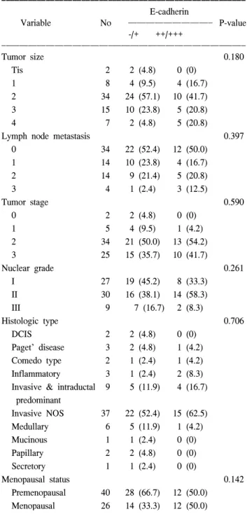
모두 연관성이 있었다. E-cadherin 발현 소실여부와 생존 기간 및 사망률과는 연관성이 없었으며, E-cadherin 발현 소실 여부에 따른 종양 표지자와의 연관성 역시 통계학적 인 의의는 없었다(Table 2). 환자들의 평균생존기간은 51.97개월이었고, 이 중 사망한 31명의 평균생존기간은 39.1개월이었으며, 생존한 35명의 평균생존기간은 63.37 개월이었다(Table 3). 유방암의 각종 병리조직학적 특성과 E-cadherin 발현의 소실 및 심한 감소와는 유의한 연관관
계를 찾을 수 없었다(Table 4). 고 찰
인체의 여러 종류의 조직에서 상피세포들은 강한 세포 간의 유착으로 체계적인 구조물을 형성한다. 하지만, 암세 포들 간의 유착성이 정상적인 세포보다 약하다는 견해가 Fig. 2. Kaplan-Meier survival curve according to E-cadherin
expression of primary tumor (there is no statistically significant survival advantage).
Table 3. Treatment and prognosis
ꠚꠚꠚꠚꠚꠚꠚꠚꠚꠚꠚꠚꠚꠚꠚꠚꠚꠚꠚꠚꠚꠚꠚꠚꠚꠚꠚꠚꠚꠚꠚꠚꠚꠚꠚꠚꠚꠚꠚꠚꠚꠚꠚꠚꠚꠚꠚꠚꠚꠚꠚꠚꠚꠚꠚ
Variable No. (%)
ꠏꠏꠏꠏꠏꠏꠏꠏꠏꠏꠏꠏꠏꠏꠏꠏꠏꠏꠏꠏꠏꠏꠏꠏꠏꠏꠏꠏꠏꠏꠏꠏꠏꠏꠏꠏꠏꠏꠏꠏꠏꠏꠏꠏꠏꠏꠏꠏꠏꠏꠏꠏꠏꠏꠏ Operation method
Radical mastectomy 3 (4.5)
Auchincloss 31 (47.0)
Scanlon 12 (18.2)
Patey 16 (24.2)
Quadrantectomy+Ax 4 (6.1)
Survive
Yes 35 (53.0)
No 31 (47.0)
Survival time, month, mean (SD) 51.97 (±18.03)
Survivor 63.37 (±1.87)
Deceased 39.10 (±2.80)
ꠏꠏꠏꠏꠏꠏꠏꠏꠏꠏꠏꠏꠏꠏꠏꠏꠏꠏꠏꠏꠏꠏꠏꠏꠏꠏꠏꠏꠏꠏꠏꠏꠏꠏꠏꠏꠏꠏꠏꠏꠏꠏꠏꠏꠏꠏꠏꠏꠏꠏꠏꠏꠏꠏꠏ No. = number of patients; Ax = axillary dissection; RM = radical mastectomy.
Table 4. The expression rates of E-cadherin according to variable in breast carcinoma
ꠚꠚꠚꠚꠚꠚꠚꠚꠚꠚꠚꠚꠚꠚꠚꠚꠚꠚꠚꠚꠚꠚꠚꠚꠚꠚꠚꠚꠚꠚꠚꠚꠚꠚꠚꠚꠚꠚꠚꠚꠚꠚꠚꠚꠚꠚꠚꠚꠚꠚꠚꠚꠚꠚꠚ E-cadherin
Variable No ꠏꠏꠏꠏꠏꠏꠏꠏꠏꠏꠏꠏꠏꠏꠏꠏꠏꠏꠏ P-value
-/+ ++/+++
ꠏꠏꠏꠏꠏꠏꠏꠏꠏꠏꠏꠏꠏꠏꠏꠏꠏꠏꠏꠏꠏꠏꠏꠏꠏꠏꠏꠏꠏꠏꠏꠏꠏꠏꠏꠏꠏꠏꠏꠏꠏꠏꠏꠏꠏꠏꠏꠏꠏꠏꠏꠏꠏꠏꠏ
Tumor size 0.180
Tis 2 2 (4.8) 0 (0)
1 8 4 (9.5) 4 (16.7)
2 34 24 (57.1) 10 (41.7)
3 15 10 (23.8) 5 (20.8)
4 7 2 (4.8) 5 (20.8)
Lymph node metastasis 0.397
0 34 22 (52.4) 12 (50.0)
1 14 10 (23.8) 4 (16.7)
2 14 9 (21.4) 5 (20.8)
3 4 1 (2.4) 3 (12.5)
Tumor stage 0.590
0 2 2 (4.8) 0 (0)
1 5 4 (9.5) 1 (4.2)
2 34 21 (50.0) 13 (54.2)
3 25 15 (35.7) 10 (41.7)
Nuclear grade 0.261
I 27 19 (45.2) 8 (33.3)
II 30 16 (38.1) 14 (58.3)
III 9 7 (16.7) 2 (8.3)
Histologic type 0.706
DCIS 2 2 (4.8) 0 (0)
Paget' disease 3 2 (4.8) 1 (4.2) Comedo type 2 1 (2.4) 1 (4.2) Inflammatory 3 1 (2.4) 2 (8.3) Invasive & intraductal 9 5 (11.9) 4 (16.7) predominant
Invasive NOS 37 22 (52.4) 15 (62.5) Medullary 6 5 (11.9) 1 (4.2)
Mucinous 1 1 (2.4) 0 (0)
Papillary 2 2 (4.8) 0 (0)
Secretory 1 1 (2.4) 0 (0)
Menopausal status 0.142
Premenopausal 40 28 (66.7) 12 (50.0) Menopausal 26 14 (33.3) 12 (50.0)
ꠏꠏꠏꠏꠏꠏꠏꠏꠏꠏꠏꠏꠏꠏꠏꠏꠏꠏꠏꠏꠏꠏꠏꠏꠏꠏꠏꠏꠏꠏꠏꠏꠏꠏꠏꠏꠏꠏꠏꠏꠏꠏꠏꠏꠏꠏꠏꠏꠏꠏꠏꠏꠏꠏꠏ NS = non significant; DCIS = Ductal carcinoma in situ; NOS = not otherwise specified.
이미 1944년(4)에 제시된 바 있으며, 이 감소된 유착성으 로 인해 종양 세포의 침윤성이 획득된다. 이러한 배경 하 에, 암세포에서 상호 유착성이 감소되는 기전을 밝히고, 그것의 생물학적인 성향에 끼치는 영향을 평가하려는 시 도들이 계속되고 있다. 종양세포의 침윤과 전이는 자신이 능동적으로 혹은 주위조직의 구조변화를 매개하는 단백 분자에 의해 세포와 세포 사이, 그리고 세포와 기질 혹은 내피세포와의 부착에 의해 세포가 탈락과 부착을 하는 복 잡한 단계를 거쳐 일어난다. 세포의 유착에 관여하는 분자 들은 크게 칼슘이온 의존성 군과 칼슘이온 비의존성 군으 로 나뉘며, 그중 칼슘이온 의존성 유착분자들을 “cadherin”
이라 하고, 이것은 칼슘이온 비의존성 군보다 더 강한 세 포간의 유착을 일으킨다고 한다.(5) Cadherin은 기본구조 와 분자적 특성에 따라 신경성의 N-cadherin, 태반성의 P-cadherin, 근육성의 M-cadherin, 망막성의 R-cadherin 등이 있으며 일부는 이미 그 구조가 밝혀져 있다.(6,7) 지금까지 밝혀진 여러 종류의 cadherin 중, E(epithelial)-cadherin은 거 의 모든 상피세포에 존재하는 것으로서, 상피세포간의 유 착과 상피 조직의 항상성 유지를 관장하는 가장 중요한 매개체이다. 종양조직에서 세포부착분자인 E-cadherin의 발현이 감소하면 종양세포는 탈분화하고, 세포 부착과 극 성 배열이 소실되며, 세포와 세포 그리고 기저막과 부착 을 하지 못하여 종양세포는 원발소에서 떨어져 나와 침윤 성을 갖게 되므로 상피암종에서 E-cadherin은 침윤성과 전 이 억제인자로 중요한 역할을 하는 것으로 알려져 있다.
E-cadherin의 변성은 세포 분화의 소실을 초래하고, 세포를 원래의 위치에서 이탈시켜 림프절이나 다른 장기로 전이되 게 한다는 연구들도 보고되고 있다. 일반적으로 종양의 분 화가 좋고 종양세포의 부착이 좋을수록 E-cadherin의 발현 은 균질하게 유지되고, 분화도가 나쁜 종양에서 E-cadherin 의 발현은 불안정하거나 소실되는 것으로 알려져 있다.(8) 이러한 이론들을 토대로, 정상적인 E-cadherin 발현의 소 실과 종양의 예후 사이에 모든 상관성이 있다는 보고들 이 여러 종류의 종양을 대상으로 이루어지고 있다.(9) Behrens 등(10)은 E-cadherin이 없는 세포는 세포 배양 실 험에서 극성이 소실된 섬유모 세포와 유사한 모양으로 변하 여 침윤성을 나타내는 것을 관찰하였고, Vleminckx 등(11)은 E-cadherin 발현이 없는 분화가 나쁜 세포주에 E-cadherin cDNA를 이식하면 침윤성을 방지할 수 있고 E-cadherin 항 체를 투여하면 침윤성이 다시 생기는 것을 실험으로 증명하 였다. 한편, E-cadherin의 발현이 다양한 종류의 인체 조직과 종양에서 그 장기 특이성에 따라 국소화(localization)되어 나 타난다는 연구 결과(12)를 비롯하여 여러 가지 종양을 대 상으로 조사한 보고들에서 E-cadherin에 대한 항체를 이용 한 면역조직화학적 연구 결과, 정상적인 E-cadherin의 발 현은 세포와 세포간, 즉 막성으로 발현되는 반면, 비균질 적 발현, 세포성 발현, 그리고 발현되지 않는 경우는 비정
상적인 것으로 판단되었다. 이렇게 종양세포 표면에서 E-cadherin이 불균질하게 발현되거나 감소를 보이는 것은 cadherin 유전자의 변이나 소실에 의한 단백질의 감소 또 는 그 기능상의 장애로 기인한다거나 E-cadherin 발현 감 소 외에 매개체 역할을 하는 catenin 유전자 결손으로 인 한 catenin 발현 소실(13)이나 cadherin과 연관된 단백의 티 로신 인산화로 인한 catenin 분자의 생화학적 변형이 일어 나면 상피층의 극성화에 중요한 E-cadherin-catenin-actin 복 합체가 만들어지지 못하여 세포간 접촉이 불안정하여 신 호전달 과정에 이상이 초래되어 정상적인 막성 E-cadherin 발현이 불가능해지기 때문이라는 실험 결과들로 해석되 고 있다.(14) 뿐만 아니라, 분화 및 침윤과 같은 생물학적 인 과정은 integrin, immunoglobulin superfamily 및 CD 44와 같은 상호 유착에 관여하는 다른 많은 복합체들에 의해서 도 조절됨으로, 이러한 유착 분자들과 각각의 수용체에 의해 조절되는 역할간의 기능적인 상호 작용에 대해서도 밝혀져야 할 것이다.(15)
본 연구에서는 유방암 환자 66예 중 63.6%에서 E-cadherin 발현의 소실 및 심한 감소를 보여 Gamallo 등(16)의 54예 중 50% 및 Oka 등(9)의 120예 중 53%에 비해 빈도가 약간 높았으나 Rimm 등(17)의 64%와는 비슷한 결과를 보였다.
Bukholm 등(30)은 유방암 90예의 연구에서 E-cadherin 발 현 감소와 소실은 grade와 관련성이 없다고 하였으며, Rimm 등(17)도 같은 결과를 발표하였다. 이들의 결과는 본 연구와 일치하는 것으로 일반적으로 분화가 좋은 종양 에서 E-cadherin 발현은 잘 유지되며 분화가 나쁜 종양에 서 발현이 불안정하거나 소실되는 것으로 알려져 있으나 유방암에서는 E-cadherin 발현 감소와 소실은 grade와 관 련성이 없을 것으로 추측된다. 하지만 본 연구에서는 E-cadherin 발현의 소실이 있는 경우 및 nuclear grade가 안 좋을수록 예후가 나쁜 경향을 발견할 수 있었다. 또한 Bukholm 등(18)은 그의 연구에서 E-cadherin 발현 감소 및 소실이 림프절 전이와는 관련성이 없다고 하였고, Shiozaki 등(19)은 유방암조직의 침윤부위에서 E-cadherin 발현이 없었으나 전이된 암조직에서는 E-cadherin 발현을 보인다 고 하며 이는 세포와 세포 사이에서 부착의 발현과 안정 성은 상호 의존성이 없다는 것을 의미한다고 하였다. 그 러나 유방암에서 E-cadherin 발현 감소 및 소실이 림프절 전이와 의미 있는 관련성을 보인다고 하는 보고도 있어 보고자들에 따라 차이를 보이고 있다. 이러한 결과는 Rimm 등(17)의 유방암에 대한 연구 결과와 유사하나 그의 논문에서 지적하듯이 적은 연구대상이 의미 있는 결과를 가져오지 못한 것으로 생각되며, 본 연구에서도 더 많은 연구대상 예가 필요할 것으로 생각된다. 본 연구에서는 유방암의 각종 병리조직학적 특성과 E-cadherin 발현 유무 와는 유의한 연관관계를 찾을 수 없었으며, E-cadherin 발 현 소실여부와 생존 기간 및 사망률과의 연관성 역시 없
었다. 병리적 특성과 종양 표지자 표출에 따른 생존율을 보면 종양 병기, 종양의 크기, 액와 림프절 전이 유무 및 여성호르몬 수용체 여부는 통계학적인 의의가 있었으나 E- cadherin 발현 및 핵 등급은 통계학적인 의의는 없었다.
하지만 E-cadherin 발현의 소실이 있는 경우 및 핵 등급이 안 좋을수록 예후가 나쁜 경향을 발견할 수 있었다.
결 론
1994년부터 1998년까지 순천향대학교 천안병원에서 외 과학교실에서 조직학적으로 진단받고 수술을 시행한 환 자 중 외래추적관찰이 가능했던 66명의 유방암 환자를 대 상으로 하여 E-cadherin 발현의 소실 및 감소가 유방암의 조직학적 형태나 핵 등급, 액와 림프절 전이부와 연관성 및 생존율과 관련된 예후인자로서의 의의가 있는지를 알 아본 결과 유방암 환자들의 생존율과 관련하여 의미 있는 예후인자는 이미 알려진 종양의 크기와 액와 림프절 전이 여부, 종양 병기 및 호르몬 수용체 존재여부 등이었으며 E-cadherin은 통계학적인 의의는 없었다.
REFERENCES
1) Pignatelli M, Vessey CJ. Adhesion molecules. Novel molecular tools in tumor pathology. Hum Pathol 1994;25:849-56.
2) Damsky CHJ, Richa D, Solter K, Knudsen SK, Buck CA.
Identification and purification of a cell surface glycoprotein mediating intercellular adhesion in embryonic and adult tissue.
Cell 1983;34:455-66.
3) Mayer B, Johnson JP, Leitl F. E-cadherin expression in primary and metastatic gastric cancer: down-regulation correlates with cellular dedifferentiaton and glandular disintegration. Cancer Res 1993;53:1690-5.
4) Coman DR. Decreased mutual adhesiveness a property of cells from squamous cell carcinomas. Cancer Res 1944;4:625-9.
5) Umbas R, Schalken JA, Aalders TW. Expression of the cellular adhesion molecule E-cadherin is reduced or absent in high grade prostate cancer. Cancer Res 1992;52:5104-9.
6) Lipponen PK, Eskelinen MJ. Reduced expression of E- cadherin is related to invasive disease and frequent recurrence in bladder cancer. J Cancer Res Clin Oncol 1995;121:303-8.
7) Takeichi M. The cadherins: Cell-cell adhesion molecules con- troling animal morphogenesis. Development 1988;102:639-55.
8) Mayer B, Johnson JP, Leitl F, Jauch KW, Heiss MM, Schildberg FW, et al. E-cadherin expression in primary and metastatic gastric cancer: Down-regulation correlates with cellular de- differentiation and glandular disintegration. Cancer Res 1993;
53:1690-5.
9) Oka H, Shiozaki H, Kobayashi K, Inoue M, Tahara H, Kobayashi T. Expression of E-cadherin cell adhesion molecules in human breast cancer tissues and its relationship to metastasis. Cancer Res 1993;53:1696-701.
10) Behrens J, Mareel MM, Van Roy FM, Birchmeier W. Dis- secting tumor cell invasion: Epithelial cells acquire invasive properties after the loss of uvomorulin-mediated cell-cell adhesion. J Cell Biol 1989;108:2435-47.
11) Vleminckx K, Vakaet L, Mareel M. Genetic manipulation of E-cadherin expression by epithelial tumor cells reveals an invasion suppressor role. Cell 1991;66:107-19.
12) Eidelman S, Damsky CH, Wheelock MJ, Damjanov I. Ex- pression of the cell-cell adhesion glycoprotein cell-CAM 120/80 in normal human tissues and tumor. Am J Pathol 1989;
135:101-10.
13) Shimoyama Y, Nagafuchi A, Fujita S. Cadherin dysfunction in a human cancer cell line: Possible involvement of loss of α-catenin expression in reduced cell-cell adhesiveness. Cancer Res 1992;52:5770-4.
14) Beherens J, Vakaet L, Friis R. Loss of epithelial differentiation and gain of invasiveness correlates with tyrosine phosphory- lation of the E-cadherin/β-catenin complex in cells trans- formed with a temperature-sensitive v-src gene. J Cell Biol 1993; 120:757-66.
15) Pignatelli M. E-cadherin a biological marker of tumor dif- ferentiation. J Pathol 1993;171:81-2.
16) Gamallo C, Palacios J, Surarez A, Pizarro A, Navaro P, Guintanilla M. Correlation of E-cadherin expression with differentiation grade and histological type in breast carcinoma. Am J Pathol 1993;142:987-93.
17) Rimm DL, Sinard JH, Morrow JS. Reduced α-catenin and E- cadherin expression in breast cancer. Lab Invest 1995;72:506- 12.
18) Bukholm IK, Nesland JM, Karesen R, Jacobsen U, Borresen- Dale A-L. E-cadherin and α-, β-, and γ-catenin protein expression in relation to metastasis in human breast carcinoma.
J Pathol 1998;185:262-6.
19) Shiozaki H, Tahara H, Oka H, Miyata M, Kadowaki K, Tamura S. Expression of immunoreactive E-cadherin mole- cules in human cancers. Am J Pathol 1991;139:17-23.