Safety Evaluation of Topical Valproate Application
저자 (Authors)
Sun Young Choi, Song Yi Seop, Moo Yeol Hyun, Kwang Ho Yoo, Beom Joon Kim, Myeung Nam Kim, Jae-We Cho
출처 (Source)
Toxicological Research29(2), 2013.6, 87-90 (4 pages)
발행처 (Publisher)
한국독성학회
The Korean Society Of Toxicology
URL
http://www.dbpia.co.kr/Article/NODE02199728APA Style Sun Young Choi, Song Yi Seop, Moo Yeol Hyun, Kwang Ho Yoo, Beom Joon Kim, Myeung Nam Kim, Jae-We Cho (2013). Safety Evaluation of Topical Valproate Application.
Toxicological Research, 29(2), 87-90.
이용정보 (Accessed)
저작권 안내
DBpia에서 제공되는 모든 저작물의 저작권은 원저작자에게 있으며, 누리미디어는 각 저작물의 내용을 보증하거나 책임을 지지 않습니다.
이 자료를 원저작자와의 협의 없이 무단게재 할 경우, 저작권법 및 관련법령에 따라 민, 형사상의 책임을 질 수 있습니다.
Copyright Information
The copyright of all works provided by DBpia belongs to the original author(s). Nurimedia is not responsible for contents of each work. Nor does it guarantee the contents.
You might take civil and criminal liabilities according to copyright and other relevant laws if you publish the contents 계명대학교
114.71.5.213
2016/07/05 13:35 (KST)
Vol. 29, No. 2, pp. 87-90 (2013)
http://dx.doi.org/10.5487/TR.2013.29.2.087 plSSN: 1976-8257 eISSN: 2234-2753
Open Access
Safety Evaluation of Topical Valproate Application
Sun Young Choi1, Song Yi Seop1, Moo Yeol Hyun1, Kwang Ho Yoo1, Beom Joon Kim1, Myeung Nam Kim1 and Jae-We Cho2
1Department of Dermatology, Chung-Ang University College of Medicine, Seoul, Korea
2Department of Dermatology, Keimyung University School of Medicine, Daegu, Korea
(Received February 8, 2013; Revised March 25, 2013; Accepted June 5, 2013)
The potential role of topical valproate (VPA) in hair regrowth has been recently suggested. However, safety reports of VPA as a topical formulation are lacking. Therefore, in the present study, we investigated whether VPA causes skin irritation in humans. We first performed a cell viability test and showed that VPA did not exhibit toxicity toward HaCaT keratinocytes, fibroblasts, and RBL-3H mast cells. We then performed clinical patch test and skin irritation test through transdermal drug delivery with the help of microneedle rollers. No significant findings were obtained in the clinical patch test. In the skin irritation test, only 1 patient showed erythema at 1 hr, but the irritation reaction faded away within a few hours.
Erythema and edema were not observed at 24 hr. We concluded that VPA has minimal potential to elicit skin irritation. Therefore, we consider that VPA can safely be applied to human skin.
Key words: Valproate, Skin toxicity
INTRODUCTION
Valproic acid (VPA) is a mood stabilizer that has been commonly prescribed for the treatment of epilepsy and bipolar disorder over the last several decades (1). Antiepi- leptic drugs including VPA frequently cause cutaneous erup- tions, especially during initiation of new therapy. In addition to causing common and usually limited morbilliform and urticarial eruptions, which have often mild morbidity, antie- pileptic drugs can also induce widespread maculopapular rash, hypersensitivity syndrome, psoriatic dermatitis and occasionally severe reactions such as Steven Johnson’s syn- drome, toxic epidermal necrolysis and erythema multi- forme (2,3).
In recent studies using animal models, topical application of VPA stimulated hair re-growth (4). In an in vitro study, VPA up-regulated the Wnt/β-catenin pathway and alkaline phosphatase activity in human dermal papilla cells (4).
Activation of the Wnt/β-catenin pathway is required for ini- tiation of hair follicle formation (5), and stimulates growth and differentiation of hair by maintaining expression of genes that function at the anagen phase of the hair cycle (6).
Consequently, it was suggested that VPA could be devel- oped as a topical drug to stimulate hair re-growth (4), but there are currently no studies on the safety of VPA for skin application. The aim of the present study was to investigate whether VPA exhibits toxicity towards skin. We tested the toxicity of VPA in keratinocytes, fibroblasts and mast cells and performed skin tests in healthy human subjects using a clinical patch test and skin irritation tests through transder- mal drug delivery using microneedle rollers.
MATERIALS AND METHODS
Materials. Twenty healthy volunteers aged 21 to 43 years (mean age 30.4) were enrolled for primary skin irrita- tion and toxicity tests. Individuals were excluded if they had current or past history of underlying chronic skin dis- eases that might interfere with the evaluation of skin reac- tions. We used sodium valproate (95 mg/ml) and normal saline (as a negative control) for skin tests.
The study was approved by the relevant institutional review boards. All subjects signed informed consent forms, and the study protocol conformed to the guidelines set forth by the Declaration of Helsinki and Korean Good Clinical
Correspondence to: Beom Joon Kim, Department of Dermatol-
ogy, Chung-Ang University College of Medicine, 224-1 Heukseok- dong, Dongjak-gu, Seoul 156-755, Korea
E-mail: [email protected]
This is an Open-Access article distributed under the terms of the Creative Commons Attribution Non-Commercial License (http://
creativecommons.org/licenses/by-nc/3.0) which permits unrestricted
non-commercial use, distribution, and reproduction in any
medium, provided the original work is properly cited.
88 S.Y. Choi et al.
Practice.
Cell culture. Human HaCaT keratinocytes were obtained from the Korean Cell Line Bank (Seoul). Cells were incu- bated in DMEM supplemented with 10% FBS, 1 mM sodium pyruvate, 50
µg/ml streptomycin and 50 µg/ml pen-
icillin at 37oC in 5% CO2.Determination of cell viability. Human HaCaT kerati- nocytes were seeded in a 24-well plate at a density of 4 × 10⁵ cells per well and fibroblasts were seeded at 5 × 10⁴ cells per well. After overnight incubation, cells were treated with various concentrations of VPA ranging from 0.1
µM to
100µM and incubated in a 37
oC CO2 incubator for 24 hr.The percentage of viable human HaCaT keratinocyte cells and fibroblast cells was determined by staining the cells with MTT. The culture medium was removed from each well, and the cells were washed with phosphate-buffered saline (PBS) and treated with the MTT staining solution.
After incubation in a 37oC CO2 incubator for 3 hr, the medium was replaced with 1 ml DMSO and the absor- bance was measured at 570 nm. Cytotoxicity towards RBL- 2H3 mast cells was measured using the cell counting kit-8 (CCK-8) assay (Dojindo Laboratories, Kumamoto, Japan) according to the manufacturer’s instructions. RBL-2H3 cells were seeded in a 96-well plate at a density of 2.5 × 10⁵ cellsper well. After overnight incubation, cells were treated with various concentrations of VPA ranging from 0.1
µM to
100µM. Absorbance was measured at 450 nm.
Clinical patch test. A patch test was performed on the skin of all subjects. The test samples (0.2 ml of VPA or nor- mal saline) were placed in a Finn Chamber (Chemotech-
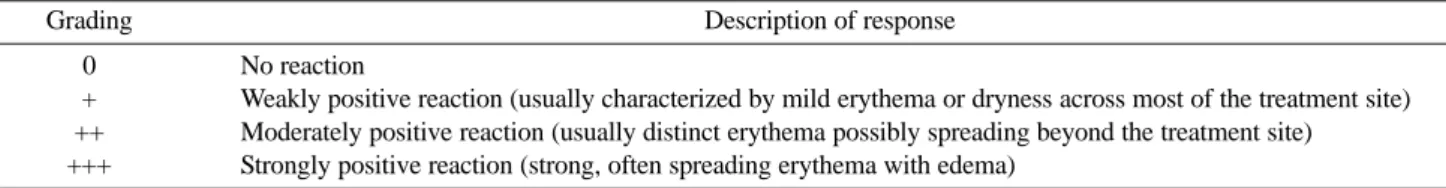
nique Diagnostics, Sweden) and applied to the ventral side of the upper arm of each subject for 48 hrs in an occlusive condition. Skin reactions were evaluated at 1 hr and 48 hrs after removing the test samples. The reaction was evalu- ated according to the International Contact Dermatitis Research Group (ISDRG) standard (Table 1) (7).
Skin irritation test. The skin irritation test was per- formed on three skin test sites of all subjects: frontal scalp, upper arm and upper back. We used the method of transder- mal drug delivery by microneedle rollers to perform the skin irritation test. The microneedle rollers used in this study were supplied by DTS Lab Inc. (Disk Microneedles Therapy System Lab, Inc., Seoul, South Korea). The microneedle roller was 0.25 mm long and had 60 circular arrays of nine needles each (a total of 540 needles) in a cylindrical assembly (20-mm diameter, 18.2-mm length).
The test samples (2.0 ml of VPA or normal saline) were applied to a 2 cm × 4 cm area of each test site and the microneedle roller was rolled four times on the skin test sites. Skin irritation test sites were examined for the pres- ence of erythema and edema according to the dermal irrita- tion scoring system (Table 2) at 1 hr and 24 hrs (8).
RESULTS AND DISCUSSION
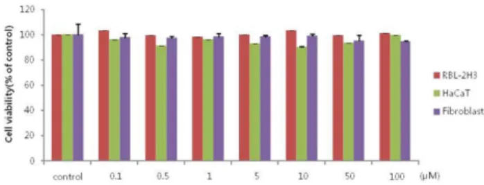
We performed cell viability tests for VPA in HaCaT kera- tinocytes, fibroblasts, and RBL-3H mast cells. After treat- ment with various concentrations of VPA, cell toxicity was not observed in RBL-2H3, HaCaT or fibroblast cells (Fig. 1).
We next performed a clinical patch test on 20 healthy vol- unteers. No significant clinical findings were observed at 1 hr and 48 hrs after removing the test samples. A skin irri- Table 1. Assessment of patch test reactions
Grading Description of response
0 No reaction
+ Weakly positive reaction (usually characterized by mild erythema or dryness across most of the treatment site) ++ Moderately positive reaction (usually distinct erythema possibly spreading beyond the treatment site) +++ Strongly positive reaction (strong, often spreading erythema with edema)
Table 2. Scoring criteria for dermal reactions
Evaluation of dermal reactions
Erythema and eschar formation Edema formation
0 No erythema 0 No edema
1 Very slight erythema (barely perceptible, edges of area not well defined)
1 Very slight edema (barely perceptible, edges of area not well defined)
2 Slight erythema (pale red in color and edges definable) 2 Slight edema (edges of area well defined by definite raising) 3 Moderate to severe erythema (defined in color and
area well defined)
3 Moderate edema (raised approximately 1 mm)
4 Severe erythema (beet to crimson red) to slight eschar formation (deep injuries)
4 Severe edema (raised more than 1 mm and extending beyond area of exposure)
tation test was also performed by transdermal drug delivery on 20 healthy volunteers. One patient appeared to have barely perceptible erythema effects on the frontal scalp and upper arm at 1 hr, but there were no erythema or edema effects in any of the patients at 24 hrs (Table 3). The time course of recovery from irritation is depicted in Fig. 2.
Erythema faded away quickly and the skin returned to nor- mal within a short period.
In our study, the skin irritation test was performed not by simple application, but by transdermal drug delivery using Fig. 1. Viability of RBL-2H3 cells, HaCaT keratinocytes and fibro-
blasts treated with VPA.
Fig. 2. Clinical signs of skin treated with valproate in patient No. 10. Slight erythema was observed at 1 hr after skin application, but
there was no erythema or edema at 24 hrs after application. N/S: normal saline, VPA: valproic acid. (a) Frontal scalp, (b) upper back.
Table 3. Results of skin reactions
Skin irritation test
Frontal scalp Upper arm Upper back
Erythema Edema Erythema Edema Erythema Edema
Patient Sex/Age 1 hr 24 hr 1 hr 24 hr 1 hr 24 hr
01 M/41 0 0 0 0 0 0
02 M/42 0 0 0 0 0 0
03 M/43 0 0 0 0 0 0
04 M/35 0 0 0 0 0 0
05 M/36 0 0 0 0 0 0
06 M/39 0 0 0 0 0 0
07 M/37 0 0 0 0 0 0
08 M/32 0 0 0 0 0 0
09 M/42 0 0 0 0 0 0
10 M/21 1 0 0 0 1 0
11 M/23 0 0 0 0 0 0
12 M/24 0 0 0 0 0 0
13 M/23 0 0 0 0 0 0
14 M/23 0 0 0 0 0 0
15 M/25 0 0 0 0 0 0
16 M/22 0 0 0 0 0 0
17 M/22 0 0 0 0 0 0
18 M/31 0 0 0 0 0 0
19 M/24 0 0 0 0 0 0
20 M/23 0 0 0 0 0 0
90 S.Y. Choi et al.
microneedle rollers. Therefore, the cause of the erythema that occurred in one patient could be irritation by the micronee- dles. Microneedle treatment offers a minimally invasive and painless route for transdermal drug application (9) but it causes superficial barrier-related skin damage. However, it has been reported that prompt recovery of barrier function occurs within 72 hrs after microneedle treatment (10).
The potential utility of VPA in the treatment of androge- netic alopecia was recently suggested. Animal studies and in vitro studies revealed that topical VPA stimulated hair re- growth (4). However, the potential of topical VPA for hair re-growth in humans had not been studied in vivo. This is the first safety study of VPA in human skin and we con- clude that VPA has minimal potential to elicit an irritation reaction. We therefore believe that VPA can be safely applied to human skin.
REFERENCES
1. Reynolds, M.F., Sisk, E.C. and Rasgon, N.L. (2007) Val- proate and neuroendocrine changes in relation to women treated for epilepsy and bipolar disorder: a review. Curr. Med.
Chem., 14, 2799-2812.
2. Tennis, P. and Stern, R.S. (1997) Risk of serious cutaneous disorders after initiation of use of phenytoin, carbamazepine, or sodium valproate: a record linkage study. Neurol., 49, 542- 546.
3. Roujeau, J.C., Kelly, J.P., Naldi, L., Rzany, B., Stern, R.S., Anderson, T., Auquier, A., Bastuji-Garin, S., Correia, O., Locati, F., et al. (1995) Medication use and the risk of Stevens-Johnson syndrome or toxic epidermal necrolysis. N.
Engl. J. Med., 333, 1600-1607.
4. Lee, S.H., Yoon, J., Shin, S.H., Zahoor, M., Kim, H.J., Park, P.J., Park, W.S., Min do, S., Kim, H.Y. and Choi, K.Y. (2012) Valproic acid induces hair regeneration in murine model and activates alkaline phosphatase activity in human dermal papilla cells. PLoS One, 7, e34152.
5. Andl, T., Reddy, S.T., Gaddapara, T. and Millar, S.E. (2002) WNT signals are required for the initiation of hair follicle development. Dev. Cell, 2, 643-653.
6. Kishimoto, J., Burgeson, R.E. and Morgan, B.A. (2000) Wnt signaling maintains the hair-inducing activity of the dermal papilla. Genes Dev., 14, 1181-1185.
7. Prausnitz, M.R. (2004) Microneedles for transdermal drug delivery. Adv. Drug Delivery Rev., 56, 581-587.
8. Draize, J.H. (1965) The Appraisal of the Safety of Chemicals in Foods, Drugs and Cosmetics, Dermal Toxicity, Association of Food and Drug Officials of the US, Topeka, KA, pp. 46-59.
9. Lachapelle, J.M. (1997) A proposed relevance scoring system for positive allergic patch test reactions: practical implications and limitations. Contact Dermatitis, 36, 39-43.
10. Han, T.Y., Park, K.Y., Ahn, J.Y., Kim, S.W., Jung, H.J. and Kim, B.J. (2012) Facial skin barrier function recovery after microneedle transdermal delivery treatment. Dermatol. Surg., 38, 1816-1822.