Introduction
Acute respiratory failure (ARF) has long been a challenge for physicians who perform bronchoscopy for diagnostic or therapeutic purposes. Hypoxaemia is aggravated when implementing bronchoalveolar lavage (BAL) or therapeutic intervention. Many physicians lose their chance to perform bronchoscopy and acquire adequate samples for appropriate treatment. Since undergoing a bronchoscopy can be hazard- ous to patients with ARF, intubation is preferred and physi- cians are reluctant to achieve BAL sample. Therefore, bron- choscopic procedures in high-risk patients, especially those with hypoxaemia, have long been a challenge in this field.
Clinical Effectiveness of High-Flow Nasal Cannula in Hypoxaemic Patients during Bronchoscopic Procedures
Sang Mi Chung, M.D. , Ju Whan Choi, M.D., Young Seok Lee, M.D., Ph.D., Jong Hyun Choi, M.D., Jee Youn Oh, M.D., Ph.D., Kyung Hoon Min, M.D., Ph.D., Gyu Young Hur, M.D., Ph.D., Sung Yong Lee, M.D., Ph.D. , Jae Jeong Shim, M.D., Ph.D. and Kyung Ho Kang, M.D., Ph.D.
Division of Respiratory and Critical Care Medicine, Korea University Guro Hospital, Seoul, Korea
Background: Bronchoscopy is a useful diagnostic and therapeutic tool. However, the clinical use of high-flow nasal cannula (HFNC) in adults with acute respiratory failure for diagnostic and invasive procedures has not been well evaluated. We present our experiences of well-tolerated diagnostic bronchoscopy as well as cases of improved saturation in hypoxaemic patients after a therapeutic bronchoscopic procedure.
Methods: We retrospectively reviewed data of hypoxaemic patients who had undergone bronchoscopy for diagnostic or therapeutic purposes from October 2015 to February 2017.
Results: Ten patients (44–75 years of age) were enrolled. The clinical purposes of bronchoscopy were for diagnosis in seven patients and for intervention in three patients. For the diagnoses, we performed bronchoalveolar lavage in six patients. One patient underwent endobronchial ultrasonography with transbronchial needle aspiration of a lymph node to investigate tumour involvement. Patients who underwent bronchoscopy for therapeutic interventions had endobronchial mass or blood clot removal with cryotherapy for bleeding control. The mean saturation (SpO
2) of pre- bronchoscopy in room air was 84.1%. The lowest and highest mean saturation with HFNC during the procedure was 95% and 99.4, respectively. The mean saturation in room air post-bronchoscopy was 87.4%, which was 3.3% higher than the mean room air SpO
2pre-bronchoscopy. Seven patients with diagnostic bronchoscopy had no hypoxic event. Three patients with interventional bronchoscopy showed improvement in saturation after the procedure. Bronchoscopy was well tolerated in all 10 cases.
Conclusion: This study suggests that the use of HFNC in hypoxaemic patients during diagnostic and therapeutic bronchoscopy procedures has clinical effectiveness.
Keywords: Bronchoscopy; Cannula; Hypoxia; Oxygen; Hypoxemia
Address for correspondence: Sung Yong Lee, M.D., Ph.D.
Division of Respiratory and Critical Care Medicine, Korea University Guro Hospital, 148 Gurodong-ro, Guro-gu, Seoul 08308, Korea
Phone: 82-2-2626-3030, Fax: 82-2-2626-1166 E-mail: [email protected]
Received: Sep. 11, 2017 Revised: Dec. 8, 2017 Accepted: Mar. 5, 2018 Published online: Jun. 19, 2018
cc
It is identical to the Creative Commons Attribution Non-Commercial License (http://creativecommons.org/licenses/by-nc/4.0/).
Copyright © 2019
The Korean Academy of Tuberculosis and Respiratory Diseases.
Conventional oxygen therapy (low flow O
2system, nonin- vasive ventilation [NIV]) was known to improve the outcome of hypoxaemic patients. Comparing with high flow nasal can- nula (HFNC) group, there were no significant differences in the reintubation rate or length of intensive care unit stay in a meta-analysis study
1.
HFNC therapy provides accurate oxygen delivery, wash out of anatomical dead space, and a low-level of positive pres- sure
2. The devices deliver heated and humidified oxygen at a maximum flow rate of up to 60 L/min via nasal cannula, and enable the maintenance of the fraction of oxygen above 95%
3,4.
Miyagi et al.
2presented five cases that underwent BAL with the use of HFNC; the patients tolerated the procedure despite hypoxia and dyspnoea. However, the shortage of cases could not confirm the clinical efficacy and safety of HFNC use dur- ing bronchoscopy and BAL. Simon et al.
5reported that NIV was superior to HFNC with regard to oxygenation before, during, and after bronchoscopy in patients with moderate to severe hypoxaemia. However, patients who were stable on HFNC tolerated the bronchoscopy well
5.
Few clinical studies have shown the effectiveness of per- forming bronchoscopy using HFNC in ARF for diagnostic purpose. Moreover, there is no clinical practice that improves oxygenation after performing therapeutic intervention with bronchoscopy. Therefore, we conducted a retrospective study to clarify the clinical effectiveness of high-flow nasal cannula in hypoxaemic patients during a diagnostic and interventional bronchoscopy.
Materials and Methods
This is a retrospective, observational, single center study ap- proved by the ethics committee of the Korea University Guro Hospital. Informed consent was waived due to the retrospec- tive study.
All patients were selected by the Pulmonology Department at a single academic hospital to undergo fiberoptic bronchos- copy and BAL fluid collection for diagnostic purposes or mass removal and cryotheraputic intervention for therapeutic pur- poses. Patients who had a peripheral arterial pulse oximetry value (SpO
2) less than 90% in room air setting were included in this study. Subjects with tracheostomy, requiring home oxygen therapy or mechanical or noninvasive ventilation, nasal or nasopharyngeal disease, not able to clearly express themselves, and pregnant individuals were excluded from the study.
The procedures for all 10 patients were conducted in the same environment. The patients were in the supine position and we administered a mixture of humidified gas warmed by a servo-controlled heated respiratory humidifier (MR730;
Fisher & Paykel, Auckland, New Zealand). Baseline room air peripheral saturation (SpO
2), heart rate, and respiratory rate
were measured before the procedure during spontaneous breathing on room air.
For local anaesthesia, 2% nebulized lidocaine was sprayed twice through the mouth and nostrils. For all patients, a rest- ing period of approximately 5 minutes was needed for the local anaesthesia to fully take effect. Conscious sedation was achieved with midazolam and propofol, which were admin- istered intravenously at maximum doses of 0.1 mg/kg body weight. Fibreoptic bronchoscopy (18-F; Olympus, Corp, To- kyo, Japan) was initiated through a nostril. HFNC was applied through both nostrils. BAL was performed by instilling and aspirating 100 to150 ml of a saline solution (NaCl 0.9%) ample fluid. The highest and lowest saturation (SpO
2), heart rate, and respiratory rate were checked during the procedure. The total duration of the bronchoscopy was measured for each patient.
At the end of the procedure, the post-bronchoscopy satura- tion (SpO
2) was checked while applying HFNC and on room air after 5 minutes.
Results
1. Patient characteristics
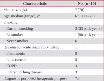
The demographic and clinical characteristics are depicted in Table 1. A total of 10 patients (3 females and 7 males), rang- ing from 44 to 75 years of age were enrolled. The clinical rea- sons for bronchoscopy were diagnostic in seven patients and interventional in three patients. Each patient had more than one comorbidity including pneumonia, chronic obstructive pulmonary disease, interstitial lung disease, and lung cancer.
Three patients did not have pulmonary function test records,
Table 1. Demographic and clinical characteristics of subjects in the study
Characteristic No. (n=10)
Male sex, n (%) 7 (70)
Age, median (range), yr 67.3 (44–75) Smoking
Current smoking 3 (33 pack-years)
Ex-smoker 1 (90 pack-years)
Never smoker 6
Reasons for acute respiratory failure
Pneumonia 5
Lung cancer 5
COPD 3
Interstitial lung disease 3
Diagnostic purpose:Therapeutic purpose 7:3
COPD: chronic obstructive pulmonary disease.
while five patients had a forced expiratory volume in the first second more than 1.0 L and the diffusing capacity of the lungs for carbon monoxide varied between 34% and 60%.
2. Clinical status during bronchoscopic procedures Seven patients were sedated during bronchoscopy, with close monitoring of heart rate, SpO
2, electrocardiogram, and respiratory rate. Baseline room air peripheral saturation (SpO
2), heart rate, and respiratory rate were measured before the procedure during spontaneous breathing in room air.
For diagnostic purposes, we performed BAL in five patients with acute pneumonia and one patient with lung cancer. One patient underwent an endobronchial ultrasound transbron- chial needle aspiration of a lymph node to evaluate for cancer metastasis. Patients who underwent therapeutic intervention did so for an endobronchial mass or blood clot removal with cryotherapy for bleeding control.
The clinical statuses of the 10 cases that underwent a bron- choscopic procedure with HFNC are shown in Table 2 and vital status before and after bronchoscopy in Table 3. All pa- tients were hypoxic at baseline; therefore, we used HFNC with different settings for each patient. In Figure 1, we compared the SpO
2/fraction of inspired oxygen (FiO
2) ratio (S/F ratio) prebronchoscopy and postbronchoscopy. Most of the patients undergoing bronchoscopy for diagnosis well tolerated during the procedure by applying HFNC. Three patients undergoing bronchoscopy for therapeutic intervention showed substan- tial improvement of oxygenation after bronchoscopy on room air after 5 minutes. The mean S/F ratio increase was 35.3.
3. Safety
We were able to discontinue HFNC in all patients after the diagnostic or interventional bronchoscopy were performed.
No patient required noninvasive positive pressure ventilation or mechanical ventilation after the completion of the bron- choscopy. HFNC was well tolerated in all 10 cases.
Discussion
Our study was the first to introduce the clinical effective- ness of HFNC for diagnostic and therapeutic interventional bronchoscopy and clarify its usefulness and safety in acute hypoxaemia. HFNC can deliver oxygen at high flow rates (40–60 L/min), permitting a high FiO
2. Mucosal injury and pa- tient discomfort is prevented by humidified and heated gas
6. Current common clinical applications are to reduce the risk of intubation in patients with moderate or severe hypoxaemia (PaO
2:FiO
2<200 mm Hg)
7.
The patients in our study can be divided into two groups;
one is the diagnostic intervention group and the other the
Ta ble 2. T h e cl inic al s ta tus of th e 10 c as es th at un der w en t br on ch os cop y w ith HFNC C as e N o. Se da tion P urp os e Sa tur ation b efor e B F (R A) (%) Sa tur ation b efor e B F (O
2s uppl y) (%) Set tin g of HFNC H ig h est sa tur ation dur in g B F (%)
L ow es t sa tur ation dur in g B F (%) Sa tur ation af ter B F (HFNC ) (%) Sa tur ation af ter B F (r oom air) (%)
Dur ation (min) Fi O
2(%) Flow (L/min) 1 No D 80 92 (r es piflow 8L 60%) 50 50 100 97 100 80 5 2 Ye s D 87 97 (5 L/min) 30 30 97 94 97 88 15 3 Ye s D 87 98 (2 L/min) 40 40 100 97 100 93 50 4 Ye s D 86 92 (10 L/min) 40 40 100 97 95 88 28 5 Ye s D 80 96 (8 L/min) 50 50 99 93 91 80 15 6 Ye s D 88 98 (5 L/min) 40 40 100 92 98 80 14 7 Ye s T 86 93 (2 L/min) 40 40 100 97 100 93 50 8 No D 77 92 (r es piflow full) 40 40 100 99 99 87 10 9 Ye s T 86 98 (r es piflow 8 L 60%) 60 60 98 94 98 91 112 10 No T 84 92 (10 L/min) 50 50 100 94 98 94 12 HFNC : high-flow nas al c annul a; BF : br onc hofib er scop e; RA: r oom air ; F iO
2: fr action of ins pir ed o xy gen; D: di ag nos tic; T : ther ap eutic.
therapeutic intervention group. Among seven patients who had undergone surveillance bronchoalveolar lavage, two pa- tients were found to be influenza A positive by a respiratory virus polymerase chain reaction test. Despite the low yield from bronchoalveolar lavage, the use of HFNC can reduce dyspnoea and discomfort during the procedure, as well as the rate of intubation and duration of hospital stay of the patient during an infection, which can further improve the outcome.
HFNC was applied to both nostrils and the flow in the nos- tril where the bronchoscope is inserted could have been influ- enced by the narrowing of the lumen. However, pre and post bronchoscopy SpO
2had no difference.
Lucangelo et al.
8presented a clinical study that determined the effects of HFNC on gas exchange and cardiovascular variables in patients undergoing bronchoscopy and BAL. The included patients had a SpO
2≥90% and a body mass index Table 3. Clinical information of 10 patients before and after bronchoscopic procedures
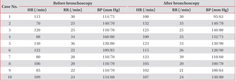
Case No. Before bronchoscopy After bronchoscopy
HR (/min) RR (/min) BP (mm Hg) HR (/min) RR (/min) BP (mm Hg)
1 113 30 114/75 100 30 95/63
2 70 25 140/70 132 35 140/70
3 120 25 110/70 125 25 140/80
4 88 34 160/80 100 25 132/75
5 130 36 120/80 123 33 130/90
6 122 22 109/83 115 26 120/90
7 80 28 110/70 123 39 110/60
8 106 20 110/70 105 20 100/70
9 85 22 110/70 102 21 100/64
10 109 24 134/60 107 24 130/80
HR: heart rate; RR: respiration rate; BP: blood pressure.
Figure 1. (A) Prebronchoscopy and postbronchoscopy saturation and SpO
2/FiO
2ratio in diagnostic bronchoscopy. (B) Prebronchoscopy and postbronchoscopy saturation and SpO
2/FiO
2ratio in therapeutic intervention. SpO
2: peripheral arterial pulse oximetry; FiO
2: fraction of inspired oxygen.
1 2 3 4 5 6 8 450
425
400
375 SpO/FiOratio22
350
Pre- and postbronchoscopy for diagnosis
A B
Case
1 2 3 4 5 6 8
Before bronchoscopy (room air) SpO (%)2
80 87 87 86 80 88 77
SpO2/FiO ratio2 380 414 414 409 380 419 366
After bronchoscopy (on room air after 5 min) SpO (%)2
80 88 93 88 80 80 87
SpO2/FiO ratio2 381 419 443 419 381 381 414
7 9 10 450
425
400
375 SpO/FiOratio22
350
Pre- and postbronchoscopy for intervention
Case
7 9 10
Before bronchoscopy (room air) SpO (%)2
86 86 84
SpO2/FiO ratio2 409 409 400
After bronchoscopy (on room air after 5 min) SpO (%)2
93 91 94
SpO2/FiO ratio2 443 443 448