ISSN 2234-3806 • eISSN 2234-3814
https://doi.org/10.3343/alm.2019.39.3.299
Chromosomal Microarray Analysis as a First-Tier
Clinical Diagnostic Test in Patients With Developmental Delay/Intellectual Disability, Autism Spectrum
Disorders, and Multiple Congenital Anomalies:
A Prospective Multicenter Study in Korea
Woori Jang, M.D.1,2, Yonggoo Kim, M.D.1,2, Eunhee Han, M.D.1,2, Joonhong Park, M.D.1,2, Hyojin Chae, M.D.1,2,
Ahlm Kwon, M.T.2, Hayoung Choi, M.T.2, Jiyeon Kim, M.T.2, Jung-Ok Son, M.T.2, Sang-Jee Lee, M.D.3, Bo Young Hong, M.D.4, Dae-Hyun Jang, M.D.5, Ji Yoon Han, M.D.6, Jung Hyun Lee, M.D.7, So Young Kim, M.D.8, In Goo Lee, M.D.6,
In Kyung Sung, M.D.6, Yeonsook Moon, M.D.9, Myungshin Kim , M.D.1,2, and Joo Hyun Park, M.D.10
1Department of Laboratory Medicine and 2Catholic Genetic Laboratory Center, College of Medicine, The Catholic University of Korea, Seoul, Korea; 3Department of Rehabilitation Medicine, Daejeon St. Mary’s Hospital, College of Medicine, The Catholic University of Korea, Daejeon, Korea; 4Department of Rehabilitation Medicine, St. Vincent’s Hospital, College of Medicine, The Catholic University of Korea, Suwon, Korea; 5Department of Rehabilitation Medicine, Incheon St.
Mary’s Hospital, College of Medicine, The Catholic University of Korea, Incheon, Korea; 6Department of Pediatrics, College of Medicine, The Catholic University of Korea, Seoul, Korea; 7Department of Pediatrics, St. Vincent’s Hospital, College of Medicine, The Catholic University of Korea, Suwon, Korea; 8Department of Pediatrics, Yeouido St. Mary’s Hospital, College of Medicine, The Catholic University of Korea, Seoul, Korea; 9Department of Laboratory Medicine, Inha University School of Medicine, Incheon, Korea; 10Department of Rehabilitation Medicine, College of Medicine, The Catholic University of Korea, Seoul, Korea Background: To validate the clinical application of chromosomal microarray analysis (CMA)
as a first-tier clinical diagnostic test and to determine the impact of CMA results on patient clinical management, we conducted a multicenter prospective study in Korean patients diagnosed as having developmental delay/intellectual disability (DD/ID), autism spectrum disorders (ASD), and multiple congenital anomalies (MCA).
Methods: We performed both CMA and G-banding cytogenetics as the first-tier tests in 617 patients. To determine whether the CMA results directly influenced treatment recom- mendations, the referring clinicians were asked to complete a 39-item questionnaire for each patient separately after receiving the CMA results.
Results: A total of 122 patients (19.8%) had abnormal CMA results, with either patho- genic variants (N=65) or variants of possible significance (VPS, N=57). Thirty-five well- known diseases were detected: 16p11.2 microdeletion syndrome was the most common, followed by Prader–Willi syndrome, 15q11-q13 duplication, Down syndrome, and Duch- enne muscular dystrophy. Variants of unknown significance (VUS) were discovered in 51 patients (8.3%). VUS of genes putatively associated with developmental disorders were found in five patients: IMMP2L deletion, PTCH1 duplication, and ATRNL1 deletion. CMA results influenced clinical management, such as imaging studies, specialist referral, and laboratory testing in 71.4% of patients overall, and in 86.0%, 83.3%, 75.0%, and 67.3%
of patients with VPS, pathogenic variants, VUS, and benign variants, respectively.
Conclusions: Clinical application of CMA as a first-tier test improves diagnostic yields and the quality of clinical management in patients with DD/ID, ASD, and MCA.
Key Words: Chromosomal microarray analysis, Pathogenic, Variant of possible significance, Variant of unknown significance, Benign, Clinical management, Developmental delay, In- tellectual disability, Autism spectrum disorders, Multiple congenital anomalies
Received: May 27, 2018 Revision received: August 6, 2018 Accepted: November 7, 2018
Corresponding author: Myungshin Kim, M.D.
https://orcid.org/0000-0001-8632-0168 Department of Laboratory Medicine, The Catholic University of Korea, 222 Banpo- daero, Seocho-gu, Seoul 06591, Korea Tel: +82-2-2258-1645
Fax: +82-2-2258-1719 E-mail: [email protected]
© Korean Society for Laboratory Medicine This is an Open Access article distributed under the terms of the Creative Commons Attribution Non-Commercial License (http://creativecom- mons.org/licenses/by-nc/4.0) which permits unrestricted non-commercial use, distribution, and reproduction in any medium, provided the original work is properly cited.
2017-03-16 https://crossmark-cdn.crossref.org/widget/v2.0/logos/CROSSMARK_Color_square.svg
INTRODUCTION
Copy number variations (CNVs) have become increasingly rec- ognized as significant contributors to human diseases [1], largely owing to technical progress of genome-wide analysis. Chromo- somal microarray analysis (CMA) is a powerful tool for the ge- nome-wide detection of invisible small chromosomal deletions or duplications.
In 2010, CMA was recommended as a first-tier diagnostic tool for patients with unexplained developmental delay/intellectual disability (DD/ID), autism spectrum disorders (ASD), and multi- ple congenital anomalies (MCA) [2, 3]. CMA results have shown perfect concordance with results from FISH or multiplex ligation- dependent probe amplification (MLPA), and provide a much higher diagnostic yield than traditional karyotyping (15–20% vs 3%) [2, 4].
However, it is not always clear if and how physicians consider genomic medicine for patient care, which is another important issue with regard to the implementation of new genetic tests in routine clinical care. The Analytic validity, Clinical validity, Clini- cal utility and associated Ethical, legal and social implications (ACCE) model provides a framework for evaluating the clinical utility of emerging genetic tests for clinical practice [5]. Recently, a few proof-of-concept studies on how CMA results affect patient management demonstrated the overall clinical utility of CMA [6- 8]. However, most of the research conducted in this field to date has been descriptive, using data from retrospective chart re- views. Therefore, we conducted a multicenter prospective study to assess the clinical application of CMA as the first-tier diagnos- tic test in Korean patients with DD/ID, ASD, and MCA, as well as the impact of CMA results on patient clinical management.
METHODS
Study populationA total of 712 individuals (617 patients and 95 family members) were referred from six Korean hospitals (Seoul St. Mary’s Hospi- tal and Yeouido St. Mary’s Hospital in Seoul, Incheon St. Mary’s Hospital and Inha University Hospital in Incheon, St. Vincent’s Hospital in Suwon, and Daejeon St. Mary’s Hospital in Daejeon) between February 2013 and January 2017 after providing in- formed consent. Patients were referred by physicians as part of clinical assessment for DD, ID, ASD, MCA, or a combination of those features with unexplained etiology. We performed both CMA and G-banding cytogenetics as the first-tier cytogenetic di- agnostic tests. When available, the origin of any imbalance was
determined through analysis of parental samples. The study pro- tocol was approved by the Institutional Review Board of Seoul St Mary’s Hospital, The Catholic University of Korea (KC17TESI0517).
Study design
The referring physicians were asked to complete a questionnaire to determine whether the CMA results had directly influenced their treatment recommendations. The questionnaire items fo- cused on the clinicians’ opinions of the following criteria: (1) de- mographic details and clinical features, such as neurodevelop- mental disorders (DD, learning disability, seizures, ID, speech delay, and ASD), congenital anomalies, dysmorphic features, abnormal growth (failure to thrive and short stature) and hypo- tonia; (2) clinical management prompted by CMA results, in- cluding pharmacological management (indication and contrain- dication for drug treatment), specialist referral, diagnostic imag- ing studies, and laboratory tests. Developmental surveillance (i.e., ongoing monitoring of development, identification of risk factors, and elicitation of parental concerns) was not included as part of direct clinical management [9, 10]. Clinicians com- pleted the questionnaire for each patient separately after receiv- ing the CMA results. Follow-up periods ranged from six to 53 months. We did not include genetic counseling, confirmatory MLPA/FISH, or parental testing results performed to clarify the inheritance of CNVs as part of clinical management because these practices should be standard after abnormal CMA results.
Banding cytogenetics
Banding cytogenetics was performed on G-banded metaphase chromosomes of cultured peripheral blood lymphocytes using routine techniques. Karyotypes were interpreted according to the International System for Human Cytogenetic Nomenclature (ISCN) 2016 [11].
Array comparative genomic hybridization and interpretation Genomic DNA was extracted from a whole blood sample col- lected in an EDTA tube. Comparative genomic hybridization (CGH) array analysis was performed with the SurePrint G3 Hu- man CGH Microarray 8X 60K kit (Agilent Technologies, Santa Clara, CA, USA), according to the manufacturer’s instructions.
Scanned images were quantified using Agilent Feature Extrac- tion software (v. 10.0). Resulting data were imported into Agilent Genomic Workbench 7.0.4.0 software for visualization. CNVs were detected using the Aberration Detection Method-2 (ADM- 2) algorithm. Genomic positions were defined according to the human reference genome hg19/GRCh37.
CNVs were classified into four groups: pathogenic, variants of possible significance (VPS), variants of unknown significance (VUS), and benign [2, 12]. We used the DGV, DECIPHER, Clin- Gen, Online Mendelian Inheritance in Man (OMIM), and dbVar databases, and peer-reviewed literature to determine clinically significant CNVs. Pathogenic variants or VPS were considered abnormal. When available, the known deletion/duplication found via CMA was confirmed by FISH or MLPA. The term “VUS” was used when the imbalance was >200 kb for deletions and >500 kb for duplications involving multiple genes that had never or rarely been reported in normal population controls or candidate genes for an inherited disease, but the significance of the imbal- ance could not be determined based on available knowledge or family studies. CNVs were considered benign when reported as a normal variant in healthy controls or detected in ≥1% of our patient population.
Statistical analysis
Differences in the frequency of clinical features (DD, learning disability, seizures, ID, speech delay, ASD, congenital anoma- lies, dysmorphic features, failure to thrive, short stature, and hy- potonia) and management (pharmacological management, spe- cialist referral, imaging studies, and laboratory testing) between groups were investigated using Fisher’s exact test for categorical variables and the Mann–Whitney U test for continuous variables.
Statistical analyses were performed using SPSS 12.0.1 for Win- dows (SPSS Inc., Chicago, IL, USA). P <0.05 (two-sided) indi- cated statistical significance.
RESULTS
Characterization of detected CNVs
Abnormal CNVs were detected in 122 of the 617 patients (patho- genic, N=65; VPS, N=57), representing overall diagnostic yield of 19.8%. VUS, excluding cases with abnormal CNVs, were found in 51 patients (8.3%), while benign CNVs were found in 444 pa- tients (72.0%) (Supplemental Data Fig. S1). The diagnostic yields of CMA were higher than those obtained with banding cytoge- netics (38/617, 6.2%, P <0.001). Aneuploidy accounted for 8.2% (10/122) of cases with abnormal results. Three patients showed an abnormal karyotype with normal CMA results, in- cluding one patient each with balanced translocation, low-level mosaicism, and marker chromosome. No incidental CNV re- sults involving cancer predisposing genes were detected in pa- tients with abnormal CNVs.
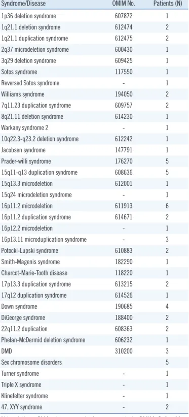
Altogether, 65 patients (10.5%) showed pathogenic variants
associated with well-known genetic diseases. Rearrangements in 15q11-q13, 16p11.2, 1q21.1, 7q11.23, and 22q11.2 were
Table 1. Classification of pathogenic CMA results identified in 65 patients with pathogenic variants
Syndrome/Disease OMIM No. Patients (N)
1p36 deletion syndrome 607872 1
1q21.1 deletion syndrome 612474 2
1q21.1 duplication syndrome 612475 2
2q37 microdeletion syndrome 600430 1
3q29 deletion syndrome 609425 1
Sotos syndrome 117550 1
Reversed Sotos syndrome - 1
Williams syndrome 194050 2
7q11.23 duplication syndrome 609757 2
8q21.11 deletion syndrome 614230 1
Warkany syndrome 2 - 1
10q22.3-q23.2 deletion syndrome 612242 1
Jacobsen syndrome 147791 1
Prader-willi syndrome 176270 5
15q11-q13 duplication syndrome 608636 5
15q13.3 microdeletion 612001 1
15q24 microdeletion syndrome - 1
16p11.2 microdeletion 611913 6
16p11.2 duplication syndrome 614671 2
16p12.2 microdeletion - 1
16p13.11 microduplication syndrome - 3
Potocki-Lupski syndrome 610883 2
Smith-Magenis syndrome 182290 1
Charcot-Marie-Tooth disease 118220 1
17p13.3 duplication syndrome 613215 2
17q12 duplication syndrome 614526 1
Down syndrome 190685 4
DiGeorge syndrome 188400 2
22q11.2 duplication 608363 2
Phelan-McDermid deletion syndrome 606232 1
DMD 310200 3
Sex chromosome disorders 5
Turner syndrome - 1
Triple X syndrome - 1
Klinefelter syndrome - 1
47, XYY syndrome - 2
Abbreviations: CMA, chromosomal microarray analysis; OMIM, Online Men- delian Inheritance in Man; DMD, Duchenne muscular dystrophy.
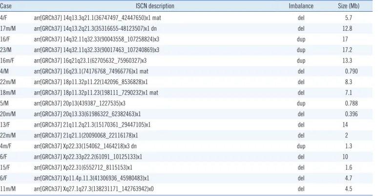
Table 2. CMA results identified in 57 patients with variant of possible significance
Case ISCN description Imbalance Size (Mb)
24m/F arr[GRCh37] 1q22q24.1(156132786_166047765)x3 dup 9.9
5/M arr[GRCh37] 1q25.2q31.3(177898011-198465186)x1 del 21
12/F arr[GRCh37] 1q43q44(240039421_249212668)x1 mat, 18q21.31q23(54037167_77982126)x3 mat del/dup* 9.2/23.9
4/M arr[GRCh37] 2p25.3p25.1(42444_7304259)x3 dup 7.3
29m/M arr[GRCh37] 2q22.1q22.3(142036895_145533609)x1 del 3.4
10/F arr[GRCh37] 2q11.1q12.3(95529039_108083956)x3 mat, 18p11.32p11.31(142096_5853122)x1 dn dup/del 12.6/5.7
neo/M arr[GRCh37] 2q32.1(186763813_188960123)x3 dn dup 2.2
3/M arr[GRCh37] 3p26.3(270649_1125759)x3 dup 0.855
11m/F arr[GRCh37] 3p26.3p26.1(93949_4994502)x1, 15q25.1q26.3(80190103_102465355)x3 del/dup 4.9/22
11m/M arr[GRCh37] 3p11.2p13(76026268_90254062)x1 del 14.2
1m/F arr[GRCh37] 4q35.1q35.2(185274461_190469337)x1 pat, 10p15.3p11.23(148206_29975521)x3 pat del/dup* 5.2/30
35m/M arr[GRCh37] 5q13.3(73470360_74032634)x1 del 0.562
14m/F arr[GRCh37] 5q21.3(106716799_108175671)x3 dup 1.4
9/M arr[GRCh37] 5q31.2(137260366_138206885)x3 dup 0.946
3/M arr[GRCh37] 5q35.2(175437847_176491972)x1 del 1.1
5m/M arr[GRCh37] 6p25.3p25.2(170426_2794740)x1 mat del 2.6
26m/M arr[GRCh37] 6p25.3p25.1(170426_5431448)x1 del 5.3
4/F arr[GRCh37] 6q14.3q15(86185546_88051322)x1 del 1.9
9m/F arr[GRCh37] 6q26q27(163357909_170890108)x1 del 7.5
5/F arr[GRCh37] 6q27(166754981_167569353)x1 del 0.814
19m/F arr[GRCh37] 6q12(66205374_67257639)x1 pat del 1.1
6/M arr[GRCh37] 7q36.1q36.3(149128443_159088636)x3 dn, 9p24.3(271257_2183334)x1 dn dup/del 10/1.9
5/F arr[GRCh37] 7q36.2q36.3(153933437_158909738)x1 del 5
20m/F arr[GRCh37] 8p23.3p23.1(221611_6914076)x1, 8p23.1p12(12583259_29936174)x3 del/dup 6.7/17.4 18m/M arr[GRCh37] 8p23.3p23.1(221611_7753583)x1 dn, 12p13.33p13.31(230421_8238072)x3 dn del/dup 7.5/8.0
8m/M arr[GRCh37] 8q21.11q21.13(76069471_81532974)x1 dn del 5.5
42/F arr[GRCh37] 8q23(113498500_114173066)x1, 12p13.33p13.32(230421_3394129)x1 del/del 0.674/3.2
23m/M arr[GRCh37] 9p24.3p13.3(271257_35163255)x3 dup 35
15m/M arr[GRCh37] 9p13.3p13.1(33414184_39156954)x1 dn del 5.7
18m/F arr[GRCh37] 9q33.2q33.3(124664562_127176303)x1 dn del 2.5
9m/M arr[GRCh37] 10p15.3p15.1(193492_6135095)x3 dup 5.9
4/M arr[GRCh37] 11p14.3p14.1(24063998_30323839)x1 del 6.3
16/F arr[GRCh37] 11q24.2q24.3(126830381_128391970)x3, 11q24.3q25(106396480_106513022)x1 dup/del 1.6/6.4
12/F arr[GRCh37] 12p13.33p13.32(230421_3394129)x1 del 3.2
2m/M arr[GRCh37] 12p13.33p11.1(450479_34345585)x2-3 dup 34
26/M arr[GRCh37] 12p13.33p11.23(230421_27768451)x3, 18p11.32(142096_1038964)x1 dup/del 27.5/0.897
3/M arr[GRCh37] 13q12.3(30656355_31905182)x3 dup 1.2
4/M arr[GRCh37] 13q31.1q31.2(85888171_87980615)x1 mat del 2.1
4/F arr[GRCh37] 13q33.3q34(109683987_115059020)x1 del 5.4
6m/M arr[GRCh37] 14q13.2q13.3(35316655_37777710)x1 dn del 2.5
(Continued to the next page)
frequently found (Table 1 and Supplemental Data Fig. S2A). Al- though cancer was not present at diagnosis, four patients were diagnosed as having syndromes in which cancer is a reported feature (Sotos, Warkany, and DiGeorge syndromes). The 67 ab- errations classified as VPS, detected in 57 patients, did not over- lap with the CNVs previously identified to be related to known syndromes, but they were large in size and found in gene-rich areas, implicating their contribution to the abnormal phenotype (Table 2 and Supplemental Data Fig. S2B). VPS were mutually exclusive except for two siblings with a 14q32.11-q32.33 dupli- cation. With the exception of the 10 aneuploidy cases, the size of the pathogenic variants ranged from 142 kb (exons 45–57 of the DMD gene) to 10.2 Mb (supernumerary marker chromo- some containing a duplication of 15q11.1-q13.2), and the ma- jority (45/55, 81.8%) were less than 5 Mb. The size of the VPS ranged from 396 kb to 35 Mb, and approximately a half of the VPS (36/67, 53.7%) were larger than 5 Mb (Supplemental Data Table S1).
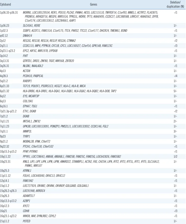
Excluding cases with abnormal CNVs, VUS were discovered in 51 patients (8.3%, 51/617), including 47 with one VUS and four with two VUS. Another three patients with VUS also had a concurrent pathogenic variant (15q11.1q13.1 duplication) or
VPS (4q35.2 duplication and 5q13.3 deletion) (Table 3). The most promising result was five patients with gene dose altera- tions associated with putatively developmental disorders. Three patients showed a microdeletion in 7q31.1 encompassing the IMMP2L (MIM 605977) gene. Among them, one patient had a maternally inherited small supernumerary marker chromosome of 15q11.1-q13.1 as well as a VUS. The other two patients had a microduplication, including the PTCH1 (MIM 601309) gene and a microdeletion in the ATRNL1 (MIM 612869) region, re- spectively.
Patient characteristics and clinical features according to the detected CNVs
The general demographic features of the patients are summa- rized in Supplemental Data Table S2. Overall, 77% (472/617) of the patients were 0–5 years old, and the percentage of males was greater than that of females (60.3% vs 39.7%, P <0.001).
At least one symptom of neurodevelopmental disorders was de- tected in most patients (95.1%), and DD and speech delay were common (91.2% and 78.7%, respectively).
The mean±SD number of clinical features was 4.4±1.7 among patients with pathogenic variants, and was 4.8±1.8, 4.0±1.9,
Case ISCN description Imbalance Size (Mb)
4/F arr[GRCh37] 14q13.3q21.1(36747497_42447650)x1 mat del 5.7
17m/M arr[GRCh37] 14q13.2q21.3(35316655-48123507)x1 dn del 12.8
16/F arr[GRCh37] 14q32.11q32.33(90043558_107258824)x3 dup 17
23/M arr[GRCh37] 14q32.11q32.33(90017463_107240869)x3 dup 17.2
16m/F arr[GRCh37] 16q21q23.1(62705632_75960327)x3 dup 13.3
4/M arr[GRCh37] 16q23.1(74176768_74966776)x1 mat del 0.790
22m/M arr[GRCh37] 18p11.32p11.22(142096_8536828)x1 del 8.3
18m/M arr[GRCh37] 18p11.32p11.23(198111_7290232)x1 mat del 7.1
5/M arr[GRCh37] 20p13(439387_1227535)x3 dup 0.788
20m/M arr[GRCh37] 20q13.33(61986322_62382463)x1 del 0.396
13/F arr[GRCh37] 21q11.2q21.3(15170361_29447105)x1 del 14
22m/M arr[GRCh37] 21q21.1(20090068_22116178)x1 del 2
4m/F arr[GRCh37] Xp22.33(154062_1464218)x3 dn dup 1.3
6/F arr[GRCh37] Xp22.33p22.2(61091_10125133)x1 del 10
15/F arr[GRCh37] Xp22.31(6552712_8115153)x1 del 1.6
6/F arr[GRCh37] Xp11.4p.11.3(41306936_45980483)x1 del 4.7
11m/M arr[GRCh37] Xq27.1q27.3(138231171_142763942)x0 del 4.5
*Two patients had a concurrent deletion and duplication in two different chromosomal regions inherited from parents with a balanced translocation.
Abbreviations: ISCN, International System for Human Cytogenetic Nomenclature; m, months; M, Male; F, Female; neo, neonate; CMA, chromosomal micro- array analysis; Mb, megabase; mat, maternal origin; dn, de novo; pat, paternal origin; del, deletion; dup, duplication.
Table 2. Continued
Table 3. CMA results identified in patients with variants of unknown significance
Cytoband Genes Deletion/
duplication (N) 1p36.33-p36.31 MORN1, LOC100129534, RER1, PEX10, PLCH2, PANK4, HES5, LOC115110, TNFRSF14, C1orf93, MMEL1, ACTRT2, FLJ42875,
PRDM16, ARHGEF16, MEGF6, MIR551A, TPRG1L, WDR8, TP73, KIAA0495, CCDC27, LOC388588, LRRC47, KIAA0562, DFFB, C1orf174, LOC100133612, LOC284661, AJAP1
-/1
1p36.23 SLC45A1, RERE 1/-
1p32.3 SSBP3, ACOT11, FAM151A, C1orf175, TTC4, PARS2, TTC22, C1orf177, DHCR24, TMEM61, BSND -/1
q42.12 DNAH14 1/-
2p12 REG3G, REG1B, REG1A, REG1P, REG3A, CTNNA2 -/2
2q21.1 CCDC115, IMP4, PTPN18, CFC1B, CFC1, LOC150527, C2orf14, GPR148, FAM123C -/3
2q23.1-q23.2 EPC2, KIF5C, MIR1978, LYPD6B -/1
3p14.2 FHIT 1/-
3q13.31 QTRTD1, DRD3, ZNF80, TIGIT, MIR568, ZBTB20 1/-
3q26.31 NLGN1, NAALADL2 -/1
4p13 KCTD8 1/-
4q28.3 PCDH10, PABPC4L -/4
5q21.2 RAB9P1 1/-
6p21.33 TCF19, POU5F1, PSORS1C3, HCG27, HLA-C, HLA-B, MICA 1/-
6p21.32 HLA-DRB6, HLA-DRB1, HLA-DQA1, HLA-DQB1, HLA-DQA2, HLA-DQB2, HLA-DOB, TAP2 5/-
6q12 EYS, MCART3P 1/-
6q13 COL19A1 1/-
6q16.1 EPHA7, TSG1 -/1
7p21.3p-p21.2 ETV1, DGKB -/2
7p21.2 DGKB 1/-
7q11.21 INTS4L1, ZNF92 2/-
7q11.23 UPK3B, LOC100133091, POMZP3, PMS2L11, LOC100132832, CCDC146, FGL2 1/-
7q31.1 IMMP2L 3/-
9p23 TYRP1 1/-
9p21.2 MOBKL2B, IFNK, C9orf72 1/-
9q22.32 PTCH1, C9orf130, C9orf102 -/1
10p15.3-p15.2 PFKP, PITRM1 -/1
10q11.22 PPYR1, LOC728643, ANXA8, ANXA8L1, FAM25B, FAM25C, FAM25G, LOC642826, FAM35B2 1/2
10q23.31 RNLS, LIPJ, LIPF, LIPK, LIPN, LIPM, ANKRD22, STAMBPL1, ACTA2, FAS, CH25H, LIPA, IFIT2, IFIT3, IFIT1L, IFIT1, IFIT5, SLC16A12,
PANK1, MIR107 1/-
10q25.3 ATRNL1 1/-
11p11.12 FOLH1, LOC440040, OR4C13, OR4C12 -/1
12q14.1 FAM19A2 -/1
15q11.2 LOC727924, OR4M2, OR4N4, OR4N3P, GOLGA8D, GOLGA6L1 1/-
15q26.2-q26.3 LOC91948, ARRDC4 -/1
15q26.3 ADAMTS17 1/-
16p13.3-p13.2 A2BP1 -/1
16p12.3 XYLT1 -/1
16q21 CDH8 -/3
16q23.1-q23.2 WWOX, MAF, DYNLRB2, CDYL2 -/1
21q11.2 POTED 1/-
and 3.9±1.8 in the VPS, VUS, and benign groups, respectively (pathogenic vs VPS, P =0.167; pathogenic vs VUS, P =0.478;
pathogenic vs benign, P =0.054; VPS vs VUS, P =0.086; VPS vs benign, P =0.001, and VUS vs benign, P =0.451). Patients with pathogenic variants or VPS were considered a single group in our analysis because no significant differences were found in the rate of clinical features and management after CMA between these two groups. The frequency of clinical features associated
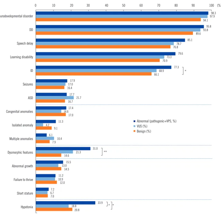
with developmental disorders, except for ASD, were the highest in the patients with abnormal variants, followed by those with VUS and those with benign variants. Frequencies of ID, dysmor- phic features, and hypotonia differed among the three groups (P =0.029, P <0.001, and P =0.006, respectively). These fea- tures were more common in patients carrying abnormal variants than in those with benign variants (ID, 77.3% vs 66.1%, P <0.001;
dysmorphic features, 31.0% vs 14.6%, P =0.016; hypotonia,
Fig. 1. Evaluation of clinical features in patients with DD/ID, ASD, and MCA. Significant differences in the frequencies of ID, dysmorphic features, and hypotonia were found among the three groups (P =0.029, P <0.001, and P =0.006, respectively).
*P <0.05; **P <0.001.
Abbreviations: DD, developmental delay; ID, intellectual disability; ASD, autism spectrum disorders; MCA, multiple congenital anomalies; VUS, variants of unknown significance.
Neurodevelopmental disorder
DD
Speech delay
Learning disability
ID
Seizures
ASD
Congenital anomalies
Isolated anomaly
Multiple anomalies
Dysmorphic features
Abnormal growth
Failure to thrive
Short stature
Hypotonia
0 10 20 30 40 50 60 70 80 90 100 (%)
97.998.3 94.1
95.8 93.8 89.6 85.1 76.878.7
79.6 73.3 70.9
77.3
*
**
* *
68.9 66.1 17.9
17.0 16.4
17.7 21.7 16.7 17.4 14.6
17.0 11.3 4.2 9.1
6.1 7.910.4
31.0 14.6 21.3
15.5 13.014.5
11.2 10.912.0
6.77.2 7.0
18.8 33.9 20.8
Abnormal (pathogenic+VPS, %) VUS (%)
Benign (%)
33.9% vs 20.8%, P =0.003) (Fig. 1).
Clinical management following CMA
Among the 581 patients available for follow-up, 415 (71.4%)
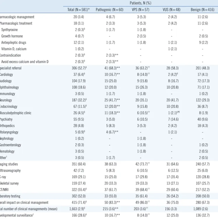
Table 4. Summary of recommendations of clinical management in response to CMA results Patients, N (%)
Total (N=581)* Pathogenic (N=60) VPS (N=57) VUS (N=48) Benign (N=416)
Pharmacologic management 20 (3.4) 4 (6.7) 3 (5.3) 2 (4.2) 11 (2.6)
Pharmacologic treatment 18 (3.1) 2 (3.3) 3 (5.3) 2 (4.2) 11 (2.6)
Synthyroxine 2 (0.3)§ 1 (1.7) 1 (1.8) - -
Growth hormone 4 (0.7) - 2 (3.5) - 2 (0.5)
Antiepileptic drugs 12 (2.1) 1 (1.7) 1 (1.8) 1 (2.1) 9 (2.2)
Vitamin D, calcium 1 (0.2) - - 1 (2.1) -
Contraindication 2 (0.3)§ 2 (3.3)** - - -
Avoid excess calcium and vitamin D 2 (0.3)§ 2 (3.3)** - - -
Specialist referral 306 (52.7)§ 41 (68.3)** 36 (63.2)†† 28 (58.3) 201 (48.3)
Cardiology 37 (6.4)§ 10 (16.7)** 8 (14.0)†† 2 (4.2)ll 17 (4.1)
Audiology 104 (17.9) 15 (25.0) 9 (15.8) 8 (16.7) 72 (17.3)
Ophthalmology 108 (18.6) 12 (20.0) 15 (26.3) 10 (20.8) 71 (17.1)
Immunology 3 (0.5) 1 (1.7) 1 (1.8) - 1 (0.2)
Neurology 187 (32.2)§ 25 (41.7)** 20 (35.1) 20 (41.7) 122 (29.3)
Endocrinology 67 (11.5)§ 12 (20.0)** 9 (15.8) 10 (20.8) 36 (8.7)
Musculodystrophic clinic 26 (4.5)§ 11 (18.3)** 6 (10.5)†† 1 (2.1)ll,¶ 8 (1.9)
Psychiatry 55 (9.5) 3 (5.0) 6 (10.5) 7 (14.6) 40 (9.6)
Orthopedics 28 (4.8) 5 (8.3) 3 (5.3) 2 (4.2) 18 (4.3)
Otolaryngology 5 (0.9)§ 4 (6.7)** - 1 (2.1) -
Nephrology 1 (0.2) - 1 (1.8) - -
Gastroenterology 2 (0.3) - 1 (1.8) - 1 (0.2)
Hematology 3 (0.5) - 1 (1.8) - 2 (0.5)
Other† 3 (0.5) 1 (1.7) - - 2 (0.5)
Imaging studies 351 (60.4) 38 (63.3) 42 (73.7)†† 31 (64.6) 240 (57.7)
Ultrasonography 42 (7.2) 5 (8.3) 6 (10.5) 6 (12.5) 25 (6.0)
X-ray 169 (29.1) 15 (25.0) 17 (29.8) 17 (35.4) 120 (28.8)
Skeletal survey 159 (27.4) 20 (33.3) 19 (33.3) 13 (27.1) 107 (25.7)
CT/MRI 322 (55.4)§ 37 (61.7) 39 (68.4)†† 29 (60.4) 217 (52.2)
Laboratory testing 302 (52.0) 33 (55.0) 35 (61.4) 26 (54.2) 208 (50.0)
Overall impact on clinical management 415 (71.4)§ 50 (83.3)** 49 (86.0)†† 36 (75.0) 280 (67.3) Total number of clinical managements (mean) 1,663 (2.9)§ 215 (3.6)** 203 (3.6)†† 156 (3.3) 1,089 (2.6)
Developmental surveillance‡ 166 (28.6)§ 10 (16.7)** 8 (14.0)†† 12 (25.0) 136 (32.7)
*Follow-up was available for 581 patients (follow-up periods: six–53 months); †Other: Urology for one patient with abnormal variants, and dermatology and general surgery for two patients with benign variants; ‡Developmental surveillance indicates ongoing monitoring of development, identification of risk factors, and elicitation of parental concerns; §P <0.05 among the four groups; llP <0.05, pathogenic vs VUS; ¶P <0.05, VPS vs VUS; **P <0.05, pathogenic vs benign;
††P <0.05, VPS vs benign.
Abbreviations: CMA, chromosomal microarray analysis; VPS, variants of possible significance; VUS, variants of unknown significance; CT, computed tomog- raphy; MRI, magnetic resonance imaging.
were given at least one recommendation of clinical manage- ment (Table 4). A total of 1,663 new management strategies were recommended, demonstrating that a mean of 2.9 new recommendations per patient were prompted by CMA results.
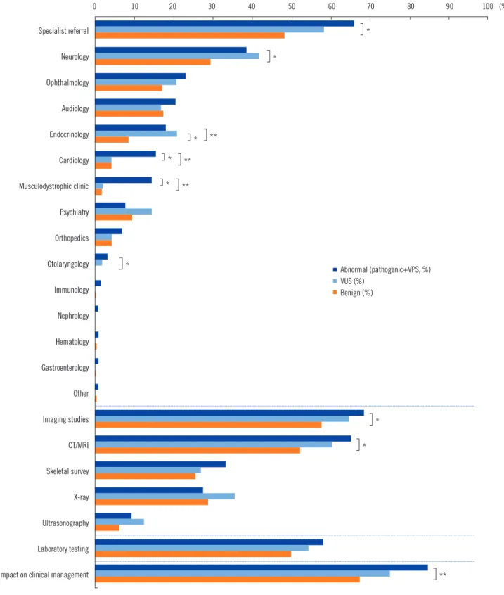
Fig. 2. Rate of clinical management recommendations following CMA.
*P <0.05; **P <0.001.
Abbreviations: CMA, chromosomal microarray analysis; VUS, variants of unknown significance; CT, computed tomography; MRI, magnetic resonance imaging.
Specialist referral
Neurology
Ophthalmology
Audiology
Endocrinology
Cardiology
Musculodystrophic clinic
Psychiatry
Orthopedics
Otolaryngology
Immunology
Nephrology
Hematology
Gastroenterology
Other
Imaging studies
CT/MRI
Skeletal survey
X-ray
Ultrasonography
Laboratory testing
Overall impact on clinical management
0 10 20 30 40 50 60 70 80 90 100 (%)
*
*
**
**
**
*
**
*
*
*
*
*
Abnormal (pathogenic+VPS, %) VUS (%)
Benign (%) Computed tomography (CT)/magnetic resonance imaging (MRI)
studies were recommended for more than half of all patients
(55.4%), and the most common CT/MRI types were of the brain.
Ultrasonography examination was recommended for 42 patients,
80% of which included echocardiogram and 20% included ab- domen and kidney ultrasound. Clinical management was not recommended after a benign CMA result in 23.4% (136/581) of all patients. Patients with abnormal variants consulted with spe- cialists for cardiology, neurology, endocrinology, and musculo- dystrophy more frequently than patients with benign CNVs did (P <0.001, P =0.040, P =0.005, and P <0.001, respectively).
CT/MRI imaging was more frequently recommended for patients with abnormal variants than for those with benign variants (P = 0.009) (Fig. 2). Pharmacological management was recommended for 20 patients (3.4%). Thyroid hormone medication was rec- ommended for treating hypothyroidism in two patients with 1p36 deletion syndrome and 13q31.1q31.2 deletion, respectively. Two patients with Williams syndrome were advised to avoid taking multivitamins with extra calcium or vitamin D to prevent hyper- calcemia, while one patient was treated with vitamin D and cal- cium supplements after excluding Williams syndrome.
DISCUSSION
The translation of research results to public health applications has been unexpectedly slow in many countries, including Ko- rea, although CMA has an enhanced diagnostic yield compared with standard karyotype analysis for patients with developmental disabilities [2, 3]. One of the main barriers to the clinical adop- tion of CMA is the lack of standardization for reporting results.
However, guidelines for the three- to five-level interpretative cat- egories of CNVs and the expansion of open-access databases of patient cohorts or healthy controls allow for the provision of pre- cise information with high reproducibility [2, 8, 11]. In our study, CMA revealed clinically relevant chromosomal imbalances in 19.8% of patients, which is similar to the previously reported di- agnostic yield of 15–20% [2]. When CMA was used as a first- tier diagnostic test [13-15], the detection rate was much higher than that obtained using CMA as a second-tier test after stan- dard karyotyping (18–30% vs 7–14%) [16, 17].
The classification and reporting of VUS remains a challenge.
Although parental testing is recommended to clarify the clinical significance of a variant detected, most disorders associated with CNVs show no clear genotype-phenotype correlation, even within the same family. In this study, 8.3% of the patients had chromo- somal imbalances of still unclear clinical relevance, which is con- sistent with rates reported in earlier studies (5–14%) [13]. How- ever, classification of VUS varies considerably across studies [17-19]. Recent analyses of variant classifications reported in ClinVar showed that among the 11% of variants with more than
one submitter, 17% showed different interpretations [20]. For example, the duplication of 8p23.2, including the CSMD1 gene has been detected in patients with speech delay, autism, and learning difficulties [18] as well as in normal individuals [19].
Duplication of the CSMD1 gene was reported as a VUS with a frequency of 3.1% (3/96) in a previous study [17], while we classified such variants as benign according to our laboratory’s current variant classification criteria (≥1% of the patient popu- lation), that is, 1.8% (11/617) of patients and 3.2% (3/95) of normal family members in our cohort.
Despite these limitations, VUS may be a good candidate gene and pathway marker for rare developmental disorders. We de- tected a 7q31.1 deletion, including IMMP2L in three unrelated patients with DD, learning disability, ID, and speech delay. In- deed, microdeletion in 7q31.1 encompassing the IMMP2L gene has been suggested as a susceptibility factor for neurodevelop- mental disorders, such as Tourette syndrome [21, 22]. Duplica- tion of the PTCH1 gene has also been reported in a family with microcephaly and DD [23]. Our patient with a PTCH1 duplica- tion exhibited not only DD and microcephaly but also polydac- tyly, tongue papilloma, and corpus callosum dysgenesis. In ad- dition, 10q25.3 deletion, including the ATRNL1 gene has been reported in a patient with cognitive impairment, autism, and dys- morphic facial features [24]. A female neonate with 10q25.3 deletion in our cohort had congenital heart defects, including an atrial septal defect and ventricular septal defect. However, be- cause of the very young age of the patient, we were not able to determine whether cognitive impairment or autism is present.
Results obtained through a “reverse genetics” approach and further collaborative efforts will help definitively characterize the role of candidate genes in pathogenesis.
Assessment of the patients’ clinical features revealed a ten- dency for a higher frequency of clinical abnormalities in the group with abnormal variants, which is consistent with the aforemen- tioned studies [6, 25]. In our study, the frequency of ID, dys- morphic features, and hypotonia were significantly higher in pa- tients with abnormal variants than in those with benign variants.
Developmental disorders and congenital anomalies or dysmor- phic features have been reported in most patients with abnor- mal CMA results [6]. Other studies reported that facial abnor- malities [26], heart defects [25] and ID and a family history of ID/MCA/ASD [14] were more common in patients with abnor- mal CMA results.
Another obstacle hindering the widespread clinical applica- tion of CMA as the first-tier cytogenetic test is related to the un- certainty of whether the testing will directly influence medical
management. Although recent studies have correlated abnor- mal CMA results with predicted clinical impact [6-8, 27], most of them were performed in a single institution, based solely on medical records [6-8]. Moreover, an appropriate control was not applied to prove that such intervention would not have occurred with patients who had not received a positive CMA result [6, 8].
Thus, we queried the referring clinicians regarding follow-up clinical management to assess the impact of CMA results. Ap- proximately 85% of patients with clinically relevant variants re- ceived more direct clinical management in our study. These re- sults support those of earlier studies [6, 8, 27, 28], demonstrat- ing that abnormal CMA results contributed to medical manage- ment in a substantial proportion (34–94%) of patients with DD/
ID, MCA, and ASD. Differences in the extent of clinical manage- ment might be attributed to the different health care systems among countries as well as the patient heterogeneity across stud- ies. Only one other study [27] has assessed medical recommen- dations following benign CMA results, finding that patients with benign CNVs received a mean of 2.7 medical recommendations.
Similarly, clinical management was recommended for patients with benign variants (mean: 2.6 recommendations) in our study.
Specifically, compared with benign CMA results, abnormal CMA variants were a significant driver of medical recommendations;
VUS results also drove recommendations, but to a lesser extent.
These results suggest that some additional diagnostic tests can be avoided in patients with negative CMA results, which could lead to tangible savings in healthcare expenditures.
Even when no specific cure is available or when some genetic diagnoses may have minimal impact on patient management, establishing a clear diagnosis through genetic testing may lead to earlier initiation of medical care and consequently improve outcomes for patients and their families who have endured a
“diagnostic odyssey.” In addition, along with the development of whole-genome analysis using genome-wide arrays, recurrent CNVs associated with ID/DD, ASD, and MCA have been labeled as novel microdeletion/duplication syndromes [29, 30]. There is now published literature supporting specific clinical management implications for at least 146 conditions potentially diagnosable by CMA [7]. Medical knowledge regarding pathogenic CNVs will also continue to progress.
Although CMA has a higher resolution than conventional karyo- typing, polyploidy, balanced translocations, inversion, low-level mosaicism, and marker chromosomes may be missed [3]. A benign CMA result does not exclude all genetic diseases. There- fore, for these patients, a next-generation sequencing approach as a subsequent diagnostic test may aid in establishing the di-
agnosis [31, 32].
Overall, this prospective multicenter study highlights the clini- cal application of CMA as a first-tier testing in patients with DD/
ID, ASD, and MCA. CMA results directly affect the subsequent clinical management strategy, and the impact is not limited to patients with abnormal or negative results. Thus, the widespread use of CMA in clinical settings has potential to improve the effi- ciency and quality of clinical management for these patients.
Authors’ Disclosures of Potential Conflicts of Interest
The authors declare that they have no competing interests.
Acknowledgements
We are grateful to the patients and parents, and to The Catholic Genetic Laboratory Center for assisting us in carrying out this study and compiling this report. We thank Samkwang Medical Laboratories for their valuable support for this project. This re- search was supported by the Bio & Medical Technology Devel- opment Program of the National Research Foundation (NRF) funded by the Ministry of Science & ICT (2018M3A9E8020866) and Research Fund of Seoul St. Mary’s Hospital, The Catholic University of Korea.
REFERENCES
1. Girirajan S, Campbell CD, Eichler EE. Human copy number variation and complex genetic disease. Annu Rev Genet 2011;45:203-26.
2. Miller DT, Adam MP, Aradhya S, Biesecker LG, Brothman AR, Carter NP, et al. Consensus statement: chromosomal microarray is a first-tier clinical diagnostic test for individuals with developmental disabilities or congenital anomalies. Am J Hum Genet 2010;86:749-64.
3. Manning M, Hudgins L, Professional Practice and Guidelines Commit- tee. Array-based technology and recommendations for utilization in medi- cal genetics practice for detection of chromosomal abnormalities. Genet Med 2010;12:742-5.
4. Sanmann JN, Pickering DL, Golden DM, Stevens JM, Hempel TE, Althof PA, et al. Assessing the utility of confirmatory studies following identifi- cation of large-scale genomic imbalances by microarray. Genet Med 2015;17:875-9.
5. Haddow JE and Palomaki GE. ACCE: a model process for evaluating data on emerging genetic tests. In: Khoury MJ, Little J, Burke W, eds.
Human genome epidemiology: a scientific foundation for using genetic information to improve health and prevent disease. New York: Oxford University Press, 2004:217-33.
6. Henderson LB, Applegate CD, Wohler E, Sheridan MB, Hoover-Fong J, Batista DA. The impact of chromosomal microarray on clinical manage- ment: a retrospective analysis. Genet Med 2014;16:657-64.
7. Riggs ER, Wain KE, Riethmaier D, Smith-Packard B, Faucett WA, Hopp-
man N, et al. Chromosomal microarray impacts clinical management.
Clin Genet 2014;85:147-53.
8. Coulter ME, Miller DT, Harris DJ, Hawley P, Picker J, Roberts AE, et al.
Chromosomal microarray testing influences medical management. Gen- et Med 2011;13:770-6.
9. Tonelli M, Parkin P, Brauer P, Leduc D, Pottie K, Jaramillo Garcia A, et al.
Recommendations on screening for developmental delay. CMAJ 2016;
188:579-87.
10. Council on Children with Disabilities, Section on Developmental Behav- ioral Pediatrics, Bright Futures Steering Committee, Medical Home Ini- tiatives for Children With Special Needs Project Advisory Committee.
Identifying infants and young children with developmental disorders in the medical home: an algorithm for developmental surveillance and screening. Pediatrics 2006;118:405-20.
11. McGowan-Jordan J, Simons A, Schmid M. eds. ISCN : an international system for human cytogenomic nomenclature (2016). Basel: Karger, 2016.
12. Kearney HM, Thorland EC, Brown KK, Quintero-Rivera F, South ST, Work- ing Group of the American College of Medical Genetics Laboratory Qual- ity Assurance Committee. American College of Medical Genetics stan- dards and guidelines for interpretation and reporting of postnatal consti- tutional copy number variants. Genet Med 2011;13:680-5.
13. Bartnik M, Wiśniowiecka-Kowalnik B, Nowakowska B, Smyk M, Kędzior M, Sobecka K, et al. The usefulness of array comparative genomic hy- bridization in clinical diagnostics of intellectual disability in children. Dev Period Med 2014;18:307-17.
14. Cappuccio G, Vitiello F, Casertano A, Fontana P, Genesio R, Bruzzese D, et al. New insights in the interpretation of array-CGH: autism spectrum disorder and positive family history for intellectual disability predict the detection of pathogenic variants. Ital J Pediatr 2016;42:39.
15. Ozyilmaz B, Kirbiyik O, Koc A, Ozdemir TR, Kaya OO, Guvenc MS, et al.
Experiences in microarray-based evaluation of developmental disabili- ties and congenital anomalies. Clin Genet 2017;92:372-9.
16. Xiang B, Zhu H, Shen Y, Miller DT, Lu K, Hu X, et al. Genome-wide oli- gonucleotide array comparative genomic hybridization for etiological di- agnosis of mental retardation: a multicenter experience of 1499 clinical cases. J Mol Diagn 2010;12:204-12.
17. Shin S, Yu N, Choi JR, Jeong S, Lee KA. Routine chromosomal microar- ray analysis is necessary in Korean patients with unexplained develop- mental delay/mental retardation/autism spectrum disorder. Ann Lab Med 2015;35:510-8.
18. Glancy M, Barnicoat A, Vijeratnam R, de Souza S, Gilmore J, Huang S, et al. Transmitted duplication of 8p23.1-8p23.2 associated with speech delay, autism and learning difficulties. Eur J Hum Genet 2009;17:37-43.
19. Harada N, Takano J, Kondoh T, Ohashi H, Hasegawa T, Sugawara H, et al. Duplication of 8p23.2: a benign cytogenetic variant? Am J Med Gen-
et 2002;111:285-8.
20. Rehm HL, Berg JS, Brooks LD, Bustamante CD, Evans JP, Landrum MJ, et al. ClinGen–the Clinical Genome Resource. N Engl J Med 2015;
372:2235-42.
21. Bertelsen B, Melchior L, Jensen LR, Groth C, Glenthøj B, Rizzo R, et al.
Intragenic deletions affecting two alternative transcripts of the IMMP2L gene in patients with Tourette syndrome. Eur J Hum Genet 2014;22:
1283-9.
22. Gimelli S, Capra V, Di Rocco M, Leoni M, Mirabelli-Badenier M, Schiaffino MC, et al. Interstitial 7q31.1 copy number variations disrupting IMMP2L gene are associated with a wide spectrum of neurodevelopmental disor- ders. Mol Cytogenet 2014;7:54.
23. Derwińska K, Smyk M, Cooper ML, Bader P, Cheung SW, Stankiewicz P.
PTCH1 duplication in a family with microcephaly and mild developmen- tal delay. Eur J Hum Genet 2009;17:267-71.
24. Stark Z, Bruno DL, Mountford H, Lockhart PJ, Amor DJ. De novo 325 kb microdeletion in chromosome band 10q25.3 including ATRNL1 in a boy with cognitive impairment, autism and dysmorphic features. Eur J Med Genet 2010;53:337-9.
25. Shoukier M, Klein N, Auber B, Wickert J, Schröder J, Zoll B, et al. Array CGH in patients with developmental delay or intellectual disability: are there phenotypic clues to pathogenic copy number variants? Clin Genet 2013;83:53-65.
26. Lee CG, Park SJ, Yun JN, Ko JM, Kim HJ, Yim SY, et al. Array-based comparative genomic hybridization in 190 Korean patients with devel- opmental delay and/or intellectual disability: a single tertiary care uni- versity center study. Yonsei Med J 2013;54:1463-70.
27. Hayeems RZ, Hoang N, Chenier S, Stavropoulos DJ, Pu S, Weksberg R, et al. Capturing the clinical utility of genomic testing: medical recom- mendations following pediatric microarray. Eur J Hum Genet 2015;23:
1135-41.
28. Saam J, Gudgeon J, Aston E, Brothman AR. How physicians use array comparative genomic hybridization results to guide patient management in children with developmental delay. Genet Med 2008;10:181-6.
29. Nevado J, Mergener R, Palomares-Bralo M, Souza KR, Vallespín E, Mena R, et al. New microdeletion and microduplication syndromes: a com- prehensive review. Genet Mol Biol 2014;37:210-9.
30. Weise A, Mrasek K, Klein E, Mulatinho M, Llerena JC Jr, Hardekopf D, et al. Microdeletion and microduplication syndromes. J Histochem Cy- tochem 2012;60:346-58.
31. Trujillano D, Bertoli-Avella AM, Kumar Kandaswamy K, Weiss ME, Köster J, Marais A, et al. Clinical exome sequencing: results from 2819 sam- ples reflecting 1000 families. Eur J Hum Genet 2017;25:176-82.
32. Grozeva D, Carss K, Spasic-Boskovic O, Tejada MI, Gecz J, Shaw M, et al. Targeted next-generation sequencing analysis of 1,000 individuals with intellectual disability. Hum Mutat 2015;36:1197-204.
Supplemental Data Table S1. Size distribution of CNVs found in patients
Size (Mb)
Number of CNVs (%) Number of
pathogenic variants (N=65)
Number of VPS
(N=67) Number of VUS (N=58)
<0.5 4 (6.2) 1 (1.5) 23 (39.7)
0.5–1 6 (9.2) 8 (11.9) 16 (27.6)
1–5 35 (53.8) 22 (32.8) 18 (31.0)
5–10 9 (13.8) 20 (29.9) 1 (1.7)
>10 11 (17.0) 16 (23.9) 0 (0.0)
Abbreviations: CNV, copy number variation; VPS, variants of possible signifi- cance; VUS, variants of unknown significance.
Supplemental Data Table S2. Demographic and clinical features of patients according to CMA results Total
N (%) Pathogenic
N (%) VPS
N (%) VUS
N (%) Benign
N (%) Gender
Male 372/617 (60.3) 34/65 (52.3) 33/57 (57.9) 23/51 (45.1) 282/444 (63.5)
Female 245/617 (39.7) 31/65 (47.7) 24/57 (42.1) 28/51 (54.9) 162/444 (36.5)
Age (yr)
<2 218/617 (35.3) 22/65 (33.8) 25/57 (43.9) 20/51 (39.2) 151/444 (34.0)
2–5 254/617 (41.2) 29/65 (44.6) 18/57 (31.6) 17/51 (33.3) 190/444 (42.8)
5–10 66/617 (10.7) 7/65 (10.8) 4/57 (7.0) 6/51 (11.8) 49/444 (11.0)
>10 79/617 (12.8) 7/65 (10.8) 10/57 (17.5) 8/51 (15.7) 54/444 (12.2)
Clinical features
Neurodevelopmental disorder 561/589 (95.2) 59/61 (96.7) 57/57 (100) 47/48 (97.9) 398/423 (94.1)
Developmental delay 536/588 (91.2) 56/61 (91.8) 57/57 (98.2) 45/48 (93.8) 378/422 (89.6)
Learning disability or behavioral or psychiatric disorder 390/535 (72.9) 44/57 (77.2) 42/51 (82.4) 33/45 (73.3) 271/382 (70.9)
Seizures 98/584 (16.8) 9/61 (14.8) 12/56 (21.4) 8/47 (17.0) 69/420 (16.4)
Intellectual disability 372/542 (68.6) 42/57 (73.7) 43/53 (81.1) 31/45 (68.9) 256/387 (66.1)
Speech delay 435/553 (78.7) 49/60 (81.7) 48/54 (88.9) 37/47 (78.7) 301/392 (76.8)
Autism spectrum disorder 95/549 (17.3) 12/58 (20.7) 8/55 (14.5) 10/46 (21.7) 65/390 (16.7)
Congenital anomalies 97/580 (16.7) 9/58 (15.5) 11/57 (19.3) 7/48 (14.6) 71/418 (17.0)
Isolated anomaly 52/580 (9.0) 8/58(13.8) 5/57 (8.8) 2/48 (4.2) 38/418 (9.1)
Multiple anomalies 45/580 (7.8) 1/58 (1.7) 6/57 (10.5) 5/48 (10.4) 33/418 (7.9)
Dysmorphic features 107/580 (18.4) 17/59 (28.8) 19/57 (33.3) 10/47 (21.3) 61/417 (14.6)
Abnormal growth 82/563 (14.6) 9/60 (15.0) 9/56 (16.1) 6/46 (13.0) 58/401 (14.5)
Failure to thrive 66/562 (11.7) 6/60 (10.0) 7/56 (12.5) 5/46 (10.9) 48/400 (12.0)
Short stature 37/529 (7.0) 4/58 (6.9) 4/53 (7.5) 3/45 (6.7) 26/373 (7.0)
Hypotonia 136/584 (23.3) 19/61 (31.1) 21/57 (36.8) 9/48 (18.8) 87/418 (20.8)
Abbreviations: CMA, chromosomal microarray analysis; VPS, variants of possible significance; VUS, variants of unknown significance.
Supplemental Data Fig. S1. Overview of patient enrollment, chro- mosomal microarray analysis results, and clinical follow-up.
Abbreviations: VPS, variants of possible significance; VUS, variants of unknown significance.
All patients
(N=617) Parents/siblings (N=95)
Pathogenic variants (N=65, 10.5%)
VPS (N=57, 9.2%)
VUS (N=51, 8.3%)
Benign variants (N=444, 72.0%)
Clinical follow-up available (N=416) Clinical follow-up
available (N=48) Clinical follow-up
available (N=61)
Clinical follow-up available
(N=56)
Supplemental Data Fig. S2. Regions of chromosomal duplication and deletion in patients with (A) pathogenic variants and (B) VPS. Red bars indicate specific regions of duplication, and green bars indicate deletion of chromosomes for each patient.
Abbreviation: VPS, Variants of possible significance.
Duplication
Duplication
Deletion
Deletion A
B