미세아교세포의 염증반응에 미치는 청뇌명신환의 영향
임용균, 최영현, 황원덕
동의대학교 한의과대학 대학원
Anti-inflammatory Effects of Cheongnoimyungshin-hwan in Microglia Cells
Yong-Gyun Im, Yung-Hyun Choi, Won-Deok Hwang
Department of Oriental Medicine Graduate School, Dong-Eui University
Received: December 5, 2014 Revised: December 17, 2014 Accepted: December 18, 2014
Objectives: Activated microglia cells play an important role in inflammatory responses in the central nervous system (CNS) which are involved in neurodegenerative diseases. We attempted to determine the anti-inflammatory effects of Cheongnoimyungshin-hwan (CNMSH) in microglia cells.
Methods: We examined the effect of CNMSH on the inflammatory responses in BV2 microglia cells in- duced by lipopolysaccharide (LPS) and explored the mechanism underlying the action of CNMSH.
Results: BV2 cells treated with LPS showed an up-regulation of nitric oxide (NO), prostaglandin PGE
2(PGE
2) and interleukin 1 (IL-1) release, whereas CNMSH suppressed this up-regulation. CNMSH in- hibited the induction of COX-2, iNOS and IL-1 proteins in LPS-treated BV2 cells and blocked the LPS-induced phosphorylation and nuclear translocation of nuclear factor B (NF-B). Furthermore, CNMSH attenuated the LPS-induced phosphorylation of extracellular signal-regulated kinase and p38 mitogen activated protein kinase (MAPK), as well as the phosphoinositide 3-kinase (PI3K)/Akt signaling pathway, but did not inhibit the LPS-induced phosphorylation of c-Jun amino terminal kinase.
Conclusions: These results suggest that the inhibitory effect of CNMSH on the LPS-induced production of inflammatory mediators and cytokines in BV2 cells is associated with the suppression of the NF-B and PI3KAkt signaling pathways.
Key Words: Cheongnoimyungshin-hwan (CNMSH), Microglia cells, Neurodegenerative diseases, NF-B, PI3KAkt.
Correspondence to Won-Deok Hwang
Department of Oriental Medicine Graduate School, Dong-Eui University, Yangjeong 2-dong, Busanjin-gu, Busan, Korea.
Tel: +82-51-850-8625 Fax: +82-51-867-5162 E-mail: [email protected]
Copyright © 2014 by The Korean Society of Oriental Neuropsychiatry. All rights reserved.
CC
This is an open access article distributed under the terms of the Creative Commons Attribution Non-Commercial License (http://creativecommons.org/licenses/by-nc/3.0) which
permits unrestricted non-commercial use, distribution, and reproduction in any medium, provided the original work is properly cited.
I. 서론
미세아교세포는 신경교세포의 일종으로 중추신경계에서 식세포 역할을 담당하여 기본적인 면역방어체계의 역할을 수행하며, 중추신경계의 항상성 유지에 중요한 역할을 하는 것으로 알려져 있다1-3). 하지만 세균감염 후 lipopoly- saccharide (LPS)와 같은 독소는 미세아교세포를 과다하게 활성화시켜 신경독소, 염증매개인자 및 염증성 사이토카인 들의 분비가 증가하고, 결과적으로 신경퇴행성 질환의 원인 이 되기도 한다4-6). 따라서 신경퇴행성 질환의 예방과 치료에 있어서 미세아교세포의 과도한 활성화의 억제는 신경퇴행성 질환 퇴치 후보 약물의 중요한 표적이 될 수 있을 것이다.
청뇌명신환(淸腦明神丸)은 동의대부속한방병원 신계내과 황원덕교수에 의해 작방된 것으로 기억력증진과 치매예방을 목적으로 개발된 환약이다. 본 처방은 원대(元代) 위역림(危 亦林)이 황제께 진상한 공진단(拱辰丹)을 근간으로 하고 있 다. 위역림의 의서인 세의득효방(世醫得效方)에 최초로 언 급된 공진단에 대해 동의보감 잡병편 허로(東醫寶鑑 雜病篇 虛勞)에서는 타고난 원기를 든든히 하여 오장자화(五藏自 和), 백병불생(百病不生)하게 하는 데에 공진단을 쓴다고 하 였다7). 본 환은 공진단의 사향(麝香), 녹용(鹿茸), 당귀(當歸) 등을 주재로 하였으며, 공진단의 산수유(山茱萸)를 산약(山 藥)으로 바꾸었고, 본방에 용안육(龍眼肉), 원지(遠志), 인삼 (人蔘), 육계(肉桂), 부자(附子), 침향(沈香), 용뇌(龍腦)를 추 가하여 구성되어져 있다. 본 연구에 사용된 청뇌명신환은 황원덕교수의 위탁을 받아 저자가 임상에서 사용중인 처방 이다.
그러나 청뇌명신환의 효능에 비하여 그에 대한 실험적인 근거는 전무한 실정이다. 따라서 본 연구에서는 LPS에 의해 자극된 BV2 생쥐 미세아교세포의 염증반응 억제에 미치는 청뇌명신환의 기전을 조사하고, 신경퇴행성 질환 예방 및 치료 후보제로서의 청뇌명신환의 효능을 확인하였다.
II. 재료 및 방법
1. 시료준비
본 실험에 사용된 청뇌명신환(淸腦明神丸, Cheongnoi- myungshin-hwan, CNMSH)은 인삼(Ginseng Radix Alba), 당귀(Angelicae Gigantis Radix), 산약(Dioscoreae Rhizoma)
과 용안육(Longan Arillus)을 각 24 g, 육계(Cinnamomi Cortex) 12 g, 원지(Polygalae Radix) 6 g, 사향(Moschus) 과 침향(Aquilariae Lignum) 각 1 g, 용뇌(Borneolum, 중 국) 0.2 g 이상을 세말한 후, 여기에 녹용(cornus cervi parvum) 24 g과 부자(Aconiti Lateralis Radix Preparata) 6 g을 5시간 정도 끊인 교(膠) 24 g을 추가한 후, 꿀 160 g으로 구성된 총 300.2 g의 환약을 4 g 씩으로 75개를 만든 후, 무작위 1환을 선정하여 금박으로 입혀서, 이를 멸균된 3차 증류수를 이용하여 100 mg/ml의 농도로 만든 다음 0.2
m 필터로 여과 후 적정 농도로 배지에 희석하여 처리하였 다.
2. 항체 구입
본 실험에서 단백질 분석을 위하여 사용된 항-IL-1, 항-
-actin, 항-extracellular signal-regulated kinase (ERK), 항-p38 mitogen-activated protein kinase (MAPK) 및 항- Akt 항체는 Santa Cruz Biotechnology Inc. (Santa Cruz, CA, USA)에서 구입하였으며, TNF-, pERK, c-Jun amino terminal kinase (JNK), p-JNK, p-p38 MAPK, nuclear factor B (NF-B) (p65)-s468, NF-B (p65)-s538 항체 는 Cell Signaling Technology, Inc. (Danvers, MA, USA) 에서 구입하였다. Immunoblotting을 위해 2차 항체로 사 용된 horseradish peroxidase (HRP)-conjugated anti- mouse 및 anti-rabbit 항체는 Santa Cruz Biotechnology Inc.에서 구입하였다.
3. 세포배양
실험에 사용된 생쥐 신경소교세포주인 BV2 세포는 인제 대학교 의과대학 미생물학교실에서 분양 받아 사용하였으 며, 세포의 배양을 위해 10%의 우태아혈청 (fetal bovine serum, FBS, Gibco BRL, Grand Island, NY, USA)과 1%의 penicillin과 streptomycin이 포함된 Dulbecco’s Modified Eagle’s Medium (DMEM) 배지(Gibco BRL)를 사용하여 37oC, 5% CO2 조건 하에서 배양하였다. 세포수의 증식에 따른 과밀도 현상을 해소하기 위하여 매 48시간마다 계대배 양을 실시하여 적정수의 세포를 유지하였다.
4. MTT assay에 의한 세포 성장억제 조사
청뇌명신환 처리에 의한 세포의 성장억제 정도를 확인하
Table 1. Antibodies Used in the Present Study
Antibody Origin Company
Akt Rabbit polyclonal Santa Cruz Biotechnology pAkt Rabbit polyclonal Cell signaling
COX-2 Rabbit polyclonal Santa Cruz Biotechnology ERK Mouse monoclonal Santa Cruz Biotechnology pERK Mouse monoclonal Cell signaling
iNOS Rabbit polyclonal Santa Cruz Biotechnology JNK Mouse monoclonal Cell signaling
pJNK Mouse monoclonal Cell signaling
NF-κB(p65) Rabbit polyclonal Santa Cruz Biotechnology pNF-κB(p65)
s438
Rabbit polyclonal Cell signaling pNF-κB(p65)
s568
Rabbit polyclonal Cell signaling
β-actin raBBIT polyclonal Santa Cruz Biotechnology
기 위하여 tetrazolium bromide salt (MTT) assay를 이용 하였다. 먼저 6 well에 배양된 BV2 세포에 청뇌명신환 (0∼
500 ug/ml)과 LPS (500 ng/ml) (Sigma-Aldrich Chemical Co., St. Louis, MO, USA)를 상기 농도로 처리하고 24시간 동안 배양한 후 배지를 제거하고 MTT (Sigma-Aldrich Chemical Co.)를 0.5 mg/ml 농도로 희석하여 200 l씩 분주하고 37oC에서 3시간 동안 다시 배양하였다. 배양이 끝 난 다음 MTT 시약을 제거하고 dimethylsulfoxide (DMSO, Sigma-Aldrich Chemical Co.)를 1 ml씩 분주하여 well에 생성된 formazin을 모두 녹인 후 96 well plate에 200 l씩 옮겨서 ELISA reader (Molecular Devices, Sunnyvale, CA, USA)로 540 nm에서 흡광도를 측정하였다. 측정은 모두 세 번을 하였으며, 그에 대한 평균값과 표준 오차를 Microsoft EXCEL program을 사용하여 분석하였다.
5. Nitric Oxide (NO) 생성량의 측정
NO의 농도는 배양액 내의 nitrite농도를 Griess Reagent (Sigma-Aldrich Chemical Co.)를 이용하여 측정하였다. 이 를 위하여 BV2 세포에 각 농도별로 청뇌명신환을 1시간 선 처리한 후 500 ng/ml의 LPS를 처리하여 24시간 배양한 후 세포 배양액을 수거하였다. 배양액 100 l와 동량의 Griess Reagent를 상온에서 반응 시킨 후 ELISA reader로 540 nm 에서 흡광도를 측정하였는데, sodium nitrite (NaNO2)의 농도 별 표준곡선을 이용하여 배양액 내의 NO 농도를 결정 하였다.
6. PGE
2생성량의 측정
PGE2 생성량 측정을 위해 사용한 PGE2 enzyme im- munoassay (EIA) kit는 Cayman Chemicals Co. (Ann Arbor, MI, USA)에서 구입하였다. BV2 세포에 125, 250, 500 ug/ml 농도의 청뇌명신환을 1시간 선 처리 후, LPS (500 ng/ml)를 처리한 배지에서 24시간 동안 세포를 배양 시킨 후 상층액만 이용하여 PGE2 EIA kit에 제시된 방법에 따라 처리한 다음 ELISA reader를 이용한 420 nm의 흡광 도로 반응의 정도를 비교하였다.
7. Cytokine 생성량의 측정
세포배양액 내의 pro-inflammatory cytokine (IL-1 및 TNF-)의 양을 측정하기 위한 Quantikine enzyme-linked
immunospecific assay (ELISA)는 R&D systems (Minnea- polis, MN, USA)에서 구입하였다. 동일한 조건에서 배양된 BV2 세포의 배양액을 이용하여 IL-1 및 TNF-의 생성 양을 제시된 방법에 따라 처리한 다음 ELISA reader를 이용 한 450 nm의 흡광도로 반응의 정도를 측정하였다.
8. Western blot analysis에 의한 단백질 발현의 분석
준비된 세포에 적당량의 lysis buffer [25 mM Tris-Cl (pH 7.5), 250 mM NaCl, 5 mM EDTA, 1% NP-40, 1 mM phenylmethylsulfonyl fluoride (PMSF), 5 mM dithio- threitol (DTT)]를 첨가하여 4oC에서 1시간 동안 반응시킨 후, 14,000 rpm으로 30분간 원심 분리하여 상층액에 있는 총 단백질을 분리하였다. 상층액의 단백질 농도는 Bio-Rad 단백질 정량 시약(Bio-Rad, Hercules, CA, USA)과 그 사용 방법에 따라 정량한 다음 동량의 Laemmli sample buffer (Bio-Rad)를 섞어서 sample을 만들었다. 동량의 sample을 sodium dodecyl sulphate (SDS)-polyacrylamide gel을 이용하여 전기영동으로 분리한 후, nitrocellulose mem- brane (Schleicher and Schuell, Keene, NH, USA)으로 electroblotting에 의해 전이시켰다. 분리된 단백질이 전이 된 nitrocellulose membrane을 5% skim milk를 처리하여 비특이적인 단백질들에 대한 blocking을 실시하고 1차 항 체(Table 1)를 처리하여 상온에서 2시간 이상 또는 4oC에서 over night 시킨 다음 PBS-T로 세척(15분간 1번, 5분간 5 번)하고 처리된 1차 항체에 맞는 2차 항체(PBS-T로 1:1,500
Fig. 1. The CNMSH and/or in combination with lipopolysaccharide (LPS) on BV2 microglia viability. The cells were incubated for 24 h at the indicated concentrations of CNMSH with or without LPS (500 ng/ml). In combination treatment, CNMSH were pretreated 1 h before LPS treatment. Cell viability was determined by the MTT assay. Data are expressed as mean±SE of three independent experiments.
Fig. 2. The effects of CNMSH on NO production and iNOS expression in LPS-stimulated BV2 microglia. Cells were pre-treated with 125, 250 and 500 g/mL of CNMSH 1 h before LPS treatment for 24 h. (A) Following 24 h treatment, and the amounts of NO production were measured with the supernatants. Data are expressed as mean±SD of three independent experiments (*p<0.05 as compared with control group,
#p<0.05 as compared with LPS treated group). (B) Following 24 h treatment, the expression of iNOS were assessed by Western blot analysis. Actin was used as the internal control.
으로 희석하여 사용)를 사용하여 상온에서 1시간 정도 반응 시켰다. 반응이 끝난 후 암실에서 Enhanced Chemilumi- noesence (ECL) solution (Santa Cruz Biotechnology Inc.)을 적용시킨 다음 X-ray film에 감광시켜 특정단백질 의 발현양을 분석하였다.
9. 통계처리
다양한 실험으로부터 얻은 결과는 mean±standard error 로 기록하였고, 유의성 검증은 one-way ANOVA 분석법을 이용하여 결정하였다.
III. 결과
1. BV2 생쥐 미세아교세포 생존도에 미치는 청뇌명신환 영향
가장 먼저 생쥐 미세아교세포 BV2에서 항염증 효과에 대 한 청뇌명신환의 농도 범위를 설정하기 위해 MTT 분석을 이용하여 세포 생존도를 측정해 보았다. Fig. 1에서 보이는 바와 같이, 청뇌명신환을 0∼500 ug/ml 범위 내에서 LPS 500 ng/ml과 복합 및 단독처리 하였을 때, BV2 생쥐 미세 아교세포의 세포 생존도에는 영향을 미치지는 않는다는 것 을 확인할 수 있었다. 이에 향후 실험에서는 청뇌명신환의 최고 처리농도를 500 ug/ml으로 설정하여 진행하였다.
2. NO 생성과 inducible NO synthese (iNOS)의 발현에 미치는 청뇌명신환 영향
Fig. 1의 결과를 바탕으로 청뇌명신환의 처리 농도(125
∼500 ug/ml)를 설정 이후에 BV2 생쥐 미세아교세포에서 LPS에 의해 유도되는 NO의 생성과 iNOS 단백질 발현의 증가에 미치는 영향에 대해 조사하였다. Fig. 2A에 나타낸 바와 같이 LPS 500 ng/ml의 단독 처리에 의해 현저히 증가 된 NO의 생성이 각 청뇌명신환의 선 처리에 의해 현저히 억제 되는 것이 확인되었다. NO 뿐만 아니라 NO를 합성하 는데 영향을 미치는 iNOS 단백질을 Western blotting을 통 해 확인해 본 결과, NO 생성 억제와 유사하게 iNOS 단백질 의 발현이 청뇌명신환 처리 농도 의존적으로 억제되었음을 알 수 있었다(Fig. 2B). 이상의 결과에서 염증 반응에 중요한 매개체로 작용하는 NO의 생성을 청뇌명신환이 iNOS의 발 현을 억제함으로서 효과적으로 차단할 수 있다는 것이 확인 되었다.
Fig. 3. The effects of CNMSH on PGE
2production and COX-2 ex- pression in LPS-stimulated BV2 microglia. Cells were pre-treated with 125, 250 and 500 g/mL of CNMSH 1 h before LPS treatment for 24 h.
(A) Following 24 h treatment, and the amounts of PGE
2production were measured with the supernatants. Data are expressed as mean±
SD of three independent experiments (*p<0.05 as compared with control group,
#p<0.05 as compared with LPS treated group). (B) Following 24 h treatment, the expression of COX-2 were assessed by Western blot analysis. Actin was used as the internal control.
Fig. 4. The effects of CNMSH on IL-1 production and protein ex- pression in LPS-stimulated BV2 microglia. Cells were pre-treated with 125, 250 and 500 g/mL of CNMSH 1 h before LPS treatment for 24 h.
(A) Following 24 h treatment, and the amounts of IL-1 production were measured with the supernatants. Data are expressed as mean±
SD of three independent experiments. (*p<0.05 as compared with control group,
#p<0.05 as compared with LPS treated group). (B) Following 24 h treatment, the expression of IL-1 were assessed by Western blot analysis. Actin was used as the internal control.
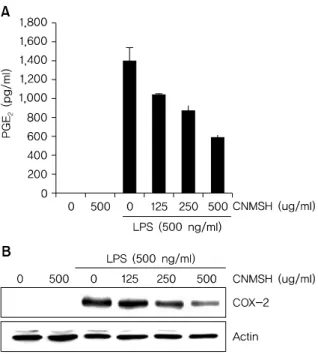
3. PGE
2의 생성과 cyclooxygenase-2 (COX-2)의 발현에 미치는 청뇌명신환 영향
BV2 생쥐 미세아교세포에 최대한 독성이 없는 농도 범위 에서 LPS에 의해 유도되는 PGE2의 생성과 COX-2 단백질 의 발현에 미치는 영향에 대해 조사하였다. Fig. 3A에서 나 타낸 바와 같이 LPS 500 ng/ml 처리에 의해 현저히 증가된 PGE2의 생성이 청뇌명신환의 선 처리에 의해 처리 농도 의 존적으로 현저히 억제 되는 것이 확인되었다. 뿐만 아니라 PGE2를 합성하는데 영향을 미치는 COX-2 단백질을 Western blotting을 통해 확인해 본 결과, PGE2 생성 억제 와 유사하게 청뇌명신환 선처리에 의해서 LPS에 의해 증가 된 COX-2 단백질의 발현이 억제되었음을 알 수 있었다 (Fig. 3B). 이상의 결과에서 염증 반응에서 발열 및 통증에 매개체로 작용하는 PGE2 생성을 청뇌명신환이 COX-2의 발현을 억제함으로써 효과적으로 차단할 수 있음을 알 수 있었다.
4. IL-1 생성과 단백질 발현에 미치는 청뇌명신환 영향
IL-1는 염증성 cytokine으로 활성화된 미세아교세포에 서 생산되어 염증 반응의 중요한 중개자 역할 뿐만 아니라 세포 증식, 분화 및 세포자살 등의 여러 가지 세포 활동에 다양하게 관여하고 있다8). 그에 따라 LPS에 의해 활성화된 BV2 미세아교세포에 유도된 IL-1 생성 및 단백질 발현 증 가에 대한 청뇌명신환의 영향을 조사해 보았다. Fig. 4에 나 타낸 바와 같이 LPS 500 ng/ml 처리에 의해 현저히 증가된 IL-1 생성과 단백질의 발현이 청뇌명신환의 선 처리에 의 해 처리 농도 의존적으로 현저히 억제되었음을 확인하였다.
이상의 결과에 따라 IL-1의 생성과 단백질 발현을 청뇌명 신환이 효과적으로 억제할 수 있다는 것을 알 수 있었다.
Fig. 5. The effects of CNMSH on TNF- production and protein ex- pression in LPS-stimulated BV2 microglia. Cells were pre-treated with 125, 250 and 500 g/mL of CNMSH 1 h before LPS treatment for 24 h.
(A) Following 24 h treatment, and the amounts of TNF- production were measured with the supernatants. Data are expressed as mean±
SD of three independent experiments (*p<0.05 as compared with control group). (B) Following 24 h treatment, the expression of IL-1
were assessed by Western blot analysis. Actin was used as the in-
ternal control. Fig. 6. The effects of CNMSH on NF-B activity in LPS-treated BV2
microglia. Cells were pre-treated with CNMSH 1 h before LPS treat- ment for an hour. (A) Cytosol (30 g) or nuclear protein (30 g) was subjected to 10% SDS-PAGE, followed by Western blotting. The NF-B p65 activity was assessed by Western blot analysis. Actin and lamin B were used as internal controls for cytoplasm and nucleus, respectively.
(B) Localization of NF-B p65 was visualized with fluorescence micro- scopy after immunofluorescence staining with NF-B p65 antibody (green). Cells were stained with DAPI to visualize nuclei (blue). These results are representatives of at least two independent experiments that showed similar patterns.
5. TNF- 생성과 단백질 발현에 미치는 청뇌명신환 영향
TNF-는 가장 대표적인 cytokine으로 면역세포를 조절 하는 역할을 한다. 하지만 TNF- 생산 조절 장애는 치매, 암을 포함한 다양한 인간 질병에 관여한다고 알려져 있다9,10). 그에 따라 LPS에 의해 활성화된 BV2 생쥐미세아교세포에 유도된 TNF-의 생성 및 단백질에 대한 청뇌명신환의 영향 을 조사해 보았다. Fig. 5에 나타낸 바와 같이 청뇌명신환 500 ug/ml의 단독처리군에서도 TNF- 생성이 어느 정도 증가되었을 뿐 아니라, LPS 처리에 의해서 증가된 TNF-
생성과 발현 억제에도 청뇌명신환은 크게 영향을 미치지 못 하는 것으로 확인 되었다. 이상의 결과에 따라 TNF-의 생 성과 단백질 발현에는 청뇌명신환이 유의적인 효과가 없음 을 알 수 있었다.
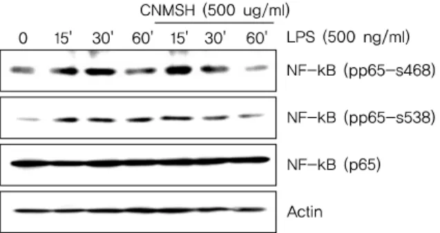
6. NF-B의 활성화 및 인산화에 미치는 청뇌명신환의 영향
활성화된 미세아교세포에서 NF-B는 앞서 확인한 iNOS, COX-2, IL-1 및 TNF- 등의 발현을 조절하는 주요 전사 조절인자로서 작용한다고 알려져 있다11-13). 이에 따라 LPS 에 의한 염증반응에서 NF-B 활성 증가에 미치는 청뇌명신 환의 영향을 확인해 보았다. 먼저 핵과 세포질의 단백질을 분리하여 NF-B의 subunit의 하나인 p65 단백질의 발현 변화를 확인해 본 결과, LPS 처리에 의해 핵 내로 축적된
Fig. 7. The effects of CNMSH on phosphorylation of NF-B in LPS-treated BV2 microglia. Cells were treated with 500 ug/ml CNMSH 1 h before LPS treatment for the indicated times. Total proteins (50 g) were separated on 10% SDS-polyacrylamide gels, followed by Western blotting using the indicated antibodies. Results are representative of those obtained from three independent experiments.
Fig. 8. The effects of CNMSH on MAPK activation in LPS-treated BV2 microglia. Cells were treated with 500 ug/ml CNMSH 1 h before LPS treatment for the indicated times. Total proteins (50 g) were separated on 10% SDS-polyacrylamide gels, followed by Western blotting using the indicated antibodies. Results are representative of those obtained from three independent experiments.
Fig. 9. The effects of CNMSH on phosphorylation of Akt in LPS-treated BV2 microglia. Cells were treated with 500 ug/ml CNMSH 1 h before LPS treatment for the indicated times. Total proteins (50 g) were sep- arated on 10% SDS-polyacrylamide gels, followed by Western blotting using the indicated antibodies. Results are representative of those ob- tained from three independent experiments.
p65 단백질이 청뇌명신환 선처리에 의해서 감소되는 것이 확인되었다(Fig. 6A). 뿐만 아니라 면역형광염색법을 이용 하여 p65 단백질의 세포 내 위치 변화를 확인 본 결과, LPS 처리에 의해 핵 내로 이동한 p65 단백질이 청뇌명신환 선처 리에 의해서 이동이 억제되었음을 알 수 있었다(Fig. 6B).
그리고 NF-B의 활성화는 인산화 과정을 동반한다고 알려 져 NF-B p65 단백질의 인산화 변화를 조사해 본 결과, LPS 처리 15분부터 증가되었던 NF-B p65의 인산화가 청 뇌명신환 선처리에 의해서 현저히 억제되었음을 알 수 있었
다(Fig. 7). 이상 결과에서 청뇌명신환의 의한 NF-B의 핵 내로의 이동 및 활성억제는 NF-B p65 인산화 억제를 통 해 이루어진다는 것을 확인할 수 있었다.
7. MAPK 신호전달계의 활성화에 미치는 청뇌명신환의 영향
다음은 세포신호전달계 중 가장 대표적인 MAPK 신호전 달계와 청뇌명신환의 항염증효과와의 상관관계를 조사하였 다. MAPK 신호전달계는 일반적으로 ERK, JNK 및 p38 MAPK 세 가지 kinase로 이루어져 있으며, 이 단백질들은 하위에 여러가지 표적 단백질을 인산화시켜 활성화를 조절 하는 것으로 보고되어 지고 있다14-16). Fig. 8에서 보이는 바 와 같이, ERK, JNK 및 p38 MAPK 모두 LPS 처리 15분 이내에 인산화형이 증가되어 활성화되었음을 알 수 있었으 며, JNK를 제외한 ERK와 p38 MAPK는 청뇌명신환 선처리 에 의하여 LPS에 의한 활성이 억제되었음을 확인하였다. 이 상의 결과에 따라 청뇌명신환의 항염증 효과에 MAPK 신호 전달계 중 ERK와 p38 MAPK가 관여하고 있음을 확인할 수 있었다.
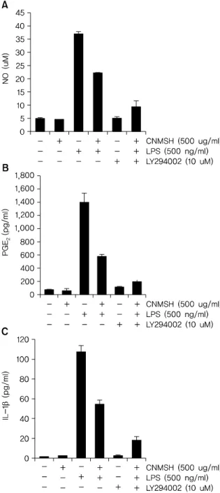
8. Phosphoinositide 3-kinase (PI3K)/Akt 신호전달계의 활성화에 미치는 청뇌명신환의 영향
PI3K/Akt 신호전달계는 NF-B의 활성을 조절할 수 있는 상위 세포 신호전달계의 중 하나이다17,18). 이에 PI3K/Akt 신호전달계와 청뇌명신환의 항염증효과와의 상관관계를 조 사하였다. Fig. 9에서 보이는 바와 같이, LPS 처리에 의해 15분 이내에 활성화되었던 PI3K의 하위 신호 단백질인 AKT가 청뇌명신환 선 처리에 의하여 활성이 억제되는 것이 확인되었다. 뿐만 아니라 PI3K의 억제제로 알려져 있는
Fig. 10. The effects of CNMSH on Akt activation in LPS-treated BV2 microglia. BV2 cells were treated with the indicated does of CNMSH alone and in combination with LY294002 for 1 h before LPS treatment for the indicated times. Following 24 h treatment, the supernatants were taken, and amounts of NO (A), PGE
2(B), IL-1 (C) were meas- ured with the supernatants. The data are shown as means±SD of three independent experiments. (*p<0.05 as compared with control group,
#p<0.05 as compared with LPS treated group).
LY294002를 청뇌명신환과 동시에 처리할 경우, 청뇌명신 환 단독 처리군에 비하여 항염증 효과가 증가되는 것을 확 인할 수 있었다(Fig. 10). 이상의 결과에 따라 청뇌명신환의
항염증 효과에 PI3K/Akt 신호전달계 역시 중요한 상위 신 호계로 작용하고 있음을 알 수 있었다.
IV. 고찰
과다 활성화된 미세아교세포에 의해 야기되는 뇌 염증은 치매, 파킨슨병, 다발성 경화증 및 근위축성 축삭 경화증을 포함한 여러 신경퇴행성 질환의 병리학적 특징이다19-21). 따 라서 과다 활성화된 미세아교세포의 조절은 많은 신경변성 질환의 치료를 위한 유망한 치료법이 될 수 있을 것이다.
본 연구에서는 청뇌명신환의 뇌세포 보호 효과 및 관련 기전의 확인을 위한 선행 연구로서, LPS에 의해 활성화된 BV2 미세아교세포에서 염증 매개 인자와 염증성 사이토카 인의 생성에 미치는 청뇌명신환의 영향을 조사하였다.
청뇌명신환은 원대 위역림이 황제께 진상한 공진단을 근 간으로 하여, 공진단의 산수유를 산약으로 바꾸었고, 본방에 인삼, 육계, 부자, 용안육, 원지, 침향, 용뇌를 추가하여 구성 된 처방이다. 이상의 약재들을 살펴보면, 현대인들의 위장운 동의 부담을 덜어 주기 위하여 공진단의 산수유를 산약으로 바꾸었고, 인삼, 육계, 부자는 전신 기능 활성화를 목적으로 추가하였으며, 용안육, 원지, 침향, 용뇌 등은 뇌의 기능을 활성화하여 현대인의 정신적인 스트레스로 생긴 기억력감 퇴 및 치매예방을 목적으로 추가하였다. 산수유의 성미(性 味)는 산(酸), 미온(微溫)하며, 정기(正氣), 진액(津液)을 수렴 하는 의미가 강하다22). 산약의 성미(性味)는 온무독(溫無毒), 고감(苦甘)하여 비신경(脾腎經)에 귀경(歸經)하여 보비익위 (補脾益胃), 조습화중(燥濕和中)하는 효능이 있다. 또한 위 장관 기능의 문제로 인한 질병 치료에 효능이 있음이 전23), Nishimura N24) 등의 연구에서 밝혀졌다. 그러므로 위장운 동의 부담을 덜어주기 위한 목적으로 산수유를 산약으로 대 체하여 본 처방에서 사용되었다. 인삼은 최근 허25), 김26), 진27) 등의 연구에 의하여 신경세포 손상 방어효과가 있음이 보고되었다. 육계는 고28)의 신생혈관 촉진 생성 작용, 김29) 의 대식세포의 염증반응에 미치는 영향 등이 보고되고 있다.
부자는 Zhu Z30)의 비위온보(脾胃溫補) 기능에 관한 연구가 보고되었다. 따라서 인삼, 육계, 부자는 여러 기전을 통해 전신 기능 활성화를 목적으로 사용되었다. 용안육은 대식세 포의 염증반응과 cytokine에 영향을 미쳐 항염증효과가 있 음이 김31)의 연구에서 보고되었다. 원지는 최신 연구에서
TNF-와 IL-1 등의 염증성 사이토카인을 억제하고 NF-B pathway에 작용하는 기전이 밝혀졌다32). 침향은 비 (脾), 폐(肺), 신(腎)의 기체(氣滯)로 인한 증상을 소통하여 치료하는 약재로서33), 최근 Kim YC44)의 과민반응 억제에 관한 연구가 보고된 바 있다. 용뇌는 항 뇌빈혈 효능이 있음 이 Kong QX34)의 연구에서 보고되었다. 이상의 연구 및 문 헌에 근거하여 용안육, 원지, 침향, 용뇌를 뇌의 기능을 활성 화하여 현대인의 정신적인 스트레스로 생긴 기억력감퇴 및 치매예방을 목적으로 사용하였다.
몇몇 연구에서는, 과다 유도된 COX-2의 활성 억제가 허 혈성 뇌손상을 줄이고 치매와 파킨슨병의 진행을 느리게 할 수 있다고 보고되어지고 있다35,36). 또한 활성화된 미세아교 세포에서 iNOS 발현 증가에 따른 NO의 과도한 생성 및 세 포 외로의 방출은 신경퇴행성질환의 유발에 중요한 매개인 자 및 촉진자로 작용한다5,37,38). 본 연구에서는 청뇌명신환 의 선 처리에 의해 COX-2와 iNOS의 단백질 발현을 현저히 억제하였을 뿐 만 아니라, PGE2와 NO 생성 역시 청뇌명신 환의 처리 농도 의존적으로 현저히 감소되었다(Fig. 2, 3).
그리고 이러한 항염증 효과가 나타난 농도 범위에서 청뇌명 신환의 세포독성은 나타내지 않았다(Fig. 1). 활성화된 미세 아교세포는 신경 염증을 유발하고, 이때 TNF-와 IL-1와 같은 염증성 사이토카인을 과도하게 생산 분비한다39,40). 이 러한 염증성 사이토카인의 과도한 분비는 신경세포의 손상 을 유도하는 것으로 밝혀져 있다. 따라서 이러한 염증성 사 이토카인의 과도한 생산을 억제하는 것 또한 퇴행성신경질 환의 예방을 위해 매우 중요하다41,42). 본 연구결과에서 청뇌 명신환의 선 처리는 LPS에 의해 활성화된 BV2 미세아교세 포에서 증가된 IL-1의 단백질 발현과 분비를 억제하였으 나(Fig. 4), TNF-의 발현에는 그다지 영향을 주지 못하는 것을 확인하였다(Fig. 5). 비록 TNF-의 발현과 생성에 대 한 추가적인 연구가 필요하지만, 청뇌명신환은 최소한 IL-1의 생성을 억제함으로서 항염증 효능을 지니고 있음 을 알 수 있었다.
이상 활성화된 미세아교세포의 염증성 매개인자와 사이 토카인의 과잉생산은 많은 세포신호전달경로를 통해 신경퇴 행성 질환을 유발하는 위험 요인이 될 수 있다. 그 중에서도, 전사조절인자인 NF-B는 염증성 유전자의 전사를 유도하는 특정 promoter 영역에 NF-B가 결합하여 염증성 유전자의
전사를 유도하는 중요한 염증반응의 조절자이다43,44). 또한 NF-B의 활성화를 통한 미세아교세포의 염증관련 유전자 들의 발현은 PI3K/AKT경로가 관련한다는 것이 보고되어지 고 있다45-47). 다양한 선행 연구 결과에 의하면 미세아교세 포에서 과도한 NF-B의 전사활성 및 PI3K/AKT 신호전달 경로의 차단은 iNOS, COX-2 및 IL-1 같은 염증성 사이토 카인과 매개인자의 발현을 억제할 수 있다고 보고되어지고
있다48,49). 따라서 NF-B와 PI3K/AKT 경로를 조절하는 것
은 많은 신경퇴행성 질환 치료를 위한 유망한 신호전달경로 로 인식되어지고 있다. 본 연구에서는 LPS에 의해 활성화된 BV2 미세아교세포에서 청뇌명신환은 NF-kB p65 단백질의 핵 내 이동을 억제하였을 뿐 아니라 NF-kB가 활성화되었음 을 의미하는 인산화 역시 억제시켰다(Fig. 6, 7). 또한 청뇌명 신환은 LPS에 의해 자극된 BV2 미세아교세포에서 AKT의 활성화를 억제하는 것으로 나타났으며, 이는 PI3K/AKT 신 호전달경로의 불활성화에 의해 LPS에 의한 NF-B 활성화 와 염증매개인자들의 증가가 청뇌명신환에 의해 억제될 수 있을 것으로 사료되는 결과이다(Fig. 9, 10). 따라서 NF-B 와 PI3K/AKT 신호 전달 경로 억제는 청뇌명신환에 의한 미세아교세포의 항염증 효과에서 중요한 조절자로서의 역 할을 수행할 것으로 예상된다.
MAPKs는 ERK, JNK 및 p38 MAPK를 포함하는 신호전 달경로로 면역 및 염증성 시스템의 중요한 신호 전달 반응 을 매개하고, 세포 분열, 세포 증식 및 유전자 발현을 포함하 여 세포 활동의 중요한 조절자로서의 역할을 수행한다50-52). 일반적으로 JNK 및 p38 MAPK는 IL-1, TNF-, 세균내독 소(LPS) 또는 세포 스트레스와 같은 염증성 사이토카인에 의해 활성화되고, ERK는 세포 분열 촉진과 같은 자극에 의 해 활성화된다53). 따라서 본 연구에서 LPS에 의하여 활성화 된 BV2 미세아교세포에서 청뇌명신환의 항염증 효과를 MAPK 활성과의 관계를 추가적으로 조사하였다. 본 실험의 결과는 LPS의해 ERK, JNK 및 p38 MAPK 등 MAPKs가 모두 인산화가 증가되었지만, 청뇌명신환의 선처리에 의해 서 ERK 및 p38 MAPK의 인산화가 억제되었으나 JNK에는 큰 영향을 미치지 못하였다(Fig. 8). 따라서 MAPK 신호 전 달 경로 역시도 청뇌명신환에 의한 미세아교세포의 항염증 효과에 관련한다는 것 확인할 수 있었다.
V. 결론
본 연구의 결과를 요약해보면, 청뇌명신환이 PI3K/Akt신 호전달계의 활성억제를 통하여 NF-B의 활성을 차단함으 로써 염증 매개 유전자인 COX-2, iNOS 및 염증성사이토카 인 IL-1와 그 산물인 PGE2, NO, IL-1의 생성을 억제하였 으며, 이는 청뇌명신환이 강력한 항염증 효능을 가지고 있 음을 의미한다. 따라서 청뇌명신환은 미세아교세포에 대한 항염증효과는 퇴행성 뇌질환의 중요한 원인이 되는 만성적 인 신경계 염증을 억제함으로써 신경퇴행성 질환 등 다양한 신경계 질환에서 신경보호역할을 할 것으로 기대된다.
REFERENCES
1. Amor S, Puentes F, Baker D, van der Valk P. Inflammation in neurodegenerative diseases. Immunology. 2010;129:
154-69.
2. Griffiths MR, Gasque P, Neal JW. The multiple roles of the innate immune system in the regulation of apoptosis and inflammation in the brain. J Neuropathol Exp Neurol.
2009;8:217-26.
3. O'Brien K, Fitzgerald DC, Naiken K, Alugupalli KR, Rostami AM, Gran B. Role of the innate immune system in autoimmune inflammatory demyelination. Curr Med Chem. 2008;15:1105-15.
4. L. Meda, M.A. Cassatella, G.I. Szendrei, L. Otvos Jr., P.
Baron, M. Villalba, D. Ferrari, F. Rossi. Activation of micro- glial cells by -amyloid protein and interferon-γ. Nature.
1995;373:647-50.
5. P. Dandona, H. Ghanim, A. Chaudhuri, S. Dhindsa, S.S.
Kim. Macronutrient intake induces oxidative and in- flammatory stress: potential relevance to atherosclerosis and insulin resistance. Experiment and Molecular Medicine. 2010;42:245-53.
6. F. Gonzalez-Scarano, G. Baltuch. Microglia as mediators of inflammatory and degenerative diseases. Annual Review of Neuroscience. 1999;22:219-40.
7. Heo J. Dong ui bo gam. Seoul: Yeogang Publisher. 2001:
1750.
8. Bours V, Bonizzi G, Bentires-Alj M, Bureau F, Piette J, Lekeux P, Merville M. NF-kappaB activation in response to toxical and therapeutical agents: role in inflammation and cancer treatment. Toxicology. 2000;16:27-38.
9. Brynskov J, Nielsen OH, Ahnfelt-Rønne I, Bendtzen K.
Cytokines (immunoinflammatory hormones) and their natural regulation in inflammatory bowel disease (Crohn's disease and ulcerative colitis): a review. Dig Dis. 1994;
12:290-304.
10. Locksley RM, Killeen N, Lenardo MJ. The TNF and TNF receptor superfamilies: integrating mammalian biology.
Cell. 2001;23:487-501.
11. Mariotto S, Suzuki Y, Persichini T, Colasanti M, Suzuki H, Cantoni O. Cross-talk between NO and arachidonic acid in inflammation. Curr Med Chem. 2007;14:1940-44.
12. Mattson MP, Barger SW, Furukawa K, Bruce AJ, Wyss- Coray T, Mark RJ, Mucke L. Cellular signaling roles of TGF beta, TNF alpha and beta APP in brain injury responses and Alzheimer's disease. Brain Res Rev. 1997;23:47-61.
13. Lee SJ, Lee S. Toll-like receptors and inflammation in the CNS. Curr Drug Targets Inflamm Allergy. 2000;1:181-91.
14. Saklatvala J, Dean J, Finch A. Protein kinase cascades in intracellular signalling by interleukin-I and tumour ne- crosis factor. Biochem Soc Symp. 1999;64:63-77.
15. Arbabi S, Maier RV. Mitogen-activated protein kinases.
Crit Care Med. 2002;30:74-9.
16. Kyriakis JM, Avruch J. Mammalian mitogen-activated protein kinase signal transduction pathways activated by stress and inflammation. Physiol Rev. 2001;81:807-69.
17. Love S. Apoptosis and brain ischaemia. Prog Neuropsy- chopharmacol Biol Psychiatry. 2003;27:267-82.
18. Saponaro C, Cianciulli A, Calvello R, Dragone T, Iacobazzi F, Panaro MA. The PI3K/Akt pathway is re- quired for LPS activation of microglial cells. Immunophar- macol Immunotoxicol. 2012;34:858-65.
19. McGeer PL, McGeer EG, The inflammatory response system of brain: implication for therapy of Alzheimer and other neurodegenerative diseases. Brain Res. Rev.
1995;2:195-218.
20. McGeer PL, Yasojima K, McGeer EG. Inflammation in Parkinson's disease. Adv. Neurol. 2001;86:83-89.
21. Liu B, Hong JS. Role of microglia in inflammation-mediated neurodegenerative diseases: mechanisms and strat- egies for therapeutic intervention. J. Pharmacol. Exp. Ther.
2003;304:1-7.
22. Choi MI, Lee HJ, Yun JB, Kim JY, Kang KS, Shin CG, JU YS. The approach of properties-flavours thoery and the study of morphological standard in Corni fructus. The Korea Association of Herbology. 2006;21(2):109-19.
23. Jeon JR, Lee JS, Lee CH, Kim JY, Kim SD, Nam DH. Effect of ethanol extract of dried Chinese yam(Dioscorea bata- tas) flour containing dioscin on gastrointestinal function in rat model. Arch pharm Res 2006;29(5):348-53.
24. Nishimura N, Tanabe H, Yamamoto T, Fukushima M. Raw Chinese yam (Dioscorea opposita) promotes cecal fer- mentation and reduces plasma non-HDL cholesterol concentration in rats. J Nutr Sci Vitaminol (Tokyo). 2011;
57(5):340-7.
25. Hu YS, Lee SG, Chai YG, Jung KH, Kim JH. Gene ex- pression profiling of SH-SY5Y cells in neuroprotective ef- fect of total ginsenosides on H2O2 induced neurotoxicity.
J. of Oriental Neuropsychiatry. 2005;18(1):95-110.
26. Kin JS. Mechanism of cytotoxicity indused by 2,2',5,5'-tet-
rachlorobiphenyl in human neuronal SK-N-MC cells and protective effect of Korean Ginseng. College or Oriental medicine, Graduate school of Wonkwang Univ. 2001.
27. Jeon BH, Kang, IH, Moon BS. Effect of Korean red gin- seng on oxidant-induced neurotoxicity in cultured bovine oligodendrocytes. Korean J. Oriental Pathology. 1998;
12(1):96-101.
28. Ko JM, Lee SH, Baek YH, Choi DY. Stimulatory effect of Cinnamomum cassia extract on angiogenesis through regulation of VEGF. The journal of Korean Acupuncture &
Moxibustion Society. 2009;26(1):153-62.
29. Kim HS, Roh JD. Effects of Cinnamomi cortex pharmaco- puncture on LPS-induced inflammatory response in macrophage. The journal of Korean Acupuncture &
Moxibustion Society. 2012;29(1):15-24.
30. Zhu Z, Shen Y, Zhang M, Chen G, Ma S. Pharmacological study on spleen-stomach warming and analgesic ac- tions of Aconitum carmichaeli Debx. Zhongguo Zhong Yao Za Zhi. 1992;17(4):238-41.
31. Kim MR, Lim EM. Effects of Arillus water extract on in- flammatory response and cytokines in mouse macro- phage cells. J Korean Obstet Gynecol. 2014;27(2):1-11.
32. Oh HS, Kim BW. Anti-inflammatory of the water extract of Polygala tenuifolia wild. Korean J Orient Int Med 2013;
34(2):204-14.
33. Kim YC, Lee EH, Lee YM, Kim HK, Song BK, Lee EJ, Kim HM. Effect of the aqueous extract of Aquilaria agallocha stems on the immediate hypersensitivity reactions. J Ethnopharmacol. 1997;58(1):31-8.
34. Kong QX, Wu ZY, Chu X, Liang RQ, Xia M, Li L. Study on the anti-cerebral ischemia effect of borneol and its mechanism. Afr J Tradit Complement Altern Med. 2013;
11(1):161-4.
35. Levi G, Minghetti L, Aloisi F. Regulation of prostanoid syn- thesis in microglial cells and effects of prostaglandin E2 on microglial functions. Biochimie 1998;80:899-904.
36. Giovannini MG, Scali C, Prosperi C, Bellucci A, Pepeu G, Casamenti G. Experimental brain inflammation and neu- rodegeneration as model of Alzheimer’s disease: pro- tective effects of selective COX-2 inhibitors. Int. J. Immu- nopathol. Pharmacol. 2003;16:31-40.
37. Del Zoppo G, Ginis I, Hallenbeck JM, Iadecola C, Wang X, Feuerstein GZ. Inflammation and stroke: putative role for cytokines, adhesion molecules and iNOS in brain re- sponse to ischemia. Brain Pathol. 2000;10(1):95-112.
38. Iadecola C, Ross ME. Molecular pathology of cerebral is- chemia: delayed gene expression and strategies for neuro- protection. Ann N Y Acad Sci. 1997;19;835:203-17.
39. Hunot S, Dugas N, Faucheux B, Hartmann A, Tardieu M, Debré P, Agid Y, Dugas B, Hirsch EC. FcepsilonRII/CD23 is expressed in Parkinson's disease and induces, in vitro, production of nitric oxide and tumor necrosis factor-alpha in glial cells. J. Neurosci. 1999;19:3440-7.
40. Boka G, Anglade P, Wallach D, Javoy-Agid F, Agid Y,
Hirsch EC. Immunocytochemical analysis of tumor ne- crosis factor and its receptors in Parkinson's disease.
Neurosci. Lett. 1994;172:151-4.
41. De Nardin E. The role of inflammatory and immunological mediators in periodontitis and cardiovascular disease.
Ann. Periodontol. 2001;6:30-40.
42. Viscido A, Aratari A, Maccioni F, Signore A, Caprilli R.
Inflammatory bowel diseases: clinical update of practical guidelines. Nucl. Med. Commun. 2005;26:649-55.
43. Lee JW, Lee MS, Kim TH, Lee HJ, Hong SS, Noh YH, Hwang BY, Ro JS, Hong JT. Inhibitory effect of inflexinol on nitric oxide generation and iNOS expression via inhibition of NF-kappaB activation. Mediators Inflamm. 2007;2007:
93148.
44. Baima ET, Guzova JA, Mathialagan S, Nagiec EE, Hardy MM, Song LR, Bonar SL, Weinberg RA, Selness SR, Woodard SS. Chrencik J, Hood WF, Schindler JF, Kishore N, Mbalaviele G, Novel insights into the cellular mecha- nisms of the anti-inflammatory effects of NF-B essential modulator binding domain peptides. J Biol Chem.
2010;285:13498-506.
45. Madrid LV, Wang CY, Guttridge DC, Schottelius AJ, Baldwin AS Jr, Mayo MW. Akt suppresses apoptosis by stimulating the transactivation potential of the RelA/p65 subunit of NF-kappaB. Mol. Cell. Biol. 2000;20:1626-38.
46. Lee JY, Jhun BS, Oh YT, Lee JH, Choe W, Baik HH, Ha J, Yoon KS, Kim SS, Kang I. Activation of adenosine A3 re- ceptor suppresses lipopolysaccharide-induced TNF-al- pha production through inhibition of PI3-kinase/Akt and NF-kappaB activation in murine BV2 microglial cells.
Neurosci. Lett. 2006;396:1-6.
47. Wei J, Feng J. Signaling pathways associated with in- flammatory bowel disease. Recent Pat. Inflamm. Allergy Drug Discov. 2010;4:105-17.
48. Baldwin Jr AS. The NF-kappaB and I-kappaB proteins:
new discoveries and insights. Ann. Rev. Immunol.
1996;14:649-83.
49. Moon DO, Choi YH, Kim ND, Park YM, Kim GY. Anti-in- flammatory effects of beta-lapachone in lipopolysaccha- ride-stimulated BV2 microglia. Int. Immunopharmacol.
2007;7:506-14.
50. Garrington TP, Johnson GL. Organization and regulation of mitogen-activated protein kinase signaling pathways.
Curr. Opin. Cell Biol. 1999;11:211-8.
51. Johnson GL, Lapadat R. Mitogen-activated protein kin- ase pathways mediated by ERK, JNK, and p38 protein kinases. Science. 2002;298:1911-2.
52. Kim EK, Choi EJ. Pathological roles of MAPK signaling pathways in human diseases. Biochim. Biophys. Acta.
2010;1802:396-405.
53. Borsello T, Forloni G. JNK signalling: a possible target to prevent neurodegeneration. Curr. Pharm. Des. 2007;13:
1875-86.