ABSTRACT
Objectives: This study aimed to assess the presence of pulp stones through an examination of cone beam computed tomography images and correlate their prevalence with age, sex, dental arch and side, tooth type, and restoration type and depth.
Materials and Methods: Cone beam computed tomography images obtained from 673 patients and archival data on 11,494 teeth were evaluated. The associations of pulp stones with age, sex, dental arch and side, tooth type, and restoration type and depth were noted. All the measurements were subjected to a χ2 test and one sample χ2 test (p < 0.05).
Results: In the study group, 163 (24.2%) patients and 379 (3.3%) teeth had at least one pulp stone. The pulp stone frequency in those aged 30–39 years was significantly greater than in those aged 18–29 and ≥ 60 years, and the frequency was higher in females than in males (p <
0.05). The highest prevalence of pulp stones was found in maxillary dental arches and molar teeth (p < 0.05). Pulp stones were significantly more common in medium-depth restorations (p < 0.05).
Conclusions: Maxillary molar teeth, medium-depth restorations, individuals aged 30–39 years and females had a greater percentage of pulp stones.
Keywords: Amalgam; Cone-beam computed tomography; Composite; Dental pulp stone;
Dental restoration
INTRODUCTION
Calcifications in dental pulp tissue may be seen as diffuse calcifications or pulp stones [1].
Pulp stones are discrete calcified bodies with an organic matrix found in both the coronal and radicular parts of the dental pulp cavity. They are also found in pulp tissue or attached or embedded in dentin [2]. The radioopacity and density of the pulp stones resemble dentine.
The stones include 2 essential chemical elements: calcium and phosphorus [3]. Additional elements that may be found in pulp stones are fluorine, magnesium, potassium, sodium, zinc, iron and copper [3,4]. In terms of their etiology, formation of pulp stones are unclear.
However, some proposed factors associated with the formation of pulp stones include pulp degeneration, periodontal disease, aging, genetics, elevated oxidative stress and alkaline phosphatase in addition to deep caries and restorations, which can cause irritation and
Research Article
Received: Dec 10, 2020 Revised: Jan 28, 2021 Accepted: Mar 10, 2021
Sezgin GP, Sönmez Kaplan S, Kaplan T
*Correspondence to
Sema Sönmez Kaplan, DDS, PhD Assistant Professor, Department of Endodontics, Faculty of Dentistry, Biruni University, 10. Yıl Caddesi Protokol Yolu No: 45, Topkapı, İstanbul 34010, Turkey.
E-mail: [email protected]
†All authors contributed significantly and equally to this study.
Copyright © 2021. The Korean Academy of Conservative Dentistry
This is an Open Access article distributed under the terms of the Creative Commons Attribution Non-Commercial License (https://
creativecommons.org/licenses/by-nc/4.0/) which permits unrestricted non-commercial use, distribution, and reproduction in any medium, provided the original work is properly cited.
Conflict of Interest
No potential conflict of interest relevant to this article was reported.
Author Contributions
Conceptualization: Sezgin GP. Data curation:
Sezgin GP, Kaplan T. Formal analysis: Sezgin GP, Kaplan T, Sönmez Kaplan S. Investigation:
Sezgin GP, Kaplan T, Sönmez Kaplan S, Methodology: Sezgin GP, Sönmez Kaplan S.
Project administration: Sezgin GP, Kaplan T, Sönmez Kaplan S . Resources: Sezgin GP, Kaplan T, Sönmez Kaplan S. Software:
Sezgin GP, Kaplan T, Sönmez Kaplan S.
Güzide Pelin Sezgin ,† Sema Sönmez Kaplan ,*† Tuna Kaplan †
Department of Endodontics, Faculty of Dentistry, Biruni University, İstanbul, Turkey
Evaluation of the relation between the pulp stones and direct
restorations using cone beam
computed tomography in a Turkish
subpopulation
Supervision: Sezgin GP, Kaplan T, Sönmez Kaplan S Validation: Sezgin GP, Kaplan T, Sönmez Kaplan S. Visualization: Sezgin GP, Kaplan T, Sönmez Kaplan S. Writing - original draft: Sezgin GP, Kaplan T, Sönmez Kaplan S.
Writing - review & editing: Sezgin GP, Kaplan T, Sönmez Kaplan S.
ORCID iDs Güzide Pelin Sezgin
https://orcid.org/0000-0002-1786-1929 Sema Sönmez Kaplan
https://orcid.org/0000-0003-2099-4826 Tuna Kaplan
https://orcid.org/0000-0003-0554-4341
inflammation, trauma and atherosclerosis [5-7]. Cardiovascular diseases and kidney diseases, as well as gallstones and salivary gland stones, are potential risk factors for the formation of pulp stones [8]. However, pulp stones can be found in all age groups, irrespective of health status [9,10].
Pulp stones may be associated with impacted teeth. They are most commonly associated with first molars, followed by maxillary and mandibular second molars [2,9]. Pulp stones are least commonly associated with incisors and canines [2]. Pulp stones may make it difficult to identify root canals due to the obstruction of the root canal orifices and increase the risk of instrument fractures during endodontic therapy [11].
There is no agreement in the literature on the prevalence of pulp stones, with studies reporting a prevalence ranging from 8% to 90% [11,12]. Histology-based research has noted a greater prevalence of pulp stones than radiographic research because calcified masses smaller than 200 µm cannot be detected on radiographs [2,13,14]. The popularity of cone- beam computed tomography (CBCT) imaging in dental practice has increased, as it can generate accurate 3-dimensional images of the anatomic structures appropriate for diagnosis and treatment planning before root canal therapy [15]. According to previous research, CBCT offers a sensitive diagnostic system to detect pulp stones as compared to 2-dimensional radiographic techniques [16].
Previous studies noted a wide discrepancy in the frequency of pulp stones in various populations. According to these studies, the variations in frequency among different populations might be owing to ethnic and geographical variations [17,18]. Some previous studies used CBCT to detect pulp stones in a Turkish population [8,17]. However, no studies have compared the prevalence of pulp stones in Turkish subpopulations according to age, sex and types/depths of coronal restorations in teeth. Hence, the aim of this study was to evaluate the presence of pulp stones using CBCT images and to correlate their prevalence with age, sex, dental arch and side, tooth type, and restoration type and depth. The null hypothesis of the present study was that there would be no significant difference in pulp stone formation between different types and depths of restorations.
MATERIALS AND METHODS
This study was performed in accordance with the guidelines of the World Medical Association Declaration of Helsinki and was reviewed and approved by the Institutional Review Board of Biruni University (2020/43-11).
Patient selection
CBCT images were obtained from 673 adult patients (363 [53.9%] females; 310 [46.1%]
males) who visited Biruni University Faculty of Dentistry for routine dental care between July 2019 and December 2019 and subsequently underwent diagnostic CBCT imaging were examined retrospectively. The mean age of the participants was 38.06 ± 12.82 years (range, 18–75 years). In total, 11,494 teeth, which had already been documented in the archives of our faculty, were examined, including all types of teeth.
The case inclusion criteria were CBCT images from patients with a minimum age of 18 years in whom the roots of all permanent teeth were fully developed and there was at least one fully
erupted permanent tooth in each CBCT image. As another inclusion criteria, radiopaque masses only in the coronal pulp chamber were examined. The exclusion criteria were teeth with root canal therapy, root resorption, indirect restorations, unerupted teeth and artifacts on CBCT images.
Data acquisition and validation
All the CBCT images were obtained with the patient in a standing position, using a Sirona Galileos Comfort Plus device (Dentsply Sirona, York, PA, USA), which was operated at 98 kVp and 6 mA, with a 36-s rotation time, voxel size of 250 µm and 150 × 150 mm field of view in accordance with the manufacturer's recommendations. The CBCT images were assessed using Sidexis software (Dentsply Sirona) in all 3 planes (axial, coronal, and sagittal) on a 28- inch Samsung LU28H750UQMXUF monitor (Samsung Electronics, Seoul, Korea) with a pixel resolution of 3,840 × 2,160. For optimum visualization, the brightness and contrast of the CBCT images were adjusted using the image-processing tool of the software.
Two endodontists, who had at least 5 years of experience, evaluated the CBCT images. Before the evaluation, each endodontist assessed a series of randomly chosen 50 images that were not associated with the present study for calibration between the examiners. Cohen's kappa coefficient was used to measure the agreement between the 2 examiners. The values for intra- examiner and inter-examiner consensus were excellent (0.93 and 0.91, respectively). After calibration, the observers assessed the CBCT images, and any disagreement in the evaluation was resolved through a discussion until reaching an agreement.
Image examination of pulp stones and direct restorations
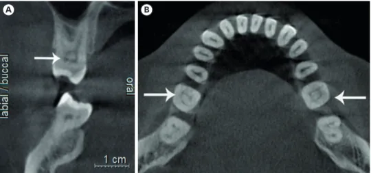
The presence of pulp stones was investigated according to age, sex, dental type and location and the presence and type (composite or amalgam) of restorations. A pulp stone was considered a clear radiopaque mass (round or oval shape) in the coronal pulp chamber (Figures 1 and 2). The depths of the restorations (shallow: up to one-third of the dentin affected; medium: one-third to two-thirds of the dentin affected; deep: two-thirds to all of the dentin affected without exposure of pulp) were also recorded according to the method described by da Silva et al. [9] (Figures 2 and 3). All the restorations were assessed in the sagittal plane, and the deepest point of the restorations was recorded.
A B
Figure 1. Teeth with pulp stones on different sections of cone-beam computed tomography (CBCT). (A) Coronal CBCT section of maxillary first molar. (B) Axial CBCT section of mandibular first molar.
Statistical analysis
Statistical analysis was performed using IBM SPSS Statistics 22 (IBM SPSS, Istanbul, Turkey).
A χ2 test was used to evaluate the number and prevalence of pulp stones according to age and sex. One sample χ2 test was used to evaluate the relationship between study parameters.
Cohen's kappa coefficient was used to measure the inter-examiner agreement. The statistical significance level was set at p < 0.05.
RESULTS
The parameters assessed in association with the presence or absence of pulp stones are reported in Table 1. One hundred and 63 patients (24.2%) had at least 1 pulp stone. Of the 11,494 teeth assessed, there were 379 (3.3%) teeth with pulp stones. In total, pulp stones were statistically higher in females than in males (28.9% vs. 18.7%, p = 0.002). In addition, the percentage of pulp stones in those aged 30–39 years (31.1%) was significantly higher than that in the 18–29 years (18.5%) and ≥ 60 years (17.3%) age groups (p1 = 0.004; p2 = 0.049) (Table 1).
Figure 2. Sagittal cone-beam computed tomography section of maxillary molars with pulp stones and a mandibular first molar with a pulp stone and medium restoration.
Figure 3. Coronal cone-beam computed tomography section of a mandibular first molar with a pulp stone and medium restoration.
The difference in the percentage of pulp stones in the mandibular and maxillary arches was statistically significant (30.4% vs 69.4%, p < 0.05). There was no statistical significance regarding the dental arch sides (p > 0.05). In terms of tooth type, pulp stones were significantly more common in molars (92.1%) than premolars (6.1%), canines (1.3%) and anterior teeth (0.5%) (p < 0.05). In addition, pulp stones were more common in premolars than canines and anterior teeth (p < 0.05) (Table 2).
Nearly one-quarter of teeth (24.8%) with pulp stones had restorations. The percentages of pulp stones in composite and amalgam restored teeth were nearly equal (p > 0.05). The prevalence of pulp stones in teeth with medium-depth restorations was significantly higher (46.8%) than that in teeth with shallow (26.6%) and deep restorations (26.6%) (p1-2= 0.022) (Table 3).
Table 1. Number and prevalence of pulp stones according to age and sex (n = 673)
Variables Pulp stone p
Present Absent
Age 0.042*
18–29 34 (18.5) 150 (81.5)
30–39 64 (31.1) 142 (68.9)
40–49 39 (24.2) 122 (75.8)
50–59 17 (24.3) 53 (75.7)
60+ 9 (17.3) 43 (82.7)
Sex 0.002*
Female 105 (28.9) 258 (71.1)
Male 58 (18.7) 252 (81.3)
Values are presented as number (%).
χ2 test, *p < 0.05.
Table 2. Prevalence of pulp stones regarding arch, side and tooth type (n = 379)
Variables Teeth with pulp stone p
Arch 0.000*
Maxilla 263 (69.4)
Mandible 116 (30.6)
Side 0.644
Right 194 (51.2)
Left 185 (48.8)
Tooth type 0.000*
Molar 349 (92.1)
Premolar 23 (6.1)
Canine 5 (1.3)
Anterior 2 (0.5)
Total 379 (100.0)
Values are presented as number (%).
One sample χ2 test, *p < 0.05.
Table 3. Distribution of teeth with pulp stones and restorations regarding restoration type and depth (n = 94)
Variables Teeth with pulp stone and restoration p
Restoration type 0.837
Amalgam 48 (51.1)
Composite 46 (48.9)
Restoration depth 0.021*
Shallow 25 (26.6)
Medium 44 (46.8)
Deep 25 (26.6)
Total 94 (100.0)
One sample χ2 test, *p < 0.05.
DISCUSSION
Pulp stones may lead to adverse outcomes in endodontic therapy, as they can block the root canal paths. In addition to blocking the passage of files through root canals, they can cause deformation of file tips [19]. Furthermore, pulp stones may hamper disinfection/irrigation procedures [20]. Consequently, pulp stones can give rise to clinical difficulties in endodontic therapy [2].
Many previous studies have used panoramic radiography and histological analysis for the detection of pulp stones [1,21,22]. As noted earlier, calcified masses with diameters smaller than 200 µm may not be detected on panoramic radiographs. Thus, the percentage of pulp stones detected using histological analysis is higher than that using panoramic radiography, which is helpful only as an ex-vivo technique [22]. In addition, pulp stones may not be detected on periapical radiographs because of the superimposition of alveolar bone. CBCT overcomes the problem of overlapping structures and permits high-resolution images in axial, coronal and sagittal planes. As such, it is the most useful and accurate imaging technique available to detect the presence of pulp stones [15]. Thus, the present study used CBCT images to detect the presence of pulp stones and analyze the possible association between pulp stones and the following parameters: age, sex, dental arch and side, tooth type, and restoration type and depth. None of the patients in this study were exposed to additional radiation, as the data were obtained from archival CBCT image records.
We detected pulp stones in 3.3% (379/11,494) of all teeth assessed and 24.2% (163/673) of all patients assessed. In other CBCT studies, Tassoker et al. [23] and Patil et al. [24] detected pulp stones in 52% and 50.93% of patients, respectively, and da Silva et al. [9] reported pulp stones in 31.9% of patients. The present study examined pulp stones only in the coronal pulp chamber, as pulp stones tend to be more common in this region. Radiopaque masses in the radicular part were considered diffuse calcifications [25]. This may explain the difference in the percentage of pulp stones detected in the present study as compared with the findings of previous research.
Sener et al. [10] reported pulp stone prevalence as 38% in 536 patients and 4.8% in teeth by evaluating periapical and bite-wing radiographs on a Turkish subpopulation. On the other hand, Gulsahi et al. [26] reported a pulp stone prevalence in periapical radiographs of 12% in 519 patients and 5% in teeth. In this study, we detected a pulp stone prevalence of 24.2% in 673 patients and a prevalence of 3.3% in teeth. The discord in the findings can be explained by geographical differences and imaging techniques.
Tamse et al. [27] noted that the percentage of pulp stones increased with age. However, Hsieh et al. [11] reported that there was no association between pulp stones and age. In the present study, the percentage of pulp stones in the 30–39 years (31.1%) age group was significantly higher than that in the 18–19 years (18.5%) and ≥ 60 years (17.3%) age groups The conflicting results of these studies may be explained by the fact that restorative treatments, which cause chronic inflammation in pulp tissue, are performed more frequently among the middle-aged population in Turkey. In addition, older age is commonly associated with obstructions in root canals rather than pulp stones [28].
Consistent with the findings of many studies, the prevalence of pulp stones in this study was higher in females than males (28.9% vs. 18.7%), which might be related to the increased
frequency of bruxism in females [10,14]. In contrast, Hsieh et al. [11] and al-Hadi Hamasha et al. [29] found that the prevalence of pulp stones was higher in males than females. The discord in the findings may be explained by the hypothesis that calcifications, which are associated with pulpal inflammation resulting from chronic irritants, are detected mostly in men from the countries where those studies were performed [30]. Some previous studies found no statistically significant association between pulp stones and sex [9,13].
In this study, the percentage of pulp stones in the maxilla was significantly higher than that in the mandible. Similarly, Hsieh et al. [11] reported higher numbers of pulp stones in maxilla than mandible. Some studies found that the percentage of pulp stones in maxillae and mandibles was nearly equal, [23,24] whereas another research detected a higher percentage of pulp stones in mandible than maxilla [31]. Thus, further examinations with larger sample sizes are needed. Similar to our results, many studies detected a higher prevalence of pulp stones in molars [11,18,29]. The volume of molar teeth may explain this finding. Due to the large amount of pulp tissue and high blood supply that molars have, their pulp tissues calcify easier than that of other tooth groups. The early eruption time of molars as compared to premolars may be another explanation for the increased prevalence of pulp stones, with molars exposed to irritation for a longer period [27].
In the present study, 24.8% of teeth with pulp stones had restorations. In other studies, da Silva et al. [9] and Tassoker et al. [23] reported that 13% and 73.5% of teeth with pulp stones had restorations, respectively. Differences in the study populations and oral care habits may explain the discord in the findings. In the present study, the percentages of pulp stones associated with composite and amalgam restorations were nearly the same. Besides, the severity and duration of chronic irritation caused by these restorations might be critical and affect the findings of the studies. Longitudinal researches are needed to resolve this problem [10] da Silva et al. [9] and Tassoker et al. [23] reported that as the depth of a restoration increased, the possibility of pulp stones increased. One possible explanation for this finding may be the theory that chronic irritation of pulp tissue leads itself to produce pulp stones as a defense mechanism. However, it was reported that pulp stones were more commonly found in teeth with medium-depth restorations in this study. The retrospective nature of this study may explain this finding. Based on this study’s findings, the null hypothesis was rejected.
The absence of data on the age of the restorations and comparatively limited sample size can be considered as limitations of the present study. Due to the uncertainty about the restoration duration and intensity of chronic irritation in teeth with restorations, we did not assess in terms of restoration presence or absence. This may also be considered a limitation of the study. In this study, the medical conditions of the patients were ignored. Considering that some systemic diseases may cause pulp stone formation, this may be noted as another limitation of this study.
CONCLUSIONS
Within the limitations of this study, a pulp stone prevalence was detected of 24.2% in 673 patients and a prevalence of 3.3% in teeth. The presence of medium-depth restorations increased the risk of pulp stone formation. It can also be concluded that female sex and maxillary molar teeth were associated with a higher percentage of pulp stones. The percentage of pulp stones in the 30–39 years age group was the highest among the other
age groups. Dentists should be aware that pulp stones can be encountered frequently in the clinical scenario and various predisposing factors can trigger pulp stone formation.
REFERENCES
1. Bevelander G, Johnson PL. Histogenesis and histochemistry of pulpal calcification. J Dent Res 1956;35:714-722.
PUBMED | CROSSREF
2. Goga R, Chandler NP, Oginni AO. Pulp stones: a review. Int Endod J 2008;41:457-468.
PUBMED | CROSSREF
3. Milcent CPF, da Silva TG, Baika LM, Grassi MT, Carneiro E, Franco A, de Lima AAS. Morphologic, structural, and chemical properties of pulp stones in exracted human teeth. J Endod 2019;45:1504-1512.
PUBMED | CROSSREF
4. Berès F, Isaac J, Mouton L, Rouzière S, Berdal A, Simon S, Dessombz A. Comparative physicochemical analysis of pulp stone and dentin. J Endod 2016;42:432-438.
PUBMED | CROSSREF
5. Edds AC, Walden JE, Scheetz JP, Goldsmith LJ, Drisko CL, Eleazer PD. Pilot study of correlation of pulp stones with cardiovascular disease. J Endod 2005;31:504-506.
PUBMED | CROSSREF
6. VanDenBerghe JM, Panther B, Gound TG. Pulp stones throughout the dentition of monozygotic twins: a case report. Oral Surg Oral Med Oral Pathol Oral Radiol Endod 1999;87:749-751.
PUBMED | CROSSREF
7. Zeng JF, Zhang W, Jiang HW, Ling JQ. Isolation, cultivation and initial identification of Nanobacteria from dental pulp stone. Zhonghua Kou Qiang Yi Xue Za Zhi 2006;41:498-501.
PUBMED | CROSSREF
8. Nayak M, Kumar J, Prasad LK. A radiographic correlation between systemic disorders and pulp stones.
Indian J Dent Res 2010;21:369-373.
PUBMED | CROSSREF
9. da Silva EJNL, Prado MC, Queiroz PM, Nejaim Y, Brasil DM, Groppo FC, Haiter-Neto F. Assessing pulp stones by cone-beam computed tomography. Clin Oral Investig 2017;21:2327-2333.
PUBMED | CROSSREF
10. Sener S, Cobankara FK, Akgünlü F. Calcifications of the pulp chamber: prevalence and implicated factors.
Clin Oral Investig 2009;13:209-215.
PUBMED | CROSSREF
11. Hsieh CY, Wu YC, Su CC, Chung MP, Huang RY, Ting PY, Lai CK, Chang KS, Tsai YC, Shieh YS. The prevalence and distribution of radiopaque, calcified pulp stones: a cone-beam computed tomography study in a northern Taiwanese population. J Dent Sci 2018;13:138-144.
PUBMED | CROSSREF
12. Chaini K, Georgopoulou MK. General pulp calcification: literature review and case report. Endod Pract Today 2016;10:69-75.
13. Kannan S, Kannepady SK, Muthu K, Jeevan MB, Thapasum A. Radiographic assessment of the prevalence of pulp stones in Malaysians. J Endod 2015;41:333-337.
PUBMED | CROSSREF
14. Turkal M, Tan E, Uzgur R, Hamidi M, Colak H, Uzgur Z. Incidence and distribution of pulp stones found in radiographic dental examination of adult Turkish dental patients. Ann Med Health Sci Res 2013;3:572-576.
PUBMED | CROSSREF
15. Patel S, Durack C, Abella F, Shemesh H, Roig M, Lemberg K. Cone beam computed tomography in Endodontics - a review. Int Endod J 2015;48:3-15.
PUBMED | CROSSREF
16. Lari SS, Shokri A, Hosseinipanah SM, Rostami S, Sabounchi SS. Comparative sensitivity assessment of cone beam computed tomography and digital radiography for detecting foreign bodies. J Contemp Dent Pract 2016;17:224-229.
PUBMED | CROSSREF
17. Colak H, Celebi AA, Hamidi MM, Bayraktar Y, Colak T, Uzgur R. Assessment of the prevalence of pulp stones in a sample of Turkish Central Anatolian population. ScientificWorldJournal 2012;2012:804278.
PUBMED | CROSSREF
18. Ranjitkar S, Taylor JA, Townsend GC. A radiographic assessment of the prevalence of pulp stones in Australians. Aust Dent J 2002;47:36-40.
PUBMED | CROSSREF
19. Verma KG, Juneja S, Randhawa S, Dhebar TM, Raheja A. Retrieval of iatrogenically pushed pulp stone from middle third of root canal in permanent maxillary central incisor: a case report. J Clin Diagn Res 2015;9:ZD06-ZD07.
PUBMED | CROSSREF
20. Ibarrola JL, Knowles KI, Ludlow MO, McKinley IB Jr. Factors affecting the negotiability of second mesiobuccal canals in maxillary molars. J Endod 1997;23:236-238.
PUBMED | CROSSREF
21. Kayal RA. Distortion of digital panoramic radiographs used for implant site assessment. J Orthod Sci 2016;5:117-120.
PUBMED | CROSSREF
22. Moss-Salentijn L, Hendricks-Klyvert M. Calcified structures in human dental pulps. J Endod 1988;14:184-189.
PUBMED | CROSSREF
23. Tassoker M, Magat G, Sener S. A comparative study of cone-beam computed tomography and digital panoramic radiography for detecting pulp stones. Imaging Sci Dent 2018;48:201-212.
PUBMED | CROSSREF
24. Patil SR, Ghani HA, Almuhaiza M, Al-Zoubi IA, Anil KN, Misra N, Raghuram P. Prevalence of pulp stones in a Saudi Arabian subpopulation: a cone-beam computed tomography study. Saudi Endod J 2018;8:93-98.
CROSSREF
25. Arys A, Philippart C, Dourov N. Microradiography and light microscopy of mineralization in the pulp of undemineralized human primary molars. J Oral Pathol Med 1993;22:49-53.
PUBMED | CROSSREF
26. Gulsahi A, Cebeci AI, Ozden S. A radiographic assessment of the prevalence of pulp stones in a group of Turkish dental patients. Int Endod J 2009;42:735-739.
PUBMED | CROSSREF
27. Tamse A, Kaffe I, Littner MM, Shani R. Statistical evaluation of radiologic survey of pulp stones. J Endod 1982;8:455-458.
PUBMED | CROSSREF
28. Alaçam T. 31. Geriatrik endodonti. In: Endodonti. Ankara: Özyurt Matbaacılık; 2012. p1178.
29. al-Hadi Hamasha A, Darwazeh A. Prevalence of pulp stones in Jordanian adults. Oral Surg Oral Med Oral Pathol Oral Radiol Endod 1998;86:730-732.
PUBMED | CROSSREF
30. Cunha-Cruz J, Pashova H, Packard JD, Zhou L, Hilton TJfor Northwest PRECEDENT. Tooth wear:
prevalence and associated factors in general practice patients. Community Dent Oral Epidemiol 2010;38:228-234.
PUBMED | CROSSREF
31. Elvery MW, Savage NW, Wood WB. Radiographic study of the broadbeach aboriginal dentition. Am J Phys Anthropol 1998;107:211-219.
PUBMED | CROSSREF