INTRODUCTION
Desmoplastic small round cell tumor (DSRCT) is a highly ag- gressive malignant small cell neoplasm that tends to affect ado- lescents and young adults and occurs predominantly in the ab- dominal cavity, including the pelvis and omentum (1, 2). Other primary sites are rare and have included the paratesticular re- gion, pleura, posterior cranial fossa, soft tissues, bone, ovary, and kidney (2-4). DSRCT in the lung is extremely rare. This dis- tinct clinicopathological entity was first described by Gerald and Rosai (1) in 1989.
A DSRCT is composed of small round tumor cells of uncer- tain histogenesis, associated with prominent stromal desmopla- sia and polyphenotypic differentiation. This article reports bilat- eral tumors in the pleura of a 15-year-old male.
CASE REPORT
A 15-year-old male presented with a 1-month history of sharp pain in the left lower chest, which occasionally woke him. He was a nonsmoker, as were his parents. He had no serious medical or surgical history and he had grown up in an ordinary residen- tial and social environment. He had no shortness of breath or cough. There was no history of weight loss, fever, or night sweats.
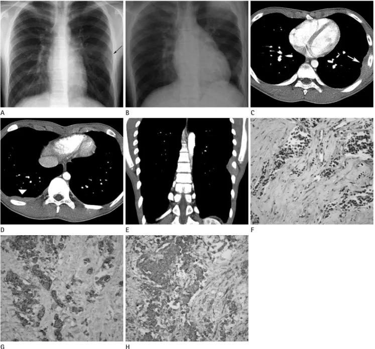
Chest radiographs showed a soft-tissue mass in the left mid hemithorax (Fig. 1A). The mass was at an obtuse angle to the chest wall. The long diameter of the mass was approximately 9 cm. There was no other abnormality in the lung parenchyma or bony thorax. In the left down decubitus view, the mass did not shift position or change contour (Fig. 1B).
Chest computed tomography (CT) revealed multifocal nodu- lar pleural thickening in the lateral, posterior aspect of the left
J Korean Soc Radiol 2015;72(4):295-299 http://dx.doi.org/10.3348/jksr.2015.72.4.295
Received October 13, 2014; Accepted December 21, 2014Corresponding author: Jai Soung Park, MD Department of Radiology, Soonchunhyang University College of Medicine, Bucheon Hospital, 170 Jomaru-ro, Wonmi-gu, Bucheon 420-767, Korea.
Tel. 82-32-621-5851 Fax. 82-32-621-5874 E-mail: [email protected]
This is an Open Access article distributed under the terms of the Creative Commons Attribution Non-Commercial License (http://creativecommons.org/licenses/by-nc/3.0) which permits unrestricted non-commercial use, distri- bution, and reproduction in any medium, provided the original work is properly cited.
Desmoplastic small round cell tumor (DSRCT) is a highly aggressive malignant small cell neoplasm occurring mainly in the abdominal cavity, but it is extremely rare in the pleura. In this case, a 15-year-old male presented with a 1-month history of left chest pain. Chest radiographs revealed pleural thickening in the left hemithorax and chest computed tomography showed multifocal pleural thickening with enhance- ment in both hemithoraces. A needle biopsy of the left pleural lesion was performed and the final diagnosis was DSRCT of the pleura. We report this unusual case aris- ing from the pleura bilaterally. The pleural involvement of this tumor supports the hypothesis that it typically occurs in mesothelial-lined surfaces.
Index terms Pleura
Thickened Pleura Neoplasm Needle Biopsy
Computed Tomography
Bilateral Presentation of Pleural Desmoplastic Small Round Cell Tumors: A Case Report
1양측 흉막에 발생한 결합조직형성 소원형세포종양의 증례 보고1
You Sun Won, MD
1, Jai Soung Park, MD
1, Sun Hye Jeong, MD
1, Sang Hyun Paik, MD
1, Heon Lee, MD
1, Jang Gyu Cha, MD
1, Eun Suk Koh, MD
2Departments of 1Radiology, 2Pathology, Soonchunhyang University College of Medicine, Bucheon Hospital, Bucheon, Korea
density was seen in the lesion in the lateral left hemithorax. The mediastinal and hilar lymph nodes were not enlarged. In the scanned portion of the abdomen, there was no mass, lymphade- nopathy, or ascites. From these imaging findings, we suspected fibromatosis of the pleura and a localized fibrous tumor because it appeared as a well-defined relatively homogeneous soft tissue hemithorax and posterior aspect of the right lower hemithorax
(Fig. 1C-E). In the pre-contrast image, the pleural lesion showed homogeneous soft tissue attenuation and there was no calcifica- tion or pleural effusion. After contrast enhancement, the thick- ened pleura generally showed homogeneous enhancement, with attenuation similar to that of the back muscles and a subtle low
Fig. 1. Chest radiographs (A, B), chest CT (C-E), and microscopic features (F-H) of desmoplastic small round cell tumor in a 15-year-old male.
A, B. Chest radiograph posteroanterior (A) and left lateral decubitus (B) views show a soft tissue mass (arrow) at the left mid hemithorax.
C, D. Transverse CT image of the chest shows multifocal pleural plaque with enhancement in both hemithoraces (left hemithorax, arrow; right hemithorax, arrowhead).
E. Coronal reformatted CT image shows a pleural plaque at the lateral aspect of the left hemithorax.
F. Photomicrograph of hematoxylin and eosin-stained specimen (× 100) shows cellular small round cell nests within dense fibrous stroma.
G, H. Positive immunoreactivity for CD56 (× 200) (G) and vimentin (× 200) (H) is seen on the tumor cell membranes.
H E B
F C
G D A
resents necrosis, hemorrhage, or fibrous components. Calcifica- tion can be seen in both primary and metastatic tumors. Due to the various fibrotic, necrotic, and calcified components, DSRCT shows inhomogeneous attenuation in pre-contrast CT. Contrast enhancement is relatively weak, probably due to the fibrous com- ponent caused by the desmoplastic reaction. MRI is useful for as- sessing tumor extent. On MRI, DSRCT often shows a heteroge- neous high signal intensity on T2-weighted images and iso to low signal intensity on T1-weighted images. After administering gadolinium, it presents as a heterogeneously enhancing mass. On positron emission tomography, DSRCT shows increased fluoro- deoxyglucose uptake (6).
The diagnosis of pleural DSRCT can be established from the clinical, histological, and immunohistochemical features. The histogenesis of DSRCT is uncertain, but its predilection for se- rosal involvement suggests a mesothelial origin.
A typical feature of DSRCT is the angulated nests of small round cells embedded in a cellular fibroblastic stroma. Grossly, areas of central low attenuation on CT might correspond to hemorrhage or necrosis. Immunohistochemistry is helpful for diagnosing DSRCT. Immunohistochemically, the co-expression of cytokeratins, vimentin and desmin is a characteristic feature.
The perinuclear dot-like staining for vimentin is unique.
The differential diagnosis of pleural DSRCT includes malig- nant lymphoma, classical neuroblastoma, rhabdomyosarcoma, rhabdoid tumor, PNET, and malignant mesothelioma. The gross pattern and radiological findings of DSRCT mostly resembled malignant mesothelioma. However, primary mesothelioma is extremely rare in children and young adults. Radiologically, ma- lignant mesothelioma is also often combined with a massive pleural effusion, but it can have a circumferential pattern of in- volvement, with disease extending along the fissural, mediasti- nal, or pericardial pleura. In our case, there was no pleural effu- sion or circumferential pattern of involvement. In addition, the patient was younger than commonly seen in mesothelioma.
Histologically, the dot-like immunostaining for vimentin and negative for CK5/6 and calretinin distinguish DSRCT from the small cell variant of mesothelioma (3).
Primitive neuroectodermal tumors are also similar to DSRCT, in the age of presentation and the imaging, histological, and cy- tological findings. Radiologically, the findings of PNET are sim- ilar to those of DSRCT from the perspective of an extraparen- attenuated mass in the pleura. We also considered a primitive
neuroectodermal tumor (PNET) because there was no calcifica- tion and it contained subtle low-density foci.
About 2 weeks later, a needle biopsy of the largest pleural le- sion in the left hemithorax was performed and we obtained two pieces of grayish elongated soft tissue, with the larger measuring 1.9 cm in length. Microscopic examination showed nests or clumps of small cells set in abundant desmoplastic stroma. The tumor cells had small hyperchromatic nuclei with little cyto- plasm and indistinct nucleoli. Mitotic figures were common (Fig. 1F). Immunohistochemically, the tumor cells were positive for CD56, desmin, synaptophysin, and vimentin, and negative for cytokeratin, epithelial membrane antigen, neuron specific enolase, and thyroid transcription factor-1 (Fig. 1G, H).
The patient refused surgery and was treated with vincristine, doxorubicin, and cyclophosphamide, alternating with ifosfamide and etoposide. The patient died from sepsis 16 months after the initial diagnosis.
DISCUSSION
A DSRCT is a rare, aggressive malignancy, typically occurring in young adults and more common in males. It is most com- monly reported in children and young adults between 15 and 35 years of age, with a male-to-female ratio of 4:1. This tumor has poor median survival rates (5).
The majority of cases occur in the abdominal serosa, includ- ing the omentum and paravesical space and this is known as an intra-abdominal desmoplastic tumor (2). There have been a few cases reported in the pleural cavity, paratesticular region, and even intracranially (2-4). The clinical signs are non-specific and vary according to the involved organ. In most cases, DSRCT presents as an abdominal mass with peritoneal seeding. A fre- quently associated symptom is crampy abdominal pain (5).
With a pleural presentation, common associated symptoms are chest pain and dyspnea (3).
Computed tomography and magnetic resonance imaging (MRI) are relatively useful tools for evaluating DSRCT, although the DSRCT image findings are nonspecific. The most common CT finding of pleural DSRCT is single or multiple soft-tissue pleural masses with combined pleural effusion. Pleural nodular- ity is another common finding. The low-density area often rep-
initial aggressive anticancer treatment (4). Therefore, the early detection of DSRCT is very important for achieving long-term survival. Consequently, DSRCT could be considered in the differ- ential diagnosis of multifocal pleural thickening containing ne- crosis or calcification with inhomogeneous enhancement and an accompanying pleural effusion, especially in adolescent males.
REFERENCES
1. Gerald WL, Rosai J. Case 2. Desmoplastic small cell tumor with divergent differentiation. Pediatr Pathol 1989;9:177- 183
2. Biswas G, Laskar S, Banavali SD, Gujral S, Kurkure PA, Muckaden M, et al. Desmoplastic small round cell tumor:
extra abdominal and abdominal presentations and the re- sults of treatment. Indian J Cancer 2005;42:78-84 3. Parkash V, Gerald WL, Parma A, Miettinen M, Rosai J. Des-
moplastic small round cell tumor of the pleura. Am J Surg Pathol 1995;19:659-665
4. Ostoros G, Orosz Z, Kovács G, Soltész I. Desmoplastic small round cell tumour of the pleura: a case report with un- usual follow-up. Lung Cancer 2002;36:333-336
5. Lal DR, Su WT, Wolden SL, Loh KC, Modak S, La Quaglia MP. Results of multimodal treatment for desmoplastic small round cell tumors. J Pediatr Surg 2005;40:251-255 6. Kis B, O’Regan KN, Agoston A, Javery O, Jagannathan J,
Ramaiya NH. Imaging of desmoplastic small round cell tu- mour in adults. Br J Radiol 2012;85:187-192
7. Xu Q, Xu K, Yang C, Zhang X, Meng Y, Quan Q. Askin tu- mor: four case reports and a review of the literature. Can- cer Imaging 2011;11:184-188
8. Dynes MC, White EM, Fry WA, Ghahremani GG. Imaging manifestations of pleural tumors. Radiographics 1992;12:
1191-1201
9. Li H, Smolen GA, Beers LF, Xia L, Gerald W, Wang J, et al.
Adenosine transporter ENT4 is a direct target of EWS/WT1 translocation product and is highly expressed in desmo- plastic small round cell tumor. PLoS One 2008;3:e2353 10. Modak S, Gerald W, Cheung NK. Disialoganglioside GD2
and a novel tumor antigen: potential targets for immuno- therapy of desmoplastic small round cell tumor. Med Pedi- atr Oncol 2002;39:547-551
chymal soft-tissue-density mass with a pleural effusion, often including a necrotic portion. However, in PNET, calcification is rare and rib destruction is often combined (7). In our case, the mass contained a low-density portion and there was no calcifi- cation or pleural effusion. However, no rib destruction, which is common with PNET, was seen. Histologically, it was composed of small round, undifferentiated blue cells with scant cytoplasm.
Immunohistologically, PNET usually show unidirectional dif- ferentiation toward neural elements, while DSRCT shows multi- directional differentiation toward muscle, neural, and epithelial elements.
Malignant non-Hodgkin’s lymphoma is also similar to DSRCT.
On imaging, pleural nodules with focal or diffuse pleural thick- ening with homogeneous enhancement accompanied by pleural effusion can be seen (8). However, it often has associated medias- tinal and hilar lymphadenopathy, unlike DSRCT (8). In our case, there was no lymphadenopathy and the image findings resem- bled DSRCT more than malignant non-Hodgkin’s lymphoma.
Most neuroblastomas occur before 5 years of age, and charac- teristically present with invasion through the neural foramina, giving a dumbbell appearance due to their origin from sympa- thetic nervous tissue (7). In our case, the patient was older than most neuroblastoma patients and the tumor did not have a dumbbell shape.
The prognosis of DSRCT remains poor and the 5-year surviv- al rate is less than 15% (5). There is no recommended treatment for DSRCT. Aggressive anticancer treatment is thought to con- tribute to relatively long-term survival (4). Aggressive surgery can be used to reduce the tumor size before or after chemother- apy and complete surgical excision seems to improve survival (2). Radiation therapy can be used to treat DSRCT (2). Despite the poor overall prognosis, there are some cases of long-term sur- vival and they common involve early aggressive anticancer treat- ment (4). There are some new approaches to treating DSRCT, such as molecular targeted therapy and immunotherapy (9, 10).
In summary, DSRCT is a rare, highly aggressive malignancy that typically involves the abdominal cavity, but is rare in the pleura. The imaging findings of DSRCT in the pleura are non- specific and it is difficult to suspect DSRCT. Various treatments have been attempted for DSRCT, but the prognosis is quite poor despite aggressive combination therapy. Nevertheless, there are some reports of long-term survival and they are common with
양측 흉막에 발생한 결합조직형성 소원형세포종양의 증례 보고1
원유선
1· 박재성
1· 정선혜
1· 백상현
1· 이 헌
1· 차장규
1· 고은석
2결합조직형성 소원형세포종양은 대부분 복강 내에 발생하는 드문 질환으로 흉막에 발생하는 경우는 매우 드문 것으로 알 려져 있다. 이 증례는 15세 남자 환자가 한 달 동안 지속되는 좌측 흉통을 주소로 내원하였고 흉부 X-선 사진에서 좌측 흉막의 비후가 관찰되었으며 흉부 전산화단층촬영에서 양측에서 조영증강을 보이는 흉막 비후가 관찰되었다. 좌측 흉막 병변에 대해 바늘 생검을 시행하였으며 병리학적으로 흉막의 결합조직형성 소원형세포종양으로 확진되었다. 저자들은 이 증례가 양측 흉막에 생긴 결합조직형성 소원형세포종양으로 매우 드문 질환이며 양측 흉막에서 발생하였다는 점은 이 종 양이 중피성 표면에서 잘 발생한다는 기존의 가설을 뒷받침하는 증례라는 점에서 보고하는 바이다.
순천향대학교 의과대학 부천병원 1영상의학과, 2병리과