INTRODUCTION
D-penicillamine has been known as one of the agents that are able to induce pemphigus (1, 2). Bucillamine [N-(2-mer- capto-2-methylpropionyl)-L-cysteine] is a thiol compound contained two free sulfhydryl groups, whereas D-penicilla- mine has only one sulfhydryl radical (3). While drug-induced pemphigus was not infrequently demonstrated in associa- tion with d-penicillamine, it has rarely been reported with bucillamine and has never been reported in rheumatoid arthri- tis (RA) and polymyositis (PM) overlap syndrome. Hereby, we report a case of bucillamine-induced pemphigus vulgaris in a patient with RA and PM overlap syndrome.
CASE REPORT
A 46-yr-old woman was referred to our rheumatology de- partment due to polyarthralgia, and myalgia in May 2001.
Soft tissue swelling and tenderness were bilaterally noted at the proximal interphalangeal and metacarpophalangeal joints with morning stiffness lasting at least 1 hr. Proximal motor weakness on upper arms and legs has insidiously developed over three months and findings of magnetic resonance image on both thighs were compatible with myositis. In addition, elevated level of muscle enzymes including creatine kinase (CK), aldolase, alanine aminotransferase (AST and ALT), and lactate dehydrogenase was shown. Antinuclear antibody was
positive at 1:80 of speckled pattern, and 1:2,560 of cytoplas- mic pattern. Auto-antibodies including anti-Jo-1, anti-RNP, anti-double stranded DNA, anti-Sm, and anti-SS-A/SS-B were absent. Level of rheumatoid factor was 3,420 IU/mL, but level of C3 and C4 was 97.9 mg/dL (normal range 79- 152 mg/dL) and 30.8 mg/dL (normal range 16-38 mg/dL) respectively. Chest radiograph showed pattern of interstitial lung disease and pulmonary function test revealed signifi- cantly decreased DLCO. She was diagnosed as RA and PM overlap, and had been treated with prednisolone, azathioprine and non-steroidal anti-inflammatory drug since May 2001.
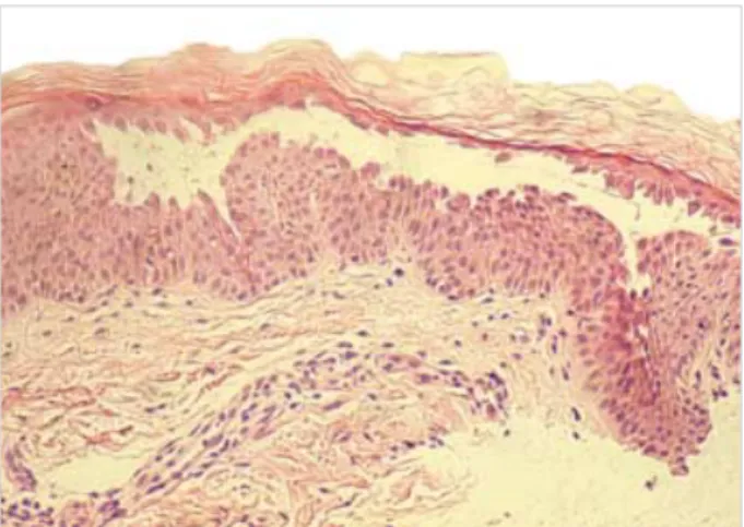
Myositis became silent over time with above regimen, how- ever she had continuous synovitis and joint pain on right knee and small joints on both hands. Her regimen was changed to cyclosporine and bucillamine. Twenty months later, the skin lesions occurred. She exhibited erythematous flaccid blisters on her chest, axillae, and back. Blisters consisted of erythematous patches and superficial erosions with turbid bullae suggesting pemphigus vulgaris (Fig. 1). Nikolsky’s sign was positive. Laboratory tests showed C-reactive protein 0.831 mg/dL (normal range <0.8 mg/dL), and erythrocyte sedimentation rate 66 mm/hr. Liver function, renal function, CK, and electrolytes were normal. A skin biopsy revealed intraepidermal vesicle formation with acantholysis and the vesicle formation is suprabasal (Fig. 2). Indirect immunoflu- orescence, performed by using serial dilutions of the patient’s serum, demonstrated no IgG, IgA and C3 staining along epidermis and papillary dermis. Bucillamine was discontin-
Jin-Wuk Hur, Chang-Woo Lee*, Dae-Hyun Yoo
Division of Rheumatology, Department of Internal Medicine, The Hospital for Rheumatic Diseases, and Department of Dermatology*, College of Medicine, Hanyang University, Seoul, Korea
Address for correspondence Dae-Hyun Yoo, M.D.
The Hospital for Rheumatic Diseases, Hanyang University Medical Center, 17 Haengdang-dong, Seongdong-gu, Seoul 133-792, Korea Tel : +82.2-2290-9202, Fax : +82.2-2298-8231 E-mail : [email protected]
585 J Korean Med Sci 2006; 21: 585-7
ISSN 1011-8934
Copyright � The Korean Academy of Medical Sciences
Bucillamine-Induced Pemphigus Vulgaris in a Patient with Rheumatoid Arthritis and Polymyositis Overlap Syndrome
Bucillamine is a disease modifying anti-rheumatic drug, structurally similar to D-peni- cillamine. Although D-penicillamine-induced pemphigus has been not infrequently demonstrated, pemphigus associated with bucillamine was rarely reported. We describe a patient complicating pemphigus vulgaris after bucillamine treatment in rheumatoid arthritis (RA) and polymyositis (PM) overlap syndrome. PM and RA overlap syndrome was diagnosed three years ago and bucillamine was adminis- trated for 20 months. Skin lesions including erythematous flaccid blisters on her chest, axillae, and back were occurred and were compatible with pemphigus vul- garis by typical pathology. Withdrawal from bucillamine and prednisolone treatment made rapid improvement of pemphigus lesions.
Key Words : bucillamine; Pemphigus; Polymyositis; Arthritis, Rheumatoid; adverse effects
Received : 7 March 2005 Accepted : 20 June 2005
586 J.-W. Hur, C.-W. Lee, D.-H. Yoo
ued and the patient was placed on oral prednisolone 20 mg daily. The skin lesions rapidly improved, and disappeared within 2 months.
DISCUSSION
D-penicillamine and bucillamine have been used for patients with rheumatoid arthritis as a disease modifying anti-rheu- matic drug. D-penicillamine can cause various autoimmune diseases including pemphigus, PM, and myasthenia gravis (4-6). Although bucillamine has a chemical structure simi- lar to D-penicillamine, autoimmune diseases are rarely asso- ciated with bucillamine. Some cases of bucillamine-induced interstitial lung disease, nephropathy, and myasthenia gravis were described (7-9). It was reported that the incidence of D-penicillamine-induced pemphigus was about 7% (5, 10) and mean duration until the onset of pemphigus was 11 to 13 months (11, 12). It frequently presents as an urticarial, annular, or moniliform erythematous prodrome. Scaly crusted patches, vesicles, and bullae are developed subsequently (5).
The mechanism of thiol drugs-induced pemphigus is still obscure. It has been suggested that the epidermal cell sur- face proteins play a role as an antigen to the host through a sulfhydryl radical and other unknown factors (1). In D-peni- cillamine-induced pemphigus, the prevalence of IgG and IgA antibodies directed against the intercellular region is lower than that in spontaneously occurring pemphigus (13). It has been shown that these antibodies might not be detected in some cases of drug-induced pemphigus like our case (13, 14).
To date, only 2 cases of bucillamine-induced pemphigus were reported in English literature. One was drug-induced pemphigus foliaceus with features of pemphigus vulgaris and the other was subcorneal pustular dermatosis-type IgA pemphigus induced by thiol drugs (12, 14).
The discontinuation of D-penicillamine may be inadequate in treatment because it frequently fails to stop disease pro-
gression. Initial dose of corticosteroid is typically about 0.75- 1 mg/kg/day. It has been suggested that high-dose or pulse therapy with steroids usually clears blisters promptly. Pred- nisolone at a lower dose of 20-30 mg/day can maintain these patients without blistering but other adjuvant immunosup- pressive drugs is need to achieve steroid-sparing effect or pre- vention of relapse (15, 16). Blisters of our patient disappeared rapidly with corticosteroid treatment and discontinuation of bucillamine. Relapse was not found during the subsequent 6 months.
In conclusion, bucillamine may cause pemphigus even at a low rate therefore we have to keep in mind that skin lesions with blisters should be carefully investigated in patients with bucillamine treatment.
REFERENCES
1. Yung CW, Hambrick GW Jr. D-Penicillamine--induced pemphigus syndrome. J Am Acad Dermatol 1982; 6: 317-24.
2. Levy RS, Fisher M, Alter JN. Penicillamine: review and cutaneous manifestations. J Am Acad Dermatol 1983; 8: 548-58.
3. Hirohata S, Lipsky PE. Regulation of B cell function by bucillamine, a novel disease-modifying antirheumatic drug. Clin Immunol Immu- nopathol 1993; 66: 43-51.
4. Ciompi ML, Marchetti G, Bazzichi L, Puccetti L, Agelli M. D-peni- cillamine and gold salt treatments were complicated by myasthenia and pemphigus, respectively, in the same patient with rheumatoid arthritis. Rheumatol Int 1995; 15: 95-7.
5. Shapiro M, Jimenez S, Werth VP. Pemphigus vulgaris induced by D-penicillamine therapy in a patient with systemic sclerosis. J Am Acad Dermatol 2000; 42: 297-9.
6. Jenkins EA, Hull RG, Thomas AL. D-penicillamine and polymyosi- tis: the significance of the anti-Jo-1 antibody. Br J Rheumatol 1993;
32: 1109-10.
7. Negishi M, Kaga S, Kasama T, Hashimoto M, Fukushima T, Yama- gata N, Tabata M, Kobayashi K, Ide H, Takahashi T. Lung injury Fig. 1.Erythematous patches and superficial erosions with pustu-
lar bullae on axillae and back of the patient.
Fig. 2.Skin biopsy shows acantholysis at the suprabasal layer, as well as at the upper layers of epidermis (H&E, ×400).
Bucillamine-induced Pemphigus 587
associated with bucillamine therapy. Ryumachi 1992; 32: 135-9.
8. Sawa N, Ubara Y, Hara S, Hideyuki K, Tagami T, Yokoyama K, Takemoto F, Yamada A, Mori T, Mikami A, Tachibana S, Nakase K. A case of rheumatoid arthritis with bucillamine-induced myasthe- nia gravis treated by immunoadsorption therapy. Ryumachi 1999;
39: 33-8.
9. Obayashi M, Uzu T, Harada T, Yamato M, Takahara K, Yamauchi A. Clinical course of bucillamine-induced nephropathy in patients with rheumatoid arthritis. Clin Exp Nephrol 2003; 7: 275-8.
10. Brenner S, Wolf R, Ruocco V. Drug-induced pemphigus. I. A survey.
Clin Dermatol 1993; 11: 501-5.
11. Ho VC, Stein HB, Ongley RA, McLeod WA. Penicillamine induced pemphigus. J Rheumatol 1985; 12: 583-6.
12. Kishimoto K, Iwatsuki K, Akiba H, Motoki Y, Kaneko F. Subcorneal pustular dermatosis-type IgA pemphigus induced by thiol drugs. Eur J Dermatol 2001; 11: 41-4.
13. Bialy-Golan A, Brenner S. Penicillamine-induced bullous dermatoses.
J Am Acad Dermatol 1996; 35: 732-42.
14. Ogata K, Nakajima H, Ikeda M, Yamamoto Y, Amagai M, Hashi- moto T, Kodama H. Drug-induced pemphigus foliaceus with features of pemphigus vulgaris. Br J Dermatol 2001; 144: 421-2.
15. Kitajima Y. Current and prospective understanding of clinical clas- sification, pathomechanisms and therapy in pemphigus. Arch Der- matol Res 2003; 295 (Suppl 1): 17-23.
16. Piamphongsant T, Ophaswongse S. Treatment of pemphigus. Int J Dermatol 1991; 30: 139-46.