pISSN: 0378-6471 eISSN: 2092-9374 http://dx.doi.org/10.3341/jkos.2013.54.4.595
= 증례보고 =
미세각막절개 백내장 수술 후 낭내 삽입한 일체형 비구면 인공수정체의 임상 결과
김윤전⋅전미현⋅고동아⋅김재용⋅김명준⋅차흥원 울산대학교 의과대학 서울아산병원 안과학교실
목적: 새로운 설계의 광학부를 가지는 비구면 인공수정체 HOYA AF-1 iSert (HOYA, Tokyo, Japan)와 HOYA AF-1 iMics (HOYA, Tokyo, Japan)를 Akreos MI60 (Bausch & Lomb, Inc., Rochester, NY, USA)의 임상 결과와 비교하여 안정성 및 광학 기능을 평가하 고자 하였다.
대상과 방법: iSert, iMics, MI60를 삽입한 55안, 50안, 60안을 대상으로, 최대교정시력, 구면대응치, 전방 깊이, 수차, 대비감도를 측정하 여 기능적 안정성, 낭내 전후 위치 안정성, 시력의 질, 수술유발난시 등을 비교하였다.
결과: 수술 전후 최대교정시력은 세 인공수정체 간 차이가 없었다. 술후 구면대응치는 MI60 군에서 유의한 근시화 경향을 보였으나, 인공수정체 전후 위치 변화는 통계적으로 유의한 차이를 보이지 않았다. 전체고위수차, 고위수차, 코마수차, 대비감도는 유의한 차이가 없었으나 구면수차는 MI60가 술후 6개월에 iSert, iMics보다 유의하게 높은 수치를 보였다.
결론: iSert와 iMics는 술후 6개월 경과 관찰에서 MI60와 대등한 안정성 및 시기능 향상을 보였다.
<대한안과학회지 2013;54(4):595-601>
■ 접 수 일: 2012년 3월 10일 ■ 심사통과일: 2012년 9월 22일
■ 게재허가일: 2013년 2월 25일
■ 책 임 저 자: 차 흥 원
서울특별시 송파구 올림픽로43길 88 서울아산병원 안과
Tel: 02-3010-3680, Fax: 02-470-6440 E-mail: [email protected]
* 이 논문의 요지는 2011년 대한안과학회 제106회 학술대회에서 구연으로 발표되었음.
백내장 수술은 전세계적인 인구의 노령화 및 개발도상국 들의 경제적 발전에 따른 새로운 수요 창출로 점차 증가 추 세에 있다. 그에 힘입어 백내장 초음파 유화술의 발달, 인공 수정체의 질적인 향상, 정확한 인공수정체 도수 계산 공식 의 개발로 최근에는 백내장 수술 후 굴절력을 정확하게 예 측할 수 있게 되었고 이에 힘입어 이제는 백내장 수술이 굴 절 이상의 교정하고 시력의 질적인 개선을 도모하는 것에 그 초점이 맞춰지고 있다.1-3즉 단순한 시력의 회복뿐만 아 니라 전반적인 시기능의 향상이 백내장 수술에서 중요한 요소가 되고 있으며, 대비감도 검사와 수차 분석이 시기능 을 수치화하여 분석하는데 이용하고 있다.4-7이와 같은 시 기능 측정의 기술적 향상으로 양의 구면수차를 가지는 기 존의 구면 인공수정체가 수술 후 높은 구면수차 값을 유발 하고 눈부심 등의 증상을 일으키는 등 시력 저하의 주요 원 인으로 작용한다는 것이 밝혀지면서 구면수차를 조절하기 위해서 비구면 인공수정체가 개발되어 그 사용이 점차 증
가하고 있다.2 그 중에서도 일체형 인공수정체는 다루기가 쉽고, injector system을 이용한 삽입이 용이하며 광학부 축 의 위치가 비교적 단기간 내에 안정화되고 각막 절개창의 크 기가 작다는 장점을 지니고 있어 널리 이용되고 있다.8,9
그러나 비구면 인공수정체들은 낭내에서 중심부 이탈이 있거나 동공의 크기가 커지는 경우 그에 따라 구면수차가 유의하게 증가되고 이로 인한 시기능의 변화가 있을 수 있 다는 단점을 지니고 있어 적응에 제한이 되어왔다.10-16 따 라서 최근 개발된 HOYA AF-1 iSert PC-60AD (HOYA Corporation, Tokyo, Japan)와 HOYA AF-1 iMics NY-60 (HOYA Corporation, Tokyo, Japan)은 제조사에 의하면, 이러한 점을 보완하고자 비구면 일체형 인공수정체를 기본 형태로 하여 광학부를 ABC (Aspheric Balanced Curve) 원 리로 설계하였다.17제조사의 설명에 따르면, -0.18 μm의 구 면수차를 가지는 두 인공수정체는 고유의 광학부 설계를 이 용하여 빛의 집중도를 높이고 홍채나 후낭의 상태에 따른 광 학적 변화를 최소화하고자 설계되었으며, 소수성 아크릴릭 재질의 광학부과 PMMA 재질의 지지부로 구성되어 있다.17
기존의 비구면 인공수정체와 다른 설계를 가지는 HOYA iSert와 iMics는 최근에 개발이 되어, 새로운 광학부의 인 공수정체의 광학 효과와 안정성에 대한 연구는 저자들이 문헌검색을 통하여 확인한 결과 현재까지 국내에서 발표된 바가 없었다. 따라서 본 연구에서는 기존의 효과와 안정성 이 입증된 일체형 비구면 인공수정체와의 비교를 통하여
새로운 설계의 광학부의 안정성 및 광학적 기능의 측면을 평가하였다. 비교를 위한 대조군으로는 현재 상용되고 있으 며, 임상적 결과가 입증된 일체형 비구면 투명 인공수정체 인 Akreos MI60 (Bausch & Lomb, Inc., Rochester, NY, USA)을 이용하였다.
대상과 방법
2010년 9월부터 2011년 2월까지 본원 안과에서 노년성 백내장으로 미세각막절개백내장 수술을 받은 137명, 165 안을 대상으로 선정하여 전향적 연구를 시행하였다. 각막 난시가 2.0 diopter를 넘지 않는 노인성 백내장 환자 중 수 술 전 각막질환이나 안구 내 염증, 녹내장, 약시, 시신경병 증 그리고 당뇨병성 망막병증의 병력이 없는 환자들을 대 상으로 하였고 술 전 백내장 정도 평가에서 LOCS (Lens Opacities Classification System) Ⅲ 분류에 의해 핵 경화 (nuclear opalescence) 정도가 5 이상이거나 술 전 전층각 막이식술이나 굴절교정수술, 녹내장 수술, 유리체-망막 수 술 등을 받은 경우, 술 중 후낭파열, 인공수정체 이탈, 중심 이탈 및 기울임이 발생한 경우는 연구 대상에서 제외하였 다. 모든 검사 과정과 분석은 Declaration of Helsinki에 의 거하여 실행되었다.
세 종류의 인공수정체 중 하나를 무작위로 배정하여 50 안은 MI60, 55안은 iSert, 60안은 iMics를 삽입하였고, 각 인공수정체의 도수 결정은 IOL master (Carl Zeiss Meditec, Dublin, CA, USA)에 내장된 3세대 공식 Haigis와 특화된 인공수정체 A constant를 이용하여 계산하였다. 백내장 수술 은 동일한 술자에 의해 점안 마취제(Alcaine, Proparacaine 0.5%)를 이용한 점안 마취 하에 동일한 방법으로 시행되었 다. 각막절개도(keratome)를 이용하여 각막곡률이 높은 면 (steep meridian)을 따라 2.0 mm 길이의 투명윤부절개창 (clear corneal incision)을 만들고 점탄물질(viscoelastic) 을 전방 내에 채워 넣은 후, cystotome을 이용하여 직경 5.0-5.5 mm 크기의 원형전낭절개(continuous curvilinear capsulorrhexis)를 시행하였다. 평형생리식염수(balanced salt solution, BSS, Alcon, USA)를 사용하여 수력분리술 과 수력분층술을 시행하고 초음파 유화기(Infinity, Alcon, USA)를 사용하여 초음파 수정체 유화술을 시행한 다음, 해 당 연성 인공수정체를 후낭에 삽입하고 전방 내에 남아있는 점탄물질은 관류 및 흡입장치(Irrigation/Aspiration device) 로 제거하였다. iMics와 MI60는 술 전 형성한 2.0 mm 크기 의 각막 절개창을 통하여 삽입하였고, iSert는 각막절개도를 이용하여 각막 절개창을 2.5 mm 크기로 넓힌 후 삽입하였다.
수술 전 모든 환자들에게 세극등현미경 검사, 안저 검사,
현성 굴절 검사, 안압 및 최대교정시력 측정 등의 기본 안 과검사와 안축장 길이 및 자동각막곡률 검사(IOL master®; Carl-Zeiss Meditec, Inc., Dublin, CA, USA), 각막지형도 검사(ORBscan® II; Bausch & Lomb, Inc.), 전안부광학단 층촬영(VisanteTM OCT; Carl-Zeiss Meditec, Inc.)을 시 행하였다. 그리고 술 후 1주, 1개월, 3개월, 6개월에 외래를 방문하여 기본 검사와 더불어 각막지형도 검사, 수차 검사와 대비감도 검사를 시행하였다. 안구의 수차는 암순응을 한 상 태에서 Hartmann-Shack 수차계인 Zywave®(Aberrometer Bausch & Lomb Inc.)를 이용하여 동공 중심부 5.0 mm 영 역을 기준으로 2회 이상 측정하였고, 구면수차(spherical aberration), 수직 및 수평 코마수차(vertical and horizontal coma aberration), 전체고위수차의 RMS (root mean square of total high order aberrations)를 측정하였다. 대비감도 검사(VCTS 6500®; Visitech Consultants Inc, Dayton, OH, USA)는 3.0 m 거리에서, 명소시(photopic condition)와 박 명시(mesopic condition) 환경에서 각각 1.5.3.6.12.18 cpd (cycle per degree)의 주파수에 대해서 측정하였다.
위의 측정치를 분석하여 경과 관찰 기간 동안의 최대교 정시력과 구면대응치(spherical equivalent)를 통하여 인공 수정체의 기능적 안정성을 평가하였으며, 구면대응치의 오 차와 각막지형도검사로 측정한 전방 깊이의 변화량을 이용 하여 인공수정체 전후 이동을 평가하고, 수직 코마수차를 이용하여 인공수정체의 중심 이탈 정도와 기울임 등 낭내 위치의 안정성을 평가하였다. 그리고 수차 검사 및 대비감 도 검사 결과를 비교하여 각 인공수정체의 시력의 질 (Quality of vision)을 평가하였고, Hill 등이 제안한 수술 시의 각막 절개방향과 수술 전후의 각막곡률 및 방향을 이 용한 벡터 분석법(vector method)을 이용하여 수술유발난 시(surgically induced astigmatism)를 계산하였다.
세 군 간의 측정치는 One way ANOVA 검정을 이용하였 고, Bonferroni 검증법을 통하여 사후분석을 시행하였다.
각 군 내에서의 수술 전후 비교에는 Paired samples t-test 을 이용하였으며, 모든 통계분석은 SPSS (version 18.0, software for Windows; SPSS Inc., Chicago, IL, USA)를 이용하였다. p-value가 0.05 미만인 경우를 통계적으로 유 의한 것으로 판단하였다.
결 과
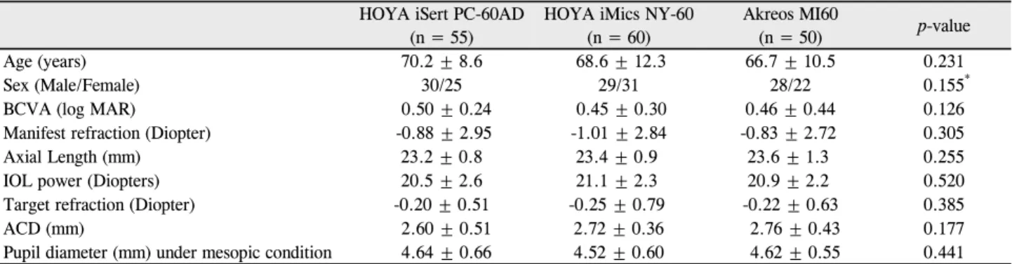
iSert 인공수정체 삽입군, iMics 인공수정체 삽입군과 MI60 인공수정체 삽입군 간의 평균 나이(p=0.231), logMAR 최 대교정시력(p=0.126), 현성 굴절 검사 구면대응치(p=0.305), 안축장 길이(p=0.255) 및 삽입한 인공수정체(p=0.520), 전
Table 1. Comparison of preoperative characteristics among three groups
HOYA iSert PC-60AD(n = 55)
HOYA iMics NY-60 (n = 60)
Akreos MI60
(n = 50) p-value
Age (years) 70.2 ± 8.6 68.6 ± 12.3 66.7 ± 10.5 0.231
Sex (Male/Female) 30/25 29/31 28/22 0.155*
BCVA (log MAR) 0.50 ± 0.24 0.45 ± 0.30 0.46 ± 0.44 0.126
Manifest refraction (Diopter) -0.88 ± 2.95 -1.01 ± 2.84 -0.83 ± 2.72 0.305
Axial Length (mm) 23.2 ± 0.8 23.4 ± 0.9 23.6 ± 1.3 0.255
IOL power (Diopters) 20.5 ± 2.6 21.1 ± 2.3 20.9 ± 2.2 0.520
Target refraction (Diopter) -0.20 ± 0.51 -0.25 ± 0.79 -0.22 ± 0.63 0.385
ACD (mm) 2.60 ± 0.51 2.72 ± 0.36 2.76 ± 0.43 0.177
Pupil diameter (mm) under mesopic condition 4.64 ± 0.66 4.52 ± 0.60 4.62 ± 0.55 0.441 Values are presented as mean ± SD.
BCVA = best corrected visual acuity; log MAR = logarithm of the minimum angle of resolution; IOL = intraocular lens; ACD = anterior chamber depth.
*ANOVA with Bonferroni post-hoc tests (except value = chi-square test).
Table 2. Comparison of postoperative total aberration, total high order aberration, spherical aberration, and coma aberration for
three different IOLs by 6 months postoperativeIOL Postop 1M p-value Postop 3M p-value Postop 6M p-value
RMS total (μm) iSert 1.09 ± 0.46 0.240 1.08 ± 0.32 0.301 1.06 ± 0.34 0.119
iMics 1.01 ± 0.61 0.99 ± 0.74 0.92 ± 0.61
MI60 1.17 ± 0.58 1.06 ± 0.42 1.02 ± 0.47
RMS HOA (μm) iSert 0.46 ± 0.31 0.144 0.42 ± 0.07 0.430 0.47 ± 0.18 0.305
iMics 0.35 ± 0.18 0.43 ± 0.10 0.40 ± 0.20
MI60 0.49 ± 0.17 0.50 ± 0.18 0.50 ± 0.22
SA (μm) iSert -0.01± 0.03 0.062 0.00 ± 0.12 0.055 0.07 ± 0.16 0.040
iMics 0.06 ± 0.16 0.02 ± 0.11 0.05 ± 0.23
MI60 0.16 ± 0.11 0.20 ± 0.07 0.22 ± 0.19
Coma aberration (μm) iSert 0.28 ± 0.09 0.101 0.30 ± 0.08 0.089 0.27 ± 0.10 0.560
iMics 0.26 ± 0.22 0.22 ± 0.16 0.24 ± 0.13
MI60 0.19 ± 0.12 0.24 ± 0.09 0.22 ± 0.13
Values are presented as mean ± SD.
IOL = intraocular lens; RMS total= root mean square of total aberrations; RMS HOA = root mean square of total higher order aberrations;
SA = spherical aberration.
방 깊이(p=0.177), 그리고 박명시의 동공 크기(p=0.441)에 는 모두 통계적으로 유의한 차이가 없었다(all analyzed with ANOVA, Table 1).
술 전(iSert 0.50 ± 0.24, iMics 0.45 ± 0.30, MI60 0.46 ±0.44; p=0.126)과 술 후 1주(iSert 0.10 ±0.13, iMics 0.10 ±0.15, MI60 0.09 ±0.19; p=0.231), 1개월 (iSert 0.06 ± 0.09, iMics 0.06 ± 0.12, MI60 0.05 ± 0.16; p=0.340), 3개월(iSert 0.04 ±0.12, iMics 0.04 ± 0.14, MI60 0.05 ±0.22; p=0.560), 6개월(iSert 0.06 ± 0.21, iMics 0.05 ±0.19, MI60 0.07 ±0.23; p=0.412)에 측정한 logMAR 최대교정시력은 세 군 사이에 유의한 차이 가 없었고(all analyzed with ANOVA), 세 인공수정체를 삽입한 군 모두 수술 전과 비교하여 수술 후 유의하게 향상 된 시력을 보였다(p<0.001, Paired samples t-test). 술 전 목표 굴절값에는 세 군 간 차이가 없었으나(iSert -0.20
±0.51D, iMics -0.25 ± 0.79D, MI60 -0.22 ±0.63D;
p=0.270, ANOVA), 술 후 현성 굴절 검사로 측정한 구면 대응치를 비교하였을 때, 수술 3개월 후부터 MI60 인공수 정체를 삽입한 군에서 다른 두 군과 비교하여 유의하게 근시화 경향을 보였다(iSert -0.04 ±0.50D, iMics -0.16 ±0.39D, MI60 -0.69 ±0.76D; p<0.001, ANOVA with Bonferroni post hoc tests).
인공수정체의 전후 위치 이동을 평가하기 위해서 시간 경과에 따른 구면대응치의 오차와 전방 깊이의 변화량 추 이를 살펴보았을 때, 시간이 경과하면서 MI60는 근시화 경 향을 보였고, iMics와 iSert는 원시화의 경향을 보였으나 술 후 6개월 동안의 절대적인 변화량은 세 군간에 통계적으로 유의한 차이를 보이지 않았다(iSert 0.12 ±0.59D, iMics 0.07 ±0.40D, MI60 -0.16 ±0.72D; p=0.520, ANOVA).
술 후 6개월의 전방 깊이 변화량 역시 세 군 간 통계적으로
0 10 20 30 40 50 60 70 80
1.5 3 6 12 18
Contrast sensitivity (photopic)
Frequency (CPD) iSert iMics MI60
0 10 20 30 40 50 60 70 80
1.5 3 6 12 18
Contrast sensitivity (mesopic)
Frequency (CPD)
iSert iMics MI60
0 10 20 30 40 50 60 70 80
1.5 3 6 12 18
Contrast sensitivity (photopic)
Frequency (CPD)
iSert iMics MI60
0 10 20 30 40 50 60 70 80
1.5 3 6 12 18
Contrast sensitivity (mesopic)
Frequency (CPD)
iSert iMics MI60
Figure 1. Comparison of postoperative contrast sensitivity in photophic and mesopic conditions (Mean). The asterisks (
*) indicate the differences in values were significant (p < 0.05) among three groups. (A) Contrast sensitivity in photopic condition at post- operative 1 month. (B) Contrast sensitivity in mesopic condition at postoperative 1 month. (C) Contrast sensitivity in photopic con- dition at postoperative 3 months. (D) Contrast sensitivity in mesopic condition at postoperative 3 months.Table 3. Comparison of postoperative surgically induced astigmatism among three IOL groups
IOL Postop 1M p-value Postop 3M p-value Postop 6M p-value
SIA iSert 0.84 ± 0.66 0.020 0.60 ± 0.31 0.159 0.64 ± 0.42 0.195
iMics 0.58 ± 0.68 0.49 ± 0.66 0.50 ± 0.60
MI60 0.57 ± 0.45 0.55 ± 0.51 0.49 ± 0.57
Values are presented as mean ± SD.
IOL = intraocular lens; SIA = surgically induced astigmatism.
유의한 차이는 보이지 않았으나(p=0.295) 그 변화량은 구 면 대응치 오차 정도와 같이 MI60 삽입군에서는 전방이 얕 아지는 경향을, iSert 삽입군과 iMics 삽입군에서는 전방이 깊어지는 경향을 보였다.
세 인공수정체 간의 수차 비교에서는, iSert 인공수정체 삽입군, iMics 인공수정체 삽입군, MI60 인공수정체 삽입 군의 술 후 6개월 동안의 전체 고위수차(Total HOA), 고위 수차(RMS HOA), 그리고 코마수차(coma aberration)에는 유의한 차이가 없었다. 그러나 구면수차(spherical aberra- tion)는 술 후 6개월째 세 인공수정체 간 차이가 있었는데 (p=0.040, ANOVA), MI60 인공수정체군이 0.22 ±0.19 µm로, iSert 인공수정체군 0.07 ± 0.16 µm과 iMics 인공 수정체군 0.05 ±0.23 µm보다 유의하게 높은 수치를 나타내 었다(MI60 vs. iSert p=0.015; MI60 vs. iMics p=0.005,
ANOVA with Bonferroni post hoc tests). 술 후 1개월과 3개월째에도 통계적으로 유의하지는않았으나 MI60 인공 수정체군이 다른 두 가지 인공수정체 삽입군과 비교하여 높은 수치를 갖는 경향을 보이고 있었다(Table 2).
인공수정체 삽입 후 시력의 질을 평가할 수 있는 또 다른 지표인 대비감도 검사의 경우 술 후 1개월과 3개월의 명소시 검사에서는 세 군 모두 대등하게 훌륭한 결과를 나타내었다 (Fig. 1A, C). 박명시 검사의 경우, 1개월 후(Fig. 1B) 고주 파 영역에서 MI60 인공수정체 삽입군에서 다소 낮은 대비 감 도를 나타내었으며(CPD 12, p=0.050), 이는 6개월 후(Fig.
1D)에도 비슷한 추세를 나타냈으나 통계적으로 유의한 차이 는 보이지 않았다(CPD 12, p=0.075; CPD 18, p=0.082).
벡터 분석법을 이용하여 계산한 수술유발난시는 술 후 1 개월에 iSert 삽입군에서 다른 두 인공수정체 삽입군과 비
A B
C D
iSert iMics MI60
iSert iMics MI60
iSert iMics MI60 iSert iMics MI60
교하여 큰 수치를 보였으나(iSert 0.84 ± 0.66D, iMics 0.58 ±0.68D, MI60 0.57 ±0.45D; p=0.020, ANOVA), 이러한 차이는 백내장 수술 후 3개월(p=0.159, ANOVA) 과 6개월(p=0.195, ANOVA) 후에는 관찰되지 않았다 (Table 3). 수술 전 후의 각막 난시 변화 정도를 비교하였 을 때, iSert 삽입군이 다른 두 인공수정체 삽입군과 비교하 여 술 전 각막 난시 양이 유의하게 컸으나(iSert 1.09 ± 0.90, iMics 0.82 ±0.90D, MI60 0.78 ±0.89D; p=0.028, ANOVA), 그 차이는 0.3D 정도로 인공수정체 삽입 후 수술 유발난시 차이로 상쇄되어, 수술 후 6개월 동안의 경과 관 찰 기간 중에는 세 군 간 각막 난시 양에 유의한 차이는 보 이지 않았다(1개월, p=0.575; 3개월, p=0.728; 6개월, p=0.316; all analyzed with ANOVA).
고 찰
백내장 수술의 굴절 교정술로서의 중요성이 더해지면서 인공수정체의 광학적 기능을 향상시키기 위해 많은 연구가 지속되고 있다. 본 연구에서는 구면 인공수정체와 비구면 인공수정체의 특성을 결합하여 동공이나 수정체낭의 편위시 영향을 덜 받을 수 있도록 새롭게 설계된 iSert와 iMics의 인 공수정체의 임상결과를 MI60와 비교하여 평가하였다.
술 후 최대교정시력과 구면대응치(spherical equivalent) 를 통한 기능적 안정성 평가에서는 세 인공수정체 모두 탁 월한 결과를 보였다. 그러나 술 후 6개월 동안 인공수정체 의 전후 위치 이동을 관찰한 결과, iSert 삽입군과 MI60 삽 입군에서 통계적으로 유의한 정도의 차이는 아니지만 다소 움직임이 큰 결과를 보였다. 이러한 각 인공수정체의 술 후 위치가 변화하는 현상에 대해서 그 이유를 정확히 알 수는 없지만 몇 가지를 추정해볼 수 있다. Wirtitsch et al18은 일 체형 인공수정체는 다중 지지부를 갖는 인공수정체와 비교 하여 술 후 위치 변화가 덜하다고 보고하고 있는데 iSert의 경우, 지지부가 모두 PMMA로 이루어져서 acrylic and PMMA로 이루어진 iMics의 지지부와 비교할 때 삼체형 인 공수정체에 가까운 특성을 띠게 되어 이에 의해 차이가 발 생한다고 생각해볼 수 있다. 또한 MI60의 경우에는, 광학 부가 기존의 인공수정체보다 30% 정도 얇아서 상대적으로 외부력에 저항하는 능력이 떨어지고, 지지부와 광학부가 이 루는 각도가 10도로 iSert나 iMics의 지지부와 광학부 사이 의 각도가 각각 5도, 0도인 것과 비교하여 큰 값을 가지기 때문에,19이러한 점이 낭내 인공수정체의 불안정성에 기여 한다고 생각한다. 수술 유발난시 비교에서는 iSert가 다른 두 인공수정체와 비교하여 수술 1개월 후 일시적으로 큰 값 을 보였는데, 미세 각막 절개창을 이용하여 인공수정체의
삽입이 가능한 MI60와 iMics와 달리 iSert는 2.5 mm까지 절개창을 넓혀야 삽입이 가능하기 때문에 나타난 차이로 추정된다.20,21
인공수정체 삽입안 전체의 고위수차의 비교에서는 구면 수차를 제외한 고위수차의 모든 항목에서 세 인공수정체는 통계적으로 유의한 차이를 보이지 않았다. 그러나 구면수차 의 비교에서는 인공수정체 삽입 6개월 후 MI60 삽입군에 서 iSert 삽입군과 iMics 삽입군과 비교하여 유의하게 높은 수치를 나타냈는데 이는 MI60의 구면수차가 0 μm인 것과 비교하여 iSert와 iMics의 구면수차가 -0.18 μm로, 각 인 공수정체가 지니는 고유의 구면수차의 차이에 기인하는 것 으로 추정된다.
대비감도 검사에서 명소시에는 세 종류의 인공수정체에 차이가 없었으나 박명시에는 MI60 삽입군과 비교하여 iMics 와iSert 삽입군에서 다소 나은 결과를 보이고 있었다. 이는 박명시 동공 크기의 증가에 따른 고위수차의 증가가 iMics 와 iSert의 고유한 광학부 특성에 의해서 보완된 것으로 추 정된다. 한편 푸른빛을 걸러내는 황색의 광학부를 가지는 인공수정체와 투명한 광학부의 인공수정체 간의 대비감도 차이에 대해서는 아직 합치되는 의견이 없는데,22-24 몇몇 연구에서 명소시에는 광학부의 색상이 대비감도에 영향을 미치지 않으나 박명시에는 황색 인공수정체가 투명 인공수 정체에 비교하여 일정 정도의 대비감도 저하와 푸른빛 계 열의 색감저하를 초래한다는 결과들이 보고된 바 있다.25 본 연구에서 서로 같은 재질의 광학부를 가지면서 색상의 차 이가 존재하는 iMics와 iSert 인공수정체 삽입군의 박명시 대비감도비교에서는 유의할 만한 차이를 보이지 않았다.
본 연구는 몇 가지 제한점을 갖는데, 우선 본 연구에 사 용된 수차계는 각막 고유의 수차를 측정할 수 없어 인공수 정체 고유의 수차 추정이 불가능하다는 점을 들 수 있다.
본 연구는 각 인공수정체 삽입군에 해당하는 대상이 통계 적으로 유의한 안과적 특성을 가지고 있으므로 각막의 수 차 역시 각 군별로 큰 차이는 없을 가정을 기반으로 진행되 었다. 그러나 백내장 수술 후 각막 수차와 안구 수차가 관 련이 있으므로 개인별 각막 고유의 구면 수차에 대한 고려 없이, 안구 전체의 구면수차만을 이용해서는 인공수정체의 효과에 대한 정확한 비교가 불가능하다. 또한 비구면 수정 체에 의한 구면 수차의 교정 정도는 개인별 동공 크기에 따 른 고유의 각막 수차에 밀접한 연관 관계를 보이므로,4,26-30 개인별 다양한 동공의 크기 역시 비구면 인공수정체의 광 학 기능 연구에서 고려해야 할 변수이다. 둘째로, 술 중 합 병증 없이 끝난 백내장 수술에서는 인공수정체의 중심이탈이 나기울임 현상이 잘 나타나지 않는다는 점을 고려할 때31,32 술 중 후낭파열이나 인공수정체 이탈, 수정체낭의 방사상
찢어짐이 나타난 경우를 대상에서 모두 제외시킨 본 연구 에는 시축의 편위나 수정체낭의 위치 이상으로 인하여 비 구면 인공수정체의 고위수차가 증가할 만한 경우는 거의 포함되지 않았다고 볼 수 있다. 그러므로 기존의 비구면 인 공수정체와 비교하여 ABC설계(Aspheric balanced curve design)를 적용한 새로운 비구면 인공수정체의 차이점이 명확하게 드러나지 않았을 것이다. 마지막으로, iSert 인공 수정체는 상대적으로 날카로운 광학부를 갖지 못하여 후발 백내장의 발생이 다소 많다는 것이 지적되어 왔으나33,34본 연구에서는 후발백내장의 발생률을 비교하기에는 경과 관 찰 기간이 짧다는 한계점이 있다.
비구면 인공수정체의 광학부 가장자리에 의해 빛이 산란되 고 대비감도가 저하되는 것을 방지하여 비구면 인공수정체의 적응을 넓히기 위해 개발된 ABC설계(Aspheric balanced curve design)를 채택하여 광학부를 설계한 iSert와 iMics 인 공수정체는 기존에 임상성적이 입증된 MI60와 비교하여 대등 한 시기능 향상을 보였다. 그러나 고유의 효능을 정확하게 평 가하기 위해서는 추후 합병증이 발생한 백내장 수술을 포함하 는 다양한 케이스에 대한 장기적인 분석이 필요할 것이다.
참고문헌
1) Alió J, Rodríguez-Prats JL, Galal A, Ramzy M. Outcomes of mi- croincision cataract surgery versus coaxial phacoemulsification.
Ophthalmology 2005;112:1997-2003.
2) Kurz S, Krummenauer F, Gabriel P, et al. Biaxial microincision versus coaxial small-incision clear cornea cataract surgery.
Ophthalmology 2006;113:1818-26.
3) Shin CJ, Lee JE, Lee JH, et al. Clinical outcomes after micro- incision cataract surgery and in-the-bag implantation of a new in- traocular lens. J Korean Ophthalmol Soc 2010;51:677-83.
4) Chantra S, Pachimkul P, Naripthaphan P. Wavefront and ocular spherical aberration after implantation of different types of asphe- ric intraocular lenses based on corneal spherical aberration. J Med Assoc Thai 2011;94 Suppl 2:S71-5.
5) Holladay JT, Piers PA, Koranyi G, et al. A new intraocular lens de- sign to reduce spherical aberration of pseudophakic eyes. J Refract Surg 2002;18:683-91.
6) Nochez Y, Favard A, Majzoub S, Pisella PJ. Measurement of cor- neal aberrations for customisation of intraocular lens asphericity:
impact on quality of vision after micro-incision cataract surgery.
Br J Ophthalmol 2010;94:440-4.
7) Kohnen T, Klaproth OK, Bühren J. Effect of intraocular lens as- phericity on quality of vision after cataract removal: an intra- individual comparison. Ophthalmology 2009;116:1697-706.
8) Nejima R, Miyai T, Kataoka Y, et al. Prospective intrapatient com- parison of 6.0-millimeter optic single-piece and 3-piece hydro- phobic acrylic foldable intraocular lenses. Ophthalmology 2006;
113:585-90.
9) Caporossi A, Casprini F, Tosi GM, Baiocchi S. Preliminary results of cataract extraction with implantation of a single-piece AcrySof
intraocular lens. J Cataract Refract Surg 2002;28:652-5.
10) Eppig T, Scholz K, Löffler A, et al. Effect of decentration and tilt on the image quality of aspheric intraocular lens designs in a model eye. J Cataract Refract Surg 2009;35:1091-100.
11) Mester U, Sauer T, Kaymak H. Decentration and tilt of a sin- gle-piece aspheric intraocular lens compared with the lens position in young phakic eyes. J Cataract Refract Surg 2009;35:485-90.
12) Casprini F, Balestrazzi A, Tosi GM, et al. Glare disability and spherical aberration with five foldable intraocular lenses: a pro- spective randomized study. Acta Ophthalmol Scand 2005;83:20-5.
13) Yoon JU, Chung JL, Hong JP, et al. Comparison of wavefront anal- ysis and visual function between monofocal and multifocal asphe- ric intraocular lenses. J Korean Ophthalmol Soc 2009;50:195-201.
14) Kang IS YI, You IC, Park YG, Yoon KC. Comparison of visual function among aspheric intraocular lenses. J Korean Ophthalmol Soc 2009;50:691-7.
15) Lee JY, Lee SH, Chung SK. Decentration, tilt and anterior chamber depth: aspheric vs spheric acrylic intraocular lens. J Korean Ophthalmol Soc 2009;50:852-7.
16) Altmann GE, Nichamin LD, Lane SS, Pepose JS. Optical perform- ance of 3 intraocular lens designs in the presence of decentration. J Cataract Refract Surg 2005;31:574-85.
17) Matsushima H. Characteristics of iMics1. Japanese J Ophthalmic Surg 2009;22:1-7.
18) Wirtitsch MG, Findl O, Menapace R, et al. Effect of haptic design on change in axial lens position after cataract surgery. J Cataract Refract Surg 2004;30:45-51.
19) Petternel V, Menapace R, Findl O, et al. Effect of optic edge design and haptic angulation on postoperative intraocular lens position change. J Cataract Refract Surg 2004;30:52-7.
20) Hayashi K, Yoshida M, Hayashi H. Postoperative corneal shape changes: microincision versus small-incision coaxial cataract surgery.
J Cataract Refract Surg 2009;35:233-9.
21) Tong N, He JC, Lu F, et al. Changes in corneal wavefront aberra- tions in microincision and small-incision cataract surgery. J Cataract Refract Surg 2008;34:2085-90.
22) Hayashi K, Hayashi H. Visual function in patients with yellow tint- ed intraocular lenses compared with vision in patients with non- tinted intraocular lenses. Br J Ophthalmol 2006;90:1019-23.
23) Neumaier-Ammerer B, Felke S, Hagen S, et al. Comparison of vis- ual performance with blue light-filtering and ultraviolet light-fil- tering intraocular lenses. J Cataract Refract Surg 2010;36:2073-9.
24) Henderson BA, Grimes KJ. Blue-blocking IOLs: a complete re- view of the literature. Surv Ophthalmol 2010;55:284-9.
25) Wang H, Wang J, Fan W, Wang W. Comparison of photochromic, yellow, and clear intraocular lenses in human eyes under photopic and mesopic lighting conditions. J Cataract Refract Surg 2010;36:
2080-6.
26) Yamaguchi T, Negishi K, Ono T, et al. Feasibility of spherical aber- ration correction with aspheric intraocular lenses in cataract sur- gery based on individual pupil diameter. J Cataract Refract Surg 2009;35:1725-33.
27) Yamaguchi T, Negishi K, Ohnuma K, Tsubota K. Correlation be- tween contrast sensitivity and higher-order aberration based on pu- pil diameter after cataract surgery. Clin Ophthalmol 2011;5:1701-7.
28) Mester U, Kaymak H. [The aspheric blue light filter IOL AcrySof IQ compared to the AcrySof SA60AT : influence of IOL power, pupil diameter, and corneal asphericity on postoperative spherical
=ABSTRACT=
Clinical Outcome of in-the-Bag Single-Piece Aspheric Intraocular Lens Implantation after Microincision Cataract Surgery
Yoon Jeon Kim, MD, Mi Hyun Cheon, MD, Dong Ah Ko, MD, Jae Yong Kim, MD, PhD, Myoung Joon Kim, MD, PhD, Hung Won Tchah, MD, PhD
Department of Ophthalmology, Asan Medical Center, University of Ulsan College of Medicine, Seoul, Korea
Purpose: To evaluate the stability and optical performance of the newly developed single-piece aspheric intraocular lens (IOL) by comparing the clinical outcome of the aspheric IOL with the new optic profile design (HOYA iSert, HOYA iMics) and the aspheric IOL (Akreos MI60), which has been proven effective and safe.
Methods: iSert, iMics, and MI60 were inserted into 55 eyes, 60 eyes, and 50 eyes, respectively, after microincision pha- coemulsification cataract surgery. Best corrected visual acuity (BCVA), refraction in spherical equivalent, anterior chamber depth (ACD), total higher order aberration (HOA), contrast sensitivity, and surgically induced astigmatism (SIA) were measured and each IOL was evaluated on the functional stability, anterior-posterior stability, centration in the capsular bag, and quality of vision.
Results: No statistical differences in preoperative and postoperative BCVA among the 3 IOL groups were observed, how- ever, MI60 showed significant myopic shift postoperatively. Anterior-posterior stability assessed with postoperative change in refractive error and ACD was slightly lower in the MI60 group. In terms of vision quality, while total aberration, to- tal HOA, coma aberration, and contrast sensitivity for the 3 IOLs were not different significantly, spherical aberration of the MI60 group was higher than the other groups at 6 months postoperative. SIA was significantly increased in eyes implanted with iSert than in eyes with iMics or MI60 at 1 month postoperatively, however, the differences were no longer evident after 3 months postoperatively.
Conclusions: The new aspheric IOLs, iSert and iMics, showed good stability and visual outcome equal to MI60 at the 6-month postoperative follow-up.
J Korean Ophthalmol Soc 2013;54(4):595-601
Key Words: Anterior-posterior stability, Aspheric intraocular lens, Contrast sensitivity, Microincision phacoemulsification cataract surgery, Spherical aberration
Address reprint requests to Hung Won Tchah, MD, PhD Department of Ophthalmology, Asan Medical Center
#88 Olympic-ro 43-gil, Songpa-gu, Seoul 138-736, Korea
Tel: 82-2-3010-3680, Fax: 82-2-470-6440, E-mail: [email protected] aberration]. Ophthalmologe 2008;105:1029-35.
29) Solomon JD. Outcomes of corneal spherical aberration-guided cat- aract surgery measured by the OPD-scan. J Refract Surg 2010;26:
863-9.
30) Negishi K, Kodama C, Yamaguchi T, et al. Predictability of ocular spherical aberration after cataract surgery determined using pre- operative corneal spherical aberration. J Cataract Refract Surg 2010;36:756-61.
31) Mutlu FM, Bilge AH, Altinsoy HI, Yumusak E. The role of capsu- lotomy and intraocular lens type on tilt and decentration of poly- methylmethacrylate and foldable acrylic lenses. Ophthalmologica
1998;212:359-63.
32) Hayashi K, Harada M, Hayashi H, et al. Decentration and tilt of polymethyl methacrylate, silicone, and acrylic soft intraocular lenses. Ophthalmology 1997;104:793-8.
33) Nanavaty MA, Spalton DJ, Boyce J, et al. Edge profile of commer- cially available square-edged intraocular lenses. J Cataract Refract Surg 2008;34:677-86.
34) Werner L, Müller M, Tetz M. Evaluating and defining the sharp- ness of intraocular lenses: microedge structure of commercially available square-edged hydrophobic lenses. J Cataract Refract Surg 2008;34:310-7.