J Korean Ophthalmol Soc 2013;54(9):1345-1352 pISSN: 0378-6471 eISSN: 2092-9374 http://dx.doi.org/10.3341/jkos.2013.54.9.1345
= 증례보고 =
생체계측치와 각막 곡률의 차이에 따른 인공수정체 삽입 후 굴절 오차의 경향
이강훈1⋅김나래1⋅서경률2
인하대학교 의과대학 안과학교실, 인하 시과학연구소1, 연세대학교 의과대학 안과학교실, 시기능 개발 연구소2
목적: 일반적인 백내장 수술에서 안축장과 전방 깊이, PentacamⓇ과 자동각막곡률계의 기기별 각막곡률의 차이에 따른 인공수정체 삽입 후 굴절 오차의 경향에 미치는 각각의 영향을 비교하고자 하였다.
대상과 방법: 84명 110안을 대상으로 수술 전 초음파 생체계측계를 통해 안축장과 전방 깊이를 측정하고 자동각막곡률계와 PentacamⓇ을 이용하여 각막곡률을 측정하였다. 수술 전 SRK/T 공식으로 도출한 예측 굴절력의 구면렌즈 대응치와 수술 2개월 후 현성 굴절 검사상 굴절력의 구면렌즈 대응치 사이의 차이를 실제오차로 간주하였고 이의 절대값을 절대오차로 간주하여 각 기기의 각막곡률을 대입하여 도출한 계산값을 비교하였다.
결과: 안축장과 전방깊이에 따른 굴절 오차는 유의한 차이가 없었고, 기기별로 측정된 각막 곡률로 계산된 평균절대 오차는 차이가 없었으나 각막 곡률의 기기간의 차이의 절대치가 1.00D 이상 차이가 날 때 인공수정체의 도수 측정의 오차가 커지는 경향을 보였다.
결론: 안축장과 전방깊이에 따른 술 후 굴절 오차는 유의한 상관관계가 없었고, 자동각막곡률계와 PentacamⓇ으로 측정된 각막곡률은 양기계간의 수치의 차이가 1.00D 이상일 때에는 인공수정체 도수 예측도의 정확성이 낮으므로 술 후 굴절 오차를 유발할 수 있어 인공 수정체 도수 결정 시 양 기기간의 수치의 차이에 유의해야 한다.
<대한안과학회지 2013;54(9):1345-1352>
■Received: 2012. 12. 15. ■ Revised: 2013. 4. 16.
■Accepted: 2013. 6. 27.
■Address reprint requests to Kyoung Yul Seo, MD, PhD Department of Ophthalmology, Severance Hospital,
#50 Yonsei-ro, Seodaemun-gu, Seoul 120-752, Korea Tel: 82-2-2228-3570, Fax: 82-2-312-0541
E-mail: [email protected]
각막곡률은각막두께, 안축장, 전방깊이등의다른생체 계측과함께각막굴절수술및백내장수술에중요시되는요 소 중하나이다. 각막굴절수술 시에는 각막곡률의정확한 계측이시력결과를좌우하며, 수술후의경과관찰시에도 진행성근시, 난시나합병증의발생등의진행을관찰할때 중요한지표가된다. 백내장수술시에는안축장, 전방깊이, 각막곡률 등이 인공수정체계산에 중요한 요소가되는데, 근래다초점인공수정체및난시교정인공수정체의개발과 더불어수술자와환자들의 결과에대한기대가커지는상 황에서정확한술후굴절력예측의중요성이더욱부각되 고있다.1,2Olsen3은백내장수술후요구되는굴절력에이 르지 못하는 이유로안축장(54%), 각막곡률(30%), 전방 깊이(20%)의부정확한 측정을들었다. 이후 많은논문들 이 안축장 및 전방깊이에 관해서는 측정기기별로 정확성 등을비교하였는데,4-8상대적으로각막곡률의영향에대한 기기별 측정값에대해서는연구가 적은편이었다.
자동각막곡률계 중의 하나인 RK-F1® (Canon Inc., Tokyo, Japan)는각막이구형이라는가정하에3.24 mm의 직경안에서카메라에의해받아지는반사상으로4-6개의 지점에서각막곡률을측정하게된다. 각막에대한대강의정 보는얻을수있으나굴절력과각막곡률이외의다른정보 특히 각막 후면의 곡률은얻기는힘들다는단점이있다.9최 근도입된Pentacam®(Oculus Optikgeräte GmbH, Wetzlar, Germany)은rotating Scheimpflug camera가360° 회전하 며2초내에각막전후면에위치한500개의점을포함하는 영상을 얻어, 각막 내 25,000개의 상승 지점을 분석한 뒤, 컴퓨터 재구성을 통해 전안부를 3차원으로 분석할 수 있다.
각막두께 및 각막 전 후면의 굴절률, 각막 지형도, 앞방깊 이, 앞방부피 측정뿐 아니라 앞방각과 수정체의 혼탁과 두 께의 정보를 추가로 얻을 수 있다.10,11
전통적인인공수정체도수계산에서각막곡률은표준각 막곡률측정계나컴퓨터비디오각막경등을이용하여측정하 였다. 많은논문들이수동각막곡률계, 자동각막곡률계그리 고컴퓨터비디오각막경등서로다른원리로측정하는각막
곡률측정의반복성과정확성에대하여보고해왔다.10-12하
지만현재까지각기기별로어떠한기기들이더정확성및 재현성이우수한지에대해서는이견이있었고, Pentacam® 과기존의자동각막곡률계의각막곡률 차이에따른인공
Table 1. The baseline demographic characteristics and bio-
metric valuesBaseline Values
Age (years) 70.76 ± 8.86
Axial length (mm) 23.10 ± 0.86
Anterior chamber depth (mm) 3.18 ± 0.44
Lens thickness (mm) 4.19 ± 0.72
Autokeratometer mean K (D) 44.67 ± 1.53
Pentacam® mean K (D) 44.71 ± 1.51
Values are presented as mean ± SD.
수정체삽입후굴절오차의경향을분석한국내보고는근 래Changet al13에의한보고가유일하나이는난시교정용 토릭(Toric) 인공수정체 삽입후난시값의변화만을관찰 하여본연구와는차이가있다. 이에저자들은술전측정 한안구축장과전방길이에따른술후굴절오차의경향 성을분석하고두 가지기기로측정된각막곡률이유의한 차이가 있는지알아보고 또차이가 나는경우, 이를 인공 수정체도수결정에대입하였을때술후굴절오차의경향 성에 미치는영향을분석하여 보고하는바이다.
대상과 방법
2011년1월부터2012년5월까지신촌 세브란스병원안 과에서한명의동일술자에의한투명각막절개를이용한 수정체유화술 및인공수정체 낭내삽입술을 받고, 동일한 인공수정체(Tecnis®, ZCB00, Abbott Medical Optics, Santa Ana, CA)를삽입한84명110안의의무기록을후향 적으로조사하였다. 당뇨나기타망막병변이있는경우, 외 상이나포도막염으로인한이차성백내장, 원발성또는속 발성 녹내장, 수정체비늘증후군(Pseudoexfoliation syn- drome)과같이전낭수축이호발하는경우나수술중각막 절개부위에봉합을시행한경우및섬모체띠용해, 수정체 이탈, 후낭파열, 초자체탈출등인공수정체의안정에영향 을미칠가능성이있는합병증이동반된경우는제외하였다.
또한이전에안과수술을받은병력이있거나Pentacam®으 로 각막 곡률을 측정할 수 없었던 경우는 제외하였다.
Pentacam®측정상 “OK” quality로출력된 결과만을 본 연구 에 포함하였다. 모든환자에서술전한 명의동일각막전문 의에의한전안부세극등검사를시행하였고의무기록상각 막과전안부의이상소견이발견된경우는환자군에서배제 하였다.
수술전 A-scan을 이용하여 모든 환자들의안축장 및 전방깊이, 수정체 두께를측정하였다. 그리고 Pentacam®, 자동각막곡률계를이용하여모든환자에서각막곡률값을 측정하였다. SRK/T 공식을 이용하여 인공수정체 도수를 계산하였으며, 자동각막곡률계의각막곡률을대입하여도 출하였고, 인공수정체의 A 상수는 인공수정체에 특화된 값을 이용하였다(Tecnis®, ZCB00, A상수: 118.8). 목표 굴절치는SRK-T 공식을이용하여계산한값에서정시에 가까운최소근시값으로하여이에적합한인공수정체도 수를 선택하여 삽입하였다. Pentacam®에서 측정한 각막 곡률값은기존의연구를참고하여자동각막곡률계와재현 성이 증명된 Front surface power의 Steep axis와 Flat axis의값의평균값을적용하여인공수정체도수를산출하
였다.10,14,15
술후2개월째현성굴절검사를시행하였으며현성굴절 검사로구면대응치(Spherical equivalent, SE)를 측정하였 다. 환자모두에서수술후2개월까지경과관찰하였을때, 인공수정체기울어짐, 중심이탈등의인공수정체관련합 병증은발생하지않았다. 수술전예측굴절력의구면렌즈 대응치와술후2개월후측정된굴절력의구면렌즈대응치 사이의차이를실제오차(numeric error)로간주하였고실 제오차의 절대값을 절대오차(absolute error)로 정의하여 비교분석하였다.
검사간평균비교를위하여paired t-test, one-way ANOVA with Bonferroni test를시행하였으며선형회귀분석을통 해변수간의상관성을추정하였다. 통계학적분석은SPSS 19.0 version (SPSS Inc., Chicago, IL, USA)을사용하였 고p값의 유의수준은0.05 미만으로 하였다.
결 과
대상인원은84명110안으로남자가26명33안(29.7%), 여자가58명77안(69.4%)이었으며평균나이는70.76 ± 8.86세(최대값-최소값, 43-90세)였다. 술전평균안축장 은23.10 ±0.86 mm (21.48-26.86 mm)였으며, 평균전 방깊이는3.18 ±0.44 mm (2.21-4.33 mm)였고, 평균수 정체두께는4.19 ±0.72 mm (2.59-5.92 mm)로측정되 었다. 이중술전평균안축장과전방깊이는양의상관관 계를보였다(상관계수: 0.49, p=0.00). 술전측정한각막 곡률의평균은Pentacam®과자동각막곡률계각각44.71 ± 1.51, 44.67 ±1.53으로측정되었고paired t-test로비교 하였을 때 양자간의 통계적으로 유의한 차이는 없었다 (p-value=0.57) (Table 1).
이미국내에도보고된바 있는Maenget al16의방법대로 술전측정한안축장과전방깊이를각각3군으로나눈뒤 평균절대오차값이안축장및전방깊이에따라달라지는 지확인하였다.17,18안축장에따라세군으로나누어(<22.5,
<24.0, ≥24.0) ANOVA test를시행한결과자동각막곡률
Table 2. Characteristics of each group according to the axial length
Group A*(n = 31)
Group B† (n = 67)
Group C‡
(n = 12) p-value
Axial length (mm) 22.26 ± 0.24 23.17 ± 0.32 24.93 ± 0.97 <0.01
Anterior chamber depth (mm) 3.03 ± 0.33 3.17 ± 0.42 3.71 ± 0.45 <0.01
Lens thickness (mm) 4.23 ± 0.68 4.21 ± 0.74 4.06 ± 0.77 0.78
Autokeratometer K applied MAE (D) 0.55 ± 0.70 0.47 ± 0.39 0.36 ± 0.25 0.51
Pentacam® K applied MAE (D) 0.46 ± 0.47 0.51 ± 0.51 0.32 ± 0.26 0.45
Values are presented as mean ± SD.
MNE = mean numeric error; MAE = mean absolute error.
*Axial length < 22.50; †22.50 ≤ Axial length < 24.00; ‡24.00 ≤ Axial length.
Table 3. Characteristics of each group according to the anterior chamber depth
Group A*(n = 37)
Group B† (n = 50)
Group C‡
(n = 23) p-value
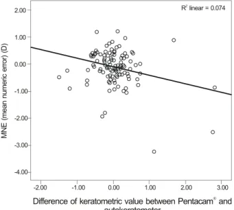
Axial length (mm) 22.72 ± 0.53 23.06 ± 0.63 22.82 ± 1.25 <0.01
Anterior chamber depth (mm) 2.70 ± 0.18 3.27 ± 0.15 3.79 ± 0.22 <0.01
Lens thickness (mm) 4.75 ± 0.55 4.10 ± 0.60 3.53 ± 0.53 <0.01
Autokeratometer K applied MAE (D) 0.42 ± 0.30 0.48 ± 0.54 0.59 ± 0.61 0.45
Pentacam® K applied MAE (D) 0.42 ± 0.40 0.49 ± 0.51 0.55 ± 0.52 0.58
Values are presented as mean ± SD.
MNE = mean numeric error; MAE = mean absolute error.
*Anterior chamber depth < 3.00; †2.50 ≤ Anterior chamber depth < 3.50; ‡3.50 ≤ Anterior chamber depth.
Table 4. The Relationship between preoperative target diopters
and postoperative refractive diopters at 2 months applied kera- tomeric values which measured by each device-Automated keratometer and Pentacam®Values
Autokeratometer K applied MNE (D) -0.12 ± 0.67 Pentacam® K applied MNE (D) -0.09 ± 0.66 Autokeratometer K applied MAE (D) 0.48 ± 0.49
Pentacam® K applied MAE (D) 0.48 ± 0.47
Values are presented as mean ± SD.
MNE = mean numeric error; MAE = mean absolute error.
MAE (D)
2.53
0.42 0.40 0.36 0.33 0.30 0.27 0.24
0.06 0.00 0.85 0.66 0.61 0.53 0.49 0.45
0.21 0.18 0.16 0.11 0.08
0.00 0.50 1.00 1.50 2.00 2.50 3.00
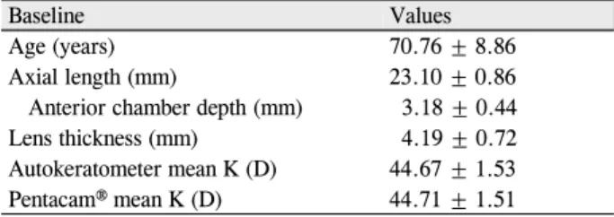
Figure 1. Relationship between mean absolute error (MAE)
and difference of keratometeric value between Pentacam® and autokeratometer. Note that the majority of large amount of MAE occurs among the group of which keratometer measured by Pentacam differed more than 1.00 diopter from autokeratometer.계와Pentacam®모두평균절대오차(mean absolute error:
MAE) 값과 안축장과의 연관성이 없는 것으로 나타났다 (Table 2). 전방깊이에따라세군으로나누어(<3.00, <3.50,
≥3.50) 분석한결과자동각막곡률계와Pentacam®모두평 균절대오차(mean absolute error: MAE) 값과전방깊이 와의 연관성이없는것으로 나타났다(Table 3).
자동각막곡률계로측정한각막곡률을SRK/T 공식에적 용하여산출한목표굴절치에대한술후2개월뒤모든환 자군의평균실제오차(mean numeric error: MNE)는-0.12 로 근시편위를 보였고 및 평균절대 오차(mean absolute error: MAE)는0.48 ±0.49로측정되었다. 이에Pentacam® 으로측정한각막곡률을대입하여다시인공수정체도수를 재측정하였고이를술후2개월뒤구면대응치와비교하였 을때평균실제오차(mean numeric error: MNE)는-0.09
으로 근시 편위를 보였고 평균절대 오차(mean absolute error: MAE)는0.48 ± 0.47로 측정되었다. 각 기기별로 측정된각막곡률로계산된인공수정체도수와술후2개 월뒤구면대응치간의차이를비교한평균절대오차(mean absolute error: MAE)는서로 통계적으로유의한 차이가 없었다(p-value=0.83) (Table 4).
양기기간의각막 곡률의차이의정도가오차의원인이
Difference of keratometric value between Pentacam® and autokeratometer
2.00
1.00
0.00
-1.00
-2.00
-3.00
-4.00
-2.00 -1.00 0.00 1.00 2.00 3.00
MNE (mean numeric error) (D)
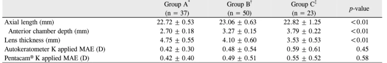
R linear = 0.0742
Figure 2. Relationship between mean numeric error (MNE)
and difference of keratometeric values between Pentacam®and autokeratometer (Pearson Correlation = -0.272, p < 0.01).
Difference of keratometric value between Pentacam® and au- tokeratometer = Pentacam® mean corneal front power kerato- metric value - Autokeratometer mean keratometric value.
Table 5. The biometry characteristics among 3 groups when divided by the difference of keratometer values between Pentacam® and
autokeratometerGroup 1* (n = 86)
Group 2† (n = 17)
Group 3‡
(n = 7) p-value
Axial length (mm) 23.06 ± 0.84 23.41 ± 1.01 22.89 ± 0.57 0.24
Anterior chamber depth (mm) 3.17 ± 0.44 3.24 ± 0.47 3.19 ± 0.44 0.85
Lens thickness (mm) 4.21 ± 0.67 4.09 ± 0.83 4.30 ± 1.08 0.77
Autokeratometer mean K (D) 44.80 ± 1.45 44.00 ± 1.91 44.74 ± 1.71 0.14
Pentacam® mean K (D) 44.81 ± 1.44 43.91 ± 1.56 45.37 ± 1.77 0.05
Autokeratometer K applied MAE (D) 0.44 ± 0.37 0.35 ± 0.23 1.32 ± 1.13 <0.01
Pentacam® K applied MAE (D) 0.42 ± 0.39 0.44 ± 0.39 1.33 ± 0.81 <0.01
Values are presented as mean ± SD.
*Absolute value of difference of keratometer between Pentacam® and autokeratometer, <0.50; †Absolute value of difference of keratometer between Pentacam® and autokeratometer, 0.50 ≤ and < 1.00; ‡Absolute value of difference of keratometer between Pentacam® and autokeratometer, ≥1.00; 1-way ANOVA test: Group 1 vs Group 3: p = 0.00; Group 2 vs Group 3: p = 0.00.
될수있다는가정하에Pentacam®으로측정된mean cor- neal front power와자동각막곡률계의각막곡률의차이를 비교해 보았다. 각각의 차이를절대값으로 환산하여평균 0.37 ±0.45 (0.02-2.83)의차이를보였다. 두기종의각 막곡률값의차이와자동각막곡률계로 측정된 평균절대 오 차(mean absolute error: MAE)와의 상관 관계를 이변량 상관분석 및 선형 회귀분석을통해분석하였고Pearson 상 관계수0.399로상호간의약한양의상관관계를나타내었 다(p-value <0.001).
평균절대 오차(mean absolute error: MAE)와 Pentacam® 으로 측정된 mean corneal front power와 자동각막곡률계
의 각막곡률의 차이 간의 경향을 산포도로 도식화하였을 때 1.00D 이상인 각 기기로 측정한 각막곡률의 차이의 절 대값이 1.00D 이상인 경우에서 주로 비교적 많은 평균절대 오차(mean absolute error: MAE)가 발생하는 것을 관찰하 였다(Fig. 1). 또 2개월 뒤 측정한 현성 굴절검사로 구면대 응치(spherical equivalent, SE)는 Pentacam®으로 측정된 mean corneal front power와 자동각막곡률계의 각막곡률 의 차이의 절대치가 클수록 목표 굴절치보다 근시화되는 경향을 나타내었고 이변량 상관분석 결과 Pearson 상관계 수 0.316으로 상호간의 약한 음의 상관관계를 나타내었다 (p-value <0.001). 또 이를 다시 각각 Pentacam®으로 측 정된 mean corneal front power가 더 클 경우와 자동각막 곡률계의 각막곡률이 더 큰 경우로 나누어 비교한 결과 Pearson 상관계수 0.272로 상호간의 약한 음의 상관관계를 나타내었고(p-value<0.001), Pentacam®으로 측정된 mean corneal front power가 더 클 경우 평균실제오차(mean numeric error: MNE)는 Pentacam®의 각막곡률 적용 시 0.45 ±0.66 D, 자동각막곡률계의 각막곡률을 적용할 시에 는 -0.22 ±0.73 D로 유의하게 자동각막곡률계의 실제 오 차가 좀더 근시화되는 결과를 보였고, 반대로 자동각막곡률 계의 각막곡률이 Pentacam®의 각막곡률보다 클 경우에는 Pentacam®의 각막곡률 적용 시 -0.24 ±0.64 D, 자동각막 곡률계의 각막곡률을 적용할 시에는 -0.04 ± 0.60 D로 Pentacam®의 각막곡률 적용 시 좀더 근시화되는 경향을 보 였다(p-value<0.001) (Fig. 2, Table 6).
Pentacam®으로측정한각막곡률과자동각막곡률계로측 정한각막곡률의차이의정도를차이의절대값에따라1군 (0-0.50 D), 2군(0.50-1.00 D), 3군(1.00 D 이상)으로3 군으로나누었고3군간의특성을각각비교하였다. 자동각 막곡률계와Pentacam®두기종모두세군간에평균절대 오차(mean absolute error: MAE) 값에유의한차이를보
Table 6. Relationship between mean numeric error (MNE), mean absolute error (MAE), and difference of keratometeric values be-
tween Pentacam® and autokeratometer
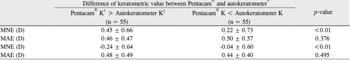
Difference of keratometric value between Pentacam® and autokeratometer*
p-value Pentacam® K† > Autokeratometer K‡
(n = 55)
Pentacam® K < Autokeratometer K (n = 55)
MNE (D) 0.45 ± 0.66 0.22 ± 0.73 <0.01
MAE (D) 0.46 ± 0.47 0.50 ± 0.57 0.376
MNE (D) -0.24 ± 0.64 -0.04 ± 0.60 <0.01
MAE (D) 0.48 ± 0.49 0.44 ± 0.40 0.495
Values are presented as mean ± SD.
*Pentacam® mean corneal front power keratometric value - Autokeratometer mean keratometric value; †Pentacam® mean corneal front power keratometric value; ‡Autokeratometer mean keratometric value.
Group
100 90 80 70 60 50 40 30 20 10 0
Percentage of patients achieving a MAE within 1.0 D of predicted value
Pentacam K Autokeratometer K
®
Figure 4. Comparison of the percentage of patients achieving
a mean absolute error (MAE) within 1.0 D of predicted value measured by autokeratometer and Pentacam®. Note that the majority of patients above the 1.0 D MAE values occurred among the group with Pentacam® photography equivalent K reading applied MAE of group 3. This was significantly high- er than any other groups as assessed by Kruskal-wallis test (p <0.01). Group 1 = Absolute value of difference of keratometer between Pentacam® and autokeratometer, < 0.50; Group 2 = Absolute value of difference of keratometer between Pentacam®
and autokeratometer, 0.50 ≤ and < 1.00; Group 3 = Absolute value of difference of keratometer between Pentacam® and au- tokeratometer, ≥ 1.00.
Keratometer differences between Pentacam® and autokeratometer
MAE (D)
1.16 1
0.59 0.50 0.47 0.42 0.39 0.36 0.33
0.15 .10 1.00 0.92 0.81 0.75 0.66
0.30 0.27 0.24 0.21 0.18 0.11 0.06 0.05 0.00
Figure 3. Comparison of mean absolute error (MAE) of IOL
calculation measured by autokeratometer and Pentacam®. IOL power prediction is more accurate measured in groups 1 and 2, than in group 3 of which keratometer values measured by Pentacam® differed more than 1.00 diopter from autokera- tometer (Group 1. vs. Group 2. p = 1.00, Group 2. vs. Group 3. p = 0.00, Group 1. vs. Group 3. p = 0.00, ANOVA test).였다. 자동각막곡률계와Pentacam®으로측정한각막곡률 값의차이가 1.00 D 이상인3군이1군및2군에비해평균 절대오차값이컸으며두기종 모두사후분석결과1군과 3군(p<0.001), 2군과 3군(p<0.001)에서 평균절대 오차 의차이를보였다(Table 5). 이를Fig. 3에서도확인할수 있다.
인공수정체의도수측정의오차가크게산출된Pentacam® 으로측정한각막곡률과자동각막곡률계로측정한각막곡 률의차이가1.00 이상인Group 3에한정하여Pentacam® 으로측정한 각막 곡률과 자동각막곡률계로 측정한 각막곡 률의 평균값을 각각 SRK/T 공식에 대입하여 인공수정체 도수 예측도의 정확성을 비교해보았고 평균절대 오차 (mean absolute error: MAE)는 각각 1.33 ± 0.81 D와 1.32 ±1.13 D로 양자간의 통계적으로 유의한 차이는 보이
지 않았다(p-value=0.40).
평균절대 오차의 수치상으로는 양자간의 유의한 차이가 없었으나 술 후 굴절 오차 즉, 평균절대 오차(mean abso- lute error: MAE)의 허용 범위를 1.0D로 기준하여 허용 범 위를 벗어난 1.0D 이상 술 후 오차를 보이는 비율을 비교하 였다.19Pentacam®으로 측정한 각막곡률을 적용한 경우 양 기기간의 측정치의 차이가 큰 그룹일수록 통계적으로 유의
하게 1.0D 이상의 술 후 굴절 오차를 보이는 비율이 증가함 을 확인하였다(p<0.001). Autokeratometer로 측정한 각 막곡률을 적용할 경우 양 기기의 차이와 무관하게 1.0D 이 상의 굴절 오차를 보이는 비율이 비슷하게 유지되었다 (p=0.09)(Fig. 4).
고 찰
근래에미세절개술등의백내장수술기법의발전과다초 점인공수정체및 난시교정인공수정체의개발그리고인 공수정체도수계산공식의발전으로술 후보다정확한굴 절력예측이가능해졌으며이에따라술후굴절상태에대 한정확한예측이환자의수술만족도에큰영향을미치게 되었다. 백내장수술후굴절력의정확히예측하기위해서 는 안축장, 각막곡률값 등의생체계측자료 및인공수정체 도수계산공식의정확성, 제조자의인공수정체질적관리의 정확성그리고술자의특성등을고려한임상적인변수등 여러 가지요소를고려해야 한다.
그중생체계측의오차가술후굴절오차에가장큰영 향을주는것으로 알려졌으며굴절오차에있어안축장길 이1 mm 오차는약2.7D의오차를야기하며, 각막곡률반 경1 mm 오차는약5.7D의오차를그리고술후전방깊이 1 mm 오차는 1.5D의 오차를 야기하는 것으로 알려졌 다.3,7,8,20
본연구에서도안축장과전방깊이의상관관계에대하여 분석하였는데기존의Maeng et al16이보고한결론에따르 면 SRK/T 공식으로 측정된 굴절 예측치가 전방 깊이가 3.50 mm 이상인경우와안축장이24.00 mm 이상인경우 에서통계적으로유의한원시편위를나타낸다고보고하였 으나, 본 연구에서는 같은분류와 공식을 적용하였음에도 불구하고평균절대오차(mean absolute error: MAE)와연 관성이없는것으로나타났다. 이는본연구에비해술전 안축장과전방깊이의편차가더커서연구표본집단의차 이가반영되었을수있고, 또본연구에서는술전안축장 과전방깊이간의상관계수가0.49로타연구에비해비교적 강한관련성을보여술후전방깊이의예측을비교적용이 하게 하였을가능성도있었을 것이다.
2006년Pentacam®의Holladay equivalent K Reading이 굴절교정수술 후 시행되는 백내장 수술에서 인공수정체의 도수예측에유용함이발표되었고이후굴절교정수술후각 막곡률값의예측에관한Pentacam®의유용성이활발하게연 구되었다.21,22하지만이전에굴절수술을받지않은정상안에 서인공수정체의도수예측에Pentacam®유용성에대한연 구는적은편이다. 기존의연구들을살펴보면Pentacam®으
로측정된각막곡률이자동각막곡률계로측정된각막곡률 에비하여인공수정체도수예측도의 정확성에있어유리 한점이없다는보고가있고,12,22,23 반면에Pentacam®으로 측정된 각막곡률이 자동각막곡률계 또는 IOL master®로 측정된각막곡률에비하여인공수정체도수예측도의정확 성에있어오히려 정확도가낮다는 보고도있다.14
본연구에서도 Pentacam®으로측정된 각막곡률이자동 각막곡률계로측정된각막곡률에비하여인공수정체도수 예측도의정확성에있어유의한차이가없음이관찰되었다.
하지만본연구에서 수술전 세극등검사와Pentacam®을 통하여전안부에이상소견이없고, 다른생체계측치의유 의한차이가없음에도불구하고, Pentacam®으로측정한각 막곡률과 자동각막곡률계로 측정한 각막 곡률의 차이가 1.00D 이상인경우유의하게인공수정체의도수예측성이 떨어지는것이관찰되었다. 또한자동각막곡률계의각막곡 률과차이가나는경우차이를유발한Pentacam®각막곡 률을인공수정체공식에대입하여도평균적인도수차이는 여전히유의하게나타나는 것을확인하였다.
최근 발표된 200안의 정상안을 대상으로 한 연구에서 Gonen et al10의보고에따르면Pentacam®으로측정된각 막곡률의경우자동각막곡률계와비교하였을때분포의구 간이더넓고, 5.5%에해당하는11안의유의하게많은이 상치가관찰되어인공수정체도수계산에사용 시유의해 야한다는보고가있고, 본 연구에서도6안의상기연구와 동일한비율의이상치가 관찰되었다.
본연구에포함된환자군은모두정상범위의각막곡률 과전안부의해부학적구조를가지고 있는바자동각막곡 률계와Pentacam®각막곡률의수치의차이를유발하는요 인으로추측할수있는것은양기기간의측정방법의차이 에따른즉, 각막중심3 mm 이내의자동각막곡률계가측 정하는4-6개지점이외의각막중심부의변화가 있는경 우이나세극등현미경또는 Pentacam®에서 다른환자와의 차이점이발견되지않아추후더 많은환자군을대상으로 이들의공통점을찾는 연구가필요할 것으로생각한다.
본연구에서는생체계측상 오차를줄이기위하여검사 상오차를유발할수있는해부학적이상또는생체계측치 에서극단적인이상치에해당하는환자를모두배제하였으 며, 도수계산및술자, 인공수정체의종류도모두통일하여 적용함으로써굴절오차에영향을미칠수있는다른요인 들을 최대한 배제하려 노력하였다. 또한 본 연구에서는 기 존의 임상 경험을 통해 표준치의 안축장에서 정확성이 증 명된 SRK/T 공식을 사용하였다.24-28
본 연구의 제한점으로 환자수가 비교적 충분하지 못한 점으로 추후 더 많은 환자를 대상으로 하는 연구가 필요할
것으로 생각한다.
결론적으로 자동각막곡률계와 Pentacam®으로 측정된 각 각의 각막 곡률 모두 인공수정체 도수 예측도의 정확성에 있어 유의한 차이가 없었다. 하지만 자동각막곡률계와 Pentacam®으로 측정된 각막곡률의 수치의 차이가 1.00D 이상일 때에는 SRK/T 공식 하에서 인공수정체 도수 예측 도의 정확성이 유의하게 낮고 나아가 Pentacam®으로 측정 한 각막 곡률을 적용할 경우 기존의 자동각막곡률계로 측 정한 각막 곡률을 적용하는 것보다 술 후 보다 큰 오차를 유발할 수 가능성이 높음을 상기하고 특히, 다초점 인공수 정체 및 난시교정 인공수정체의 삽입이 예상되는 경우 양 기기간의 수치의 차이에 더욱 유의해야 할 것이다.
REFERENCES
1) Holladay JT, Prager TC, Ruiz RS, et al. Improving the predict- ability of intraocular lens power calculations. Arch Ophthalmol 1986;104:539-41.
2) Mamalis N. Complications of foldable intraocular lenses requiring explanation or secondary intervention--1998 survey. J Cataract Refract Surg 2000;26:766-72.
3) Olsen T. Sources of error in intraocular lens power calculation. J Cataract Refract Surg 1992;18:125-9.
4) Maeng HS, Ryu EH, Chung TY, Chung ES. Effects of anterior chamber depth and axial length on refractive error after intra- ocularlens implantation. J Korean Ophthalmol Soc 2010;51:195-202.
5) Koranyi G, Lydahl E, Norrby S, Taube M. Anterior chamber depth measurement: a-scan versus optical methods. J Cataract Refract Surg 2002;28:243-7.
6) Hosny M, Alio JL, Claramonte P, et al. Relationship between ante- rior chamber depth, refractive state, corneal diameter, and axial length. J Refract Surg 2000;16:336-40.
7) Olsen T. Prediction of the effective postoperative (intraocular lens) anterior chamber depth. J Cataract Refract Surg 2006;32:419-24.
8) Norrby S. Sources of error in intraocular lens power calculation. J Cataract Refract Surg 2008;34:368-76.
9) Speicher L. Intra-ocular lens calculation status after corneal re- fractive surgery. Curr Opin Ophthalmol 2001;12:17-29.
10) Gonen T, Cosar CB, Sener B, Keskinbora KH. Comparison of ker- atometric data obtained by automated keratometer, dicon ct200, al- legro topolyzer, and pentacam. J Refract Surg 2012;28:557-61.
11) Salouti R, Nowroozzadeh MH, Zamani M, et al. Comparison of an- terior chamber depth measurements using Galilei, HR Pentacam, and Orbscan II. Optometry 2010;81:35-9.
12) Shammas HJ, Hoffer KJ, Shammas MC. Scheimpflug photography keratometry readings for routine intraocular lens power calculation.
J Cataract Refract Surg 2009;35:330-4.
13) Chang M, Kang SY, Kim HM. Which keratometer is most reliable for correcting astigmatism with toric intraocular lenses? Korean J Ophthalmol 2012;26:10-4.
14) Savini G, Barboni P, Carbonelli M, Hoffer KJ. Accuracy of Scheimpflug corneal power measurements for intraocular lens power calculation. J Cataract Refract Surg 2009;35:1193-7.
15) Módis L Jr, Szalai E, Kolozsvári B, et al. Keratometry evaluations with the Pentacam high resolution in comparison with the auto- mated keratometry and conventional corneal topography. Cornea 2012;31:36-41.
16) Maeng HS, Ryu EH, Chung TY, Chung ES. Effects of anterior chamber depth and axial length on refractive error after intraocular lens implantation. J Korean Ophthalmol Soc 2010;51:195-202.
17) Weissman JL, Beatty RL, Hirsch WL, Curtin HD. Enlarged ante- rior chamber: CT finding of a ruptured globe. AJNR Am J Neuroradiol 1995;16(4 Suppl):936-8.
18) Masket S. Alcon Restor multifocal-clinical pearls. In: Chang DF, ed. Mastering refractive IOLs: The art and science. Thorofare, NJ:
SLACK Incorporated, 2008; chap. 38.
19) Gale RP, Saldana M, Johnston RL, et al. Benchmark standards for refractive outcomes after NHS cataract surgery. Eye (Lond) 2009;23:149-52.
20) Jo DH, Oh JY, Kim MK, Wee WR. Corneal power estimation using Orbscan II videokeratography in eyes with previous corneal re- fractive surgeries. J Korean Ophthalmol Soc 2009;50:1730-4.
21) Tang Q, Hoffer KJ, Olson MD, Miller KM. Accuracy of Scheimpflug Holladay equivalent keratometry readings after corneal refractive surgery. J Cataract Refract Surg 2009;35:1198-203.
22) Kim SW, Kim EK, Cho BJ, et al. Use of the pentacam true net cor- neal power for intraocular lens calculation in eyes after refractive corneal surgery. J Refract Surg 2009;25:285-9.
23) Symes RJ, Ursell PG. Automated keratometry in routine cataract surgery: comparison of Scheimpflug and conventional values. J Cataract Refract Surg 2011;37:295-301.
24) Retzlaff JA, Sanders DR, Kraff MC. Development of the SRK/T intraocular lens implant power calculation formula. J Cataract Refract Surg 1990;16:333-40.
25) Cho YT, Lee EH. Evaluation for the accuracy of the SRK/T Formula in PCL Implanted Patients(I). J Korean Opthalmol Soc 1991;32:752-60.
26) Aristodemou P, Knox Cartwright NE, Sparrow JM, Johnston RL.
Formula choice: Hoffer Q, Holladay 1, or SRK/T and refractive outcomes in 8108 eyes after cataract surgery with biometry by par- tial coherence interferometry. J Cataract Refract Surg 2011;37:63-71.
27) Narváez J, Zimmerman G, Stulting RD, Chang DH. Accuracy of intraocular lens power prediction using the Hoffer Q, Holladay 1, Holladay 2, and SRK/T formulas. J Cataract Refract Surg 2006;
32:2050-3.
28) Sanders DR, Retzlaff JA, Kraff MC, et al. Comparison of the SRK/T formula and other theoretical and regression formulas. J Cataract Refract Surg 1990;16:341-6.
=ABSTRACT=
Comparison of the Refractive Outcomes According to the Differences of Biometry and Keratometry Reading
Kang Hoon Lee, MD1, Na Rae Kim, MD1, Kyoung Yul Seo, MD, PhD2
Department of Ophthalmology and Inha Vision Science Laboratory, Inha University School of Medicine1, Incheon, Korea The Institute of Vision Research, Department of Ophthalmology, Yonsei University College of Medicine2, Seoul, Korea
Purpose: The purpose of this study was to investigate the error tendency between preoperative expected refraction and postoperative manifest refraction based on axial length, anterior chamber depth, and keratometric data obtained by an au- tomated keratometer and Pentacam® in cataract surgery cases and to report how their differences affect determination of intraocular lens (IOL) power.
Methods: The authors retrospectively reviewed the medical records of 110 eyes of 84 patients who underwent cataract surgery. Axial length and anterior chamber depth were measured by A scan ultrasound biometry, while keratometric val- ues were obtained by an automated keratometer and Pentacam®. IOL power was calculated using the SRK/T formula.
Patients were divided into 3 groups based on the axial length, anterior chamber depth, and the difference of keratometric values between the 2 devices. Refractive error was analyzed 2 months after surgery.
Results: There were no statistically significant differences between axial length and anterior chamber depth among the groups; however, the K reading differences were statistically significant. Although the mean absolute error (MAE) of each group showed no statistical significance among the groups, the MAE was more pronounced in the group in which the kera- tometeric value measured by Pentacam® differed more than 1.00 diopter from the automated keratometer measurements.
Conclusions: There was no statistically significant difference between axial length and anterior chamber depth among the groups. A difference of 1.00 diopter or more between the keratometric values obtained by an automated keratometer and Pentacam® significantly affects the postoperative refractive error; therefore, these factors should be considered when de- termining IOL power.
J Korean Ophthalmol Soc 2013;54(9):1345-1352
Key Words: Biometry, Cataract surgery, Keratometry reading, Refractive outcomes, Scheimpflug photography
Address reprint requests to Kyoung Yul Seo, MD, PhD Department of Ophthalmology, Severance Hospital
#50 Yonsei-ro, Seodaemun-gu, Seoul 120-752, Korea
Tel: 82-2-2228-3570, Fax: 82-2-312-0541, E-mail: [email protected]