DOI 10.4167/jbv.2011.41.2.83
Adhesion Activity of Lactobacillus plantarum PM 008 Isolated from Kimchi on the Intestine of Mice
Se-Eun Jang1, Yang-Jin Hyun2, Young-Joo Oh3, Kum Boo Choi3, Taesok Kim3, Ik Hyun Yeo3, Myung Joo Han1* and Dong-Hyun Kim2*
1Department of Food and Nutrition, Kyung Hee University, Seoul; 2Department of Life and Pharmaceutical Sciences, Kyung Hee University, Seoul; 3Pulmuone Co., Seoul, Korea
Lactic acid bacteria (LAB), including L. plantarum isolated from Kimchi, are beneficial and safe microorganisms that improve disturbances of the indigenous microflora and the host's immune system. The adhesion abilities of Kimchi- derived L. plantarum PM008 and yogurt-derived L. casei were measured in vitro and in vivo. When L. plantarum or L.
casei was incubated with Caco-2 cells, these Lactobacillus strains were potently attached. When these strains were orally administered to mice, the LABs were attached on the large intestine of mice. The attachment of L. plantarum on murine intestine or Caco-2 intestinal epithelial cell lines was more potent than that of L. casei, although numbers of LAB between their feces were not different. Treatment with either L. plantarum or L. casei for 14 days suppressed fecal
-glucuronidase activity, although treatment for one day did not affect it. L. plantarum showed more potent inhibition than L. casei. In addition, L. plantarum and L. casei were stable to artificial gastric and intestinal juice. L. plantarum was more stable than L. casei. Based on these findings, the survival and adhesion effects of orally administered LAB strains in the intestine may increase numbers of LAB in intestine and express their biological activities.
Key Words: Lactic acid bacteria, Lactobacillus plantarum, Lactobacillus casei, Adhesion activity
INTRODUCTION
Normal intestinal microflora consist of an estimated 500 different bacterial species, and reach their highest concen- trations in the terminal ileum and colon (1~3). Intestinal microflora consisted of beneficial and harmful bacteria, such as Lactobacillus acidophilus, Bifidobacterium longum,
Escherichia coli and Staphylococcus aureus.
The harmful bacteria produce toxic compounds, such as gram-negative bacterial endotoxin, and harmful enzymes, such as -glucuronidase and tryptophanase, which produce cytotoxic or carcinogenic agents (4~7). Cytotoxin and endotoxins may interact at the apical intestinal surface and induce responses in the intestinal epithelial cells, which produce proinflammatory cytokines and other mediators that induce inflammatory activation of the mucosal immune system, and cause colitis, diarrhea or constipation.
Lactic acid bacteria (LAB) are beneficial and safe micro- organisms (8), that improve disturbances of the indigenous microflora (9), ameliorate the development of microflora (8), have antidiabetic and antihyperlipidemic effects (10, 11), inhibit carcinogenesis (12), have anticolitic effects (9, 13~15), and show non-specific activation of the host's immune system (12). The biological activities of the 83
Received: February 17, 2011/ Revised: March 22, 2011 Accepted: March 31, 2011
*Corresponding author: Myung Joo Han, Ph.D. Department of Food and Nutrition, Kyung Hee University, 1, Hoegi, Dongdaemun-ku, Seoul 130-701, Korea.
Phone: +82-2-961-0553, Fax: +82-2-968-0260 e-mail: [email protected]
*Corresponding author: Dong-Hyun Kim, Ph.D. Department of Life and Nanopharmaceutical Sciences and Department of Pharmaceutical Science, Kyung Hee University, 1, Hoegi, Dongdaemun-ku, Seoul 130-701, Korea.
Phone: +82-2-961-0374, Fax: +82-2-957-5030 e-mail: [email protected]
Original Article
beneficial LAB have mainly been studied for many LAB derived from yogurts and healthy human intestinal micro- flora, including Lactobacillus acidophilus, Lacobacillus casei, Bifidobacterium breve, and Bifidobacterium longum.
Although LAB derived from foods such as Kimchi has been taken in Asia countries, particularly Japan and Korea, their adhesion abilities on the intestine have not been thoroughly studied. Therefore, we investigated the adhesion ability of Kimchi-derived Lactobacillus plantarum and yogurt-derived L. casei on Caco-2 cells and mice.
MATERIALS AND METHODS Materials and bacterial strains
p-Nitrophenyl--D-glucuronide and p-nitrophenyl--D- glucopyranoside were purchased from Sigma Co. (St. Louis, MO, USA). Ex Taq polymerase chain reaction (PCR) kit was purchased from TaKaRa Co. (Tokyo, Japan). All reagents used in slot hybridization analysis and fluo- rescence in situ hybridization were purchased from Roche Diagnostics (Indianapolis, IN, USA).
L. plantarum PM008 isolated from Kimchi and L. casei were donated from Pulmuone Co. (Seoul, Korea), and grown in MRS broth at 37℃ for 24 h. The cultured Lactobacillus strains were harvested by centrifugation at 3,000 × g for 10 min. Each pelleted bacterium was then washed two times in sterile PBS and resuspended 1 × 10⁹ CFU/ml and 5 × 10⁹ CFU/ml in PBS.
Survival Stability assays in gastric and intestinal juice To evaluate the survival stabilities of orally administered L. plantarum and L. casei in intestine, they were stored in artificial gastric [pepsin (1,000 units/ml) containing MRS broth adjusted to pH 2.5 with 5 N HCl] and intestinal juices [GAM broth (pH 6.8) containing 0.1% mucin, 0.04%
pancreatin, 0.2% bile, 0.85% NaCl, 0.04% trypsin] for 3 h.
Numbers of surviving Lactobacillus strains were determined by plating serial dilutions (in PBS, pH 7.2) on MRS agar followed by incubation at 37℃ for 48 h.
Adhesion assay
Caco-2 cells, heterogeneous human epithelial colorectal adenocarcinoma cells, purchased from Korea Cell Line Bank (Seoul, Korea). The cells were cultured at 37℃ in a 5% CO2-95% air atmosphere in RPMI 1640 (Sigma Co.) supplemented with 10% heat-inactivated (30 min, 56℃) fetal calf serum (FCS, Sigma Co.). To investigate the adhesion activity of Lactobacillus strains, postconfluent Caco-2 cells were washed twice with PBS. For each adhesion assay, 500 μL of the Lactobacillus suspension [bacteria (1 × 10⁷ CFU) with broth culture supernatant]
was mixed with Dulbecco's modified Eagle's medium (DMEM) (500 μL), and then added to each well of the tissue-culture plate (24 wells), which was then incubated at 37℃ for 2 h in 10% CO2-90% air. After incubation, the cells were washed five times with sterile PBS, the cells were lysed with sterile H2O, and appropriate dilutions were then plated on MRS agar to determine the number of viable cell-associated bacteria. Results were expressed as CFU/
well of cell-associated bacteria.
Animals
Male ICR mice (20~25 g, 5 weeks old) were obtained from the Charles River Orient Experimental Animal Breeding Center (Seoul, Korea). All animals were housed in wire cages at 20~22℃, relative humidity of 50±10%, air ventilation frequency of 15~20 times/h and 12-h illumi- nation (07:00~19:00; intensity, 150~300 Lux), fed standard laboratory chow (Charles River Orient Experimental Animal Breeding Center, Seoul, Korea), and allowed water ad libitum. All experiments were performed in accordance with the NIH and Kyung Hee University guides for Laboratory Animals Care and Use and approved by the Committee for the Care and Use of Laboratory Animals in the College of Pharmacy, Kyung Hee University.
Intestinal bacterial enzyme activity assay
Mice were randomly divided into 9 groups: normal; high dosage (1.0 × 10⁹ CFU) of L. plantarum or L. casei for one day; high dosage (1.0 × 10⁹ CFU) of L. plantarum or
L. casei for 14 days; low dosage (0.2 × 10⁹ CFU) of L.
plantarum or L. casei for one day; low dosage (0.2 × 10⁹ CFU) of L. plantarum or L. casei for 14 days. Dosage was once a day with 200 μl of each LAB, and the normal group was treated with sterile PBS alone. Each group contains 12 mice, and half of each group was sacrificed 1st day after final treatment with LAB, and the rest were sacrificed 7th day after the final treatment.
Counting of bacteria in stool
Number of survival LAB were determined by plating serial dilutions (in PBS, pH 7.2) on MRS agar followed by incubation at 37℃ for 48 h.
Preparation of fecal bacterial suspension
The fresh mouse stools (0.5 g) from each group were separately collected in sterilized plastic cups, carefully suspended with 20-fold diluted anaerobic broth in a cooled tube and centrifuged at 250 × g for 5 min. The supernatant was re-centrifuged at 10,000 × g for 20 min. The resulting precipitates were used as the sources for the fecal enzyme assay. All procedures were performed at 4℃.
Assay of -glucuronidase and -glucosidase activities The reaction mixture (2.0 ml), consisting of 0.04 ml of 2 mM p-nitrophenyl--D-glucuronide (or p-nitrophenyl-- D-glucopyranoside), 0.76 ml of 0.1 M phosphate buffer (pH 7.0), and 0.2 ml of fecal suspension, was incubated for 30 min at 37℃, with the reaction stopped by the addition 1 ml of 0.5 M NaOH. The mixture was then centrifuged at 3,000 × g for 10 min and the absorbance measured at 405 nm.
Quantification of LAB in mouse intestine
Species-specific probes for L. plantarum and L. casei used the following three oligonucleotide probes: LPANG (5'-TATCATTGCCATGGTGA-3'), L. plantarum species- specific probe; and 5'-GAGATTCAACATGGAACG-3', L.
casei species-specific probe; and UP041 (5'-CTGCTGCC- TCCCGATGGAGT-3'), a universal oligonucleotide com- plementary to virtually all 16S rRNA (16, 17). These oligonucleotide probes were marked with digoxigenin at 3'-ends for immunoblot and fluorescent in situ hybridization (FISH). Total bacterial genomic DNAs were isolated and
purified from the intestine using the Quiagen Blood and tissue DNA purification kit (Qiagen GmbH, Hilden, Germany). The rDNA was amplified by using the TaKaRa Ex Taq polymerase kit (Takara Co., Tokyo, Japan), and the following primers were selected for the PCR reactions: 27F, 5'-AGAGTTTGATCCTGGCTCAG-3'; and 1492R, 5'- GGCTACCTTGTTACGACTT-3'. The PCR cycle was 4 min at 94℃; 30 sec at 92℃, 30 s at 52℃ and 1.5 min at 72℃, repeated 20 cycles; and 10 min at 72℃. The PCR products were purified using QIAquick PCR purification kit (Qiagen GmbH). To calibrate the data, PCR amplification using L. plantarum or L. casei genomic DNA as a template was also performed by following the same method.
Quantification of LAB attached in intestine by slot blot hybridization
Sections of 3.5 cm colon from each mouse were sampled and homogenized with 500 μl of sterile PBS on ice. The sludge was centrifuged at 800 rpm at 4℃ for 5 min, and then the supernatant was collected. The supernatant was centrifuged at 15,000 × g at 4℃ for 2 min to collect the pellet. Total bacterial genomic DNA was isolated and purified from the pellet using the Quiagen Blood and tissue DNA purification kit (Qiagen GmbH), according to the manufaturer's manual and amplified by PCR. PCR products (2 μg) were denatured with 0.3 M NaOH for 10 min at 98℃, and then loaded onto the positively charged nylon membrane using Minifold-II slot blotting system (Whatman Inc. Florham Park, NJ, USA). The membrane was fixed twice with UV-crosslinker. The membrane was hybridized in DIG-easy hybridization solution containing 100 pmol of LPANG for 2 h at 40℃, and then washed twice in the washing buffer (0.5 × SSC buffer containing 0.1% SDS) at 49℃. The hybrids were detected by using a CSPD-DIG detection kit (Roche), according to the manufacturer's manual. The chemiluminescent signals were detected by image analyzer, LAS-4000 mini (Fujifilm Co., Tokyo, Japan).
After the detection, the membrane was stripped with a stripping solution (0.2 M NaOH containing 0.1% SDS) and then rehybridized with the universal DIG-labeled probe, UP041, for control detection. The hybridization and washing temperatures for UP041 were 48 and 58℃, respectively,
and the other protocols were the same as those described above for LPANG. The signal intensity was converted to the mass unit (μg) by the calibration curve, and the percentage of L. plantarum or L. casei in the total microbes of the stool was calculated.
Analysis of LAB attached in intestine by FISH The colon (3.5 cm) from each mouse was sampled and homogenized with 500 μl of sterile PBS on ice. The homogenized sludge was centrifuged at 1,000 × g at 4℃
for 5 min, and the supernatant was collected. A 200 μl volume of supernatant was fixed with 600 μl of 4%
paraformaldehyde in PBS at 4℃ for 4 h. To remove the paraformaldehyde, the cell suspension was pelleted by centrifugation at 15,000 × g at 4℃ for 2 min, and resuspended in ice-cold PBS. These washing steps were repeated 3 times. The fixed pellet was finally resuspended in 200 μl of a 1:1 mixture of EtOH and PBS, and then stored at -20℃ before use. The procedure was used to identify L. plantarum by FISH method using DIG-labeled oligonucleotide (18). To view the cell smears hybridized with fluorescein-labeled anti-DIG Fab fragment, the slide was mounted with Vectashield (Vecta Laboratories, Inc., Burlingame, CA, USA), and the hybridized cells were counted visually with Axio Observer.D1 epifluorescence microscope (× 1,000, Carl Zeiss MicroImaging GmbH, Göttingen, Germany).
Statistical analysis
The data were calculated with mean values, and standard deviations (mean ± SD) were determined from triplicate trials. Statistical significance of the results was evaluated by one way ANOVA (analysis of variance) test (p < 0.05).
RESULTS AND DISCUSSION
We investigated the adhesion ability of L. plantarum isolated from Kimchi to evaluate whether the LAB can attach on the intestine (Fig. 1). When L. plantarum or L.
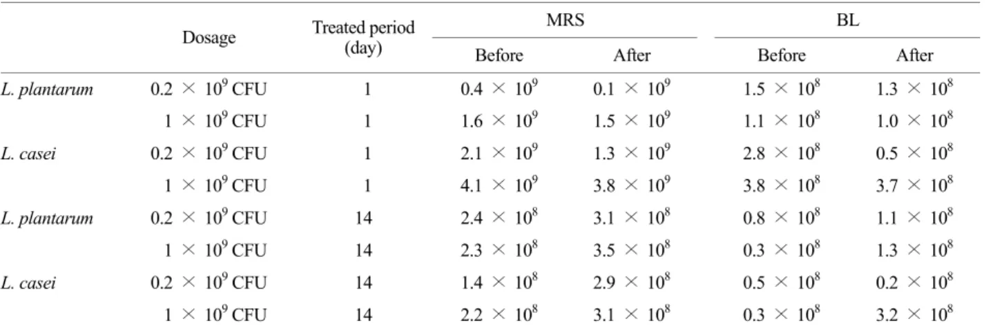
Table 1. Colony forming unit (CFU) of fecal bacteria grown in MRS and BL agar plates in mice orally treated with lactic acid bacteria
MRS BL
Dosage Treated period
(day) Before After Before After
L. plantarum 0.2 × 10⁹ CFU 1 0.4 × 10⁹ 0.1 × 10⁹ 1.5 × 10⁸ 1.3 × 10⁸
1 × 10⁹ CFU 1 1.6 × 10⁹ 1.5 × 10⁹ 1.1 × 10⁸ 1.0 × 10⁸
L. casei 0.2 × 10⁹ CFU 1 2.1 × 10⁹ 1.3 × 10⁹ 2.8 × 10⁸ 0.5 × 10⁸
1 × 10⁹ CFU 1 4.1 × 10⁹ 3.8 × 10⁹ 3.8 × 10⁸ 3.7 × 10⁸
L. plantarum 0.2 × 10⁹ CFU 14 2.4 × 10⁸ 3.1 × 10⁸ 0.8 × 10⁸ 1.1 × 10⁸
1 × 10⁹ CFU 14 2.3 × 10⁸ 3.5 × 10⁸ 0.3 × 10⁸ 1.3 × 10⁸
L. casei 0.2 × 10⁹ CFU 14 1.4 × 10⁸ 2.9 × 10⁸ 0.5 × 10⁸ 0.2 × 10⁸
1 × 10⁹ CFU 14 2.2 × 10⁸ 3.1 × 10⁸ 0.3 × 10⁸ 3.2 × 10⁸ Lactic acid bacteria (L. plantarum or L. casei) were orally administered once a day for one or 14 days. Numbers of intestinal bacteria were counted by plating serial dilutions (in diluted anaerobic broth, pH 7.2) on MRS and BL agars followed by anaerobic incubation at 37℃
for 48 h. All values are mean (n=3).
Figure 1. Adhesion ability of lactic acid bacteria to Caco-2 cells. Lactic acid bacteria (closed circle, 1 × 10⁷ L. plantarum per well; open circle, 1.0 × 10⁷ L. casei per well) were incubated with Caco-2 cells for 2 h, washed with PBS and then inoculated in MRS agar plate.
casei were incubated with Caco-2 cells, these Lactobacillus strains potently attached on Caco-2 cells. The adhesion ability of L. plantarum was superior to that of L. casei isolated from yogurt. To investigate whether these Lacto- bacillus strains are able to attach and proliferate in the intestine, we orally administered them to mice and counted numbers of LAB in the stools of mice using LAB selection media (MRS and BL agars) (Table 1). Treatment with both strains increased numbers of LAB on MRS and BL.
Treatment with L. plantarum and L. casei were increased LAB numbers, but their increases were not dependent on treatment periods.
To measure the attachment ability of orally administered LAB in intestine in vivo, we orally administered L.
plantarum or L. casei to mice and measured numbers of LAB attached to intestines using the slot blot hybridization method (Fig. 2). Numbers of LAB attached in intestines were found to be dependent on the administration period.
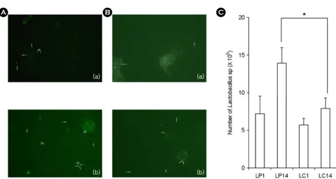
To confirm numbers of LAB directly attached to the intestines, we measured LAB numbers by fluorescent in situ hybridization (Fig. 3). Both strains were detected in the
Figure 2. Number of intestinal bacteria attached in colon membrane in Lactobacillus plantarum (A) or Lactobacillus casei (B)-treated mice by a fluorescent hybridization method. Lactic acid bacteria were orally administered once a day for one (a) or 14 days (b): LP1, treated with 1.0 × 10⁹ L. plantarum per mouse for one day; LP14, 1.0 × 10⁹ treated with L. plantarum per mouse for 14 days; LC1, 1.0
× 109 L. casei per mouse for one day; LC14, 1.0 × 10⁹ treated with L. casei per mouse for 14 days). All values are mean ± SD. (n=6).
*significantly different compared to control group.
(a)
(b) (b)
(a)
A B C
Figure 3. Number of intestinal bacteria attached in colon mem- brane in Lactobacillus plantarum (LP) or Lactobacillus casei (LC)-treated mice by a slot blot hybridization method. Lactic acid bacteria were orally administered once a day for one or 14 days:
LP1, treated with 1.0 × 10⁹ L. plantarum per mouse for one day;
LP14, 1.0 × 10⁹ treated with L. plantarum per mouse for 14 days;
LC1, 1.0 × 10⁹ L. casei per mouse for one day; LC14, 1.0 × 10⁹ treated with L. casei per mouse for 14 days). All values are mean
± SD. (n=6). *significantly different compared to control group.
intestines and the attached numbers were dependent on the administration period. When L. plantarum was orally administered to mice for one and 14 days, the numbers of L. plantarum attached to large intestine were 7.0 × 10⁶ and 1.4 × 10⁷, respectively. L. plantarum attached more efficiently to the intestine than L. casei.
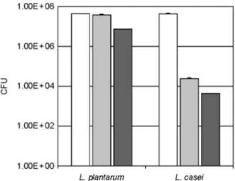
To understand the reason why L. plantarum more plen- tifully attached on the intestine than L. casei, we measured the survival ability of L. plantarum and L. casei in gastric and intestinal juices. When these Lactobacillus strains were stored in artificial gastric and intestinal juices (Fig. 4). L.
plantarum was more stable to gastric and intestinal juices than L. casei.
Next, we measured the effect of these Lactobacillus strains by their attachment on the intestine of mice. We
orally administered these Lactobacillus strains to mice and monitored the activities of the fecal enzymes, - glucuronidase and -glucosidase (Fig. 5). Treatment with either Lactobacillus strain for 14 days suppressed fecal
-glucuronidase activity, but increased fecal -glucosidase activity: these enzyme activities were gradually affected from 7th day after treatment with either LAB. L. plantarum more potently suppressed -glucuronidase activity than L.
casei, which more potently increased -glucosidase activity.
However, treatment with either LAB for one day did not affect fecal enzyme activities.
LAB is recognized beneficial microorganisms and have been demonstrated to exert preventive and therapeutic effects on diarrhea, constipation, and inflammatory diseases.
Fermented foods, such as, yogurt, cheese, and Kimchi, are Figure 4. Effect of lactic acid bacteria on fecal bacterial -glucosidase (A) and -glucuronidase activities (B) in mice. Lactic acid bacteria (LP0.2, 0.2 × 10⁹ L. plantarum per mouse; LP1.0, 1.0 × 10⁹ L. plantarum per mouse; LC0.2, 0.2 × 10⁹ L. casei per mouse;
LC1.0, 1 × 10⁹ L. casei per mouse) were orally administered once a day for one (upper) or 14 days (bottom). All values are mean ± SD.
(n=6). *significantly different compared to control group.
B A
considered to be excellent sources of microorganisms and a variety of LAB strains have been detected in these foods (19), and been shown to regulate host defense mechanisms against pathogens (20, 21). Of them, the Kimchi-derived L.
plantarum may have beneficial effects in terms of improving disturbances of indigenous bacteria in the gut and yogurt- derived L. casei, L. acidophilus, and B. longum have been reported to have similar effects. Nevertheless, the attach- ment ability of LAB isolated from Kimchi has not been thoroughly studied.
In the present study, the number of LAB detected in intestinal epithelia increased with administration period, and L. plantarum was more effective in this respect than L.
casei. Furthermore, L. plantarum was found to be resistant to gastric and intestinal juices more than L. casei. These results were supported that L. plantarum was highly tolerant for acid and alkaline stress because the arginine dihydrolase pathway and glutamate dicarboxylation (22~24). In addition, L. plantarum more potently attached to Caco-2 cells, which is intestinal cell lines, than L. casei. These results also were supported by previous reports that highest attachment ability
was observed with L. plantarum (25). When these strains were orally administered to mice for one and 14 days, they both increased LAB numbers in intestine and inhibited intestinal bacteria-producing -glucuronidase activity which is known to cause colon cancer and liver injury (7, 26).
Based on these findings, the survival and attachment of orally administered Lactobacillus strains in the intestine increases numbers of parental Lactobacillus strains residing in intestine, inhibit harmful enzyme production of intestinal microflora, and thus, ameliorate intestinal diseases, such as constipation and diarrhea.
REFERENCES
1) Hill MJ, Drasar BS. The normal colonic bacterial flora.
Gut 1975;16:318-23.
2) Simon GL, Gorbach SL. Intestinal flora in health and disease. Gastroenterology 1984;86:174-93.
3)Hattori M, Taylor TD. The human intestinal micro- biome: a new frontier of human biology. DNA Res 2009;16:1-12.
4)Chung KT, Fulk GE, Slein MW. Tryptophanase of fecal flora as a possible factor in the etiology of colon cancer. J Natl Cancer Inst 1975;54:1073-8.
5) Ganguly NK, Kingham JG, Lloyd B, Lloyd RS, Price CP, Triger DR, et al. Acid hydrolases in monocytes from patients with inflammatory bowel disease, chronic liver disease, and rheumatoid arthritis. Lancet 1978;1:
1073-5.
6) Rhodes JM, Gallimore R, Elias E, Allan RN, Kennedy JF. Faecal mucus degrading glycosidases in ulcerative colitis and Crohn's disease. Gut 1985;26:761-5.
7) Reddy BS, Weisburger JH, Wynder EL. Fecal bacterial beta-glucuronidase: control by diet. Science 1974;183:
416-7.
8)Collins MD, Gibson GR. Probiotics, prebiotics, and synbiotics: approaches for modulating the microbial ecology of the gut. Am J Clin Nutr 1999;69:1052S -1057S.
9) Campieri M, Gionchetti P. Probiotics in inflammatory bowel disease: New insight to pathogenesis or a possi- ble therapeutic alternative? Gastroenterology 1999;116:
1246-9.
Figure 5. Stability of lactic acid bacteria against artificial gastric juice and intestinal juice. Lactic acid bacteria (L. plantarum or L.
casei) were incubated in saline (white bar), artificial gastric [gray bar, pepsin (1,000 units)-contained MRS broth adjusted at pH 2.5 with 5 N HCl] or intestinal juice [black bar, GAM broth containing 0.1% mucin, 0.04% pancreatin, 0.2% bile, 0.85% NaCl, and 0.04%
trypsin (pH 6.8)] for 3 h at 37℃ and number of survival LAB were determined by plating serial dilutions (in PBS, pH 7.2) on MRS agar followed by incubation at 37℃ for 48 h. All values are mean ± SD. (n=6). *significantly different compared to saline control group.
10) Tabuchi M, Ozaki M, Tamura A, Yamada N, Ishida T, Hosoda M, et al. Antidiabetic effect of Lactobacillus GG in streptozotocin-induced diabetic rats. Biosci Biotechnol Biochem 2003;67:1421-4.
11) Taranto MP, Medici M, Perdigon G, Ruiz Holgado AP, Valdez GF. Evidence for hypocholesterolemic effect of Lactobacillus reuteri in hypercholesterolemic mice. J Dairy Sci 1998;81:2336-40.
12) Perdigon G, de Jorrat WEB, de Petrino SF, de Budeguer MV. Effect of oral administration of Lactobacillus casei on various biological functions of the host. Food Agric Immunol 1991;3:93-102.
13) Daniel C, Poiret S, Goudercourt D, Dennin V, Leyer G, Pot B. Selecting lactic acid bacteria for their safety and functionality by use of a mouse colitis model. Appl Environ Microbiol 2006;72:5799-805.
14) Han W, Mercenier A, Ait-Belgnaoui A, Pavan S, Lamine F, van Swam II, et al. Improvement of an experimental colitis in rats by lactic acid bacteria producing super- oxide dismutase. Inflamm Bowel Dis 2006;12:1044-52.
15)Peran L, Sierra S, Comalada M, Lara-Villoslada F, Bailon E, Nieto A, et al. A comparative study of the preventative effects exerted by two probiotics, Lactobacillus reuteri and Lactobacillus fermentum, in the trinitrobenzenesulfonic acid model of rat colitis. Br J Nutr 2007;97:96-103.
16)Mare L, Wolfaardt GM, Dicks LM. Adhesion of Lactobacillus plantarum 423 and Lactobacillus salivarius 241 to the intestinal tract of piglets, as recorded with fluorescent in situ hybridization (FISH), and production of plantaricin 423 by cells colonized to the ileum. J Appl Micribiol 2006;100:838-45.
17)Sheiness D, Dix K, Watanabe S, Hillier SL. High levels of Gardnerella vaginalis detected with an oligonuceotide probe combined with elevated pH as a diagnostic indicator of bacterial vaginosis. J Clin Microbiol 1992;30:642-8.
18)Amann RI, Binder BJ, Olson RJ, Chisholm SW,
Devereux R, Stahl DA. Combination of 16S rRNA- targeted oligonucleotide probes with flow cytometry for analyzing mixed microbial populations. Appl Environ Microbiol 1990;56:1919-25.
19) Cho J, Lee D, Yang C, Jeon J, Kim J, Han H. Microbial population dynamics of kimchi, a fermented cabbage product. FEMS Microbiol Lett 2006;257:262-7.
20)Lee J, Hwang KT, Heo MS, Lee JH, Park KY.
Resistance of Lactobacillus plantarum KCTC 3099 from Kimchi to oxidative stress. J Med Food 2005;8:
299-304.
21) Cho YR, Chang JY, Chang HC. Production of gamma- aminobutyric acid (GABA) by Lactobacillus buchneri isolated from kimchi and its neuroprotective effect on neuronal cells. J Microbiol Biotechnol 2007;17:104-9.
22) Parente E, Ciocia F, Ricciardi A, Zotta T, Felis GE, Torriani S. Diversity of stress tolerance in Lactobacillus plantarum, Lactobacillus pentosus and Lactobacillus paraplantarum: A multivariate screening study. Int J Food Microbiol 2010;144:270-9.
23) Arena ME, Saguir FM, Manca de Nadra MC. Arginine dihydrolase pathway in Lactobacillus plantarum from orange. Int J Food Microbiol 1999;47:203-9.
24) Siragusa S, De Angelis M, Di Cagno R, Rizzello CG, Coda R, Gobbetti M. Synthesis of gamma- amminobutyric acid by lactic acid bacteria isolated from a variety of Italian cheeses. Appl Environ Microbiol 2007;73:7283-90.
25)Maragkoudakis PA, Chingwaru W, Gradisnik L, Tsakalidou E, Cencic A. Lactic acid bacteria efficiently protect human and animal intestinal epithelial and immune cells from enteric virus infection. Int J Food Microbiol 2010;141 Suppl 1:S91-7.
26)Park HY, Bae EA, Han MJ, Choi EC, Kim DH.
Inhibitory effects of Bifidobacterium spp. isolated from a healthy Korean on harmful enzymes of human intestinal microflora. Arch Pharm Res 1998;21:54-61.