ORIGINAL ARTICLE
DOI: 10.4174/jkss.2011.80.6.412
JKSS
Journal of the Korean Surgical Society pISSN 2233-7903ㆍeISSN 2093-0488
Received November 2, 2010, Accepted March 15, 2011 Correspondence to: Yang Seok Koh
Division of Hepato-biliary-pancreatic Surgery, Department of Surgery, Chonnam National University Hwasun Hospital, Chonnam National University Medical School, 160 Ilsim-ri, Hwasun-eup, Hawsun 519-763, Korea
Tel: +82-61-379-7646, Fax: +82-61-379-7661, E-mail: [email protected]
cc Journal of the Korean Surgical Society is an Open Access Journal. All articles are distributed under the terms of the Creative Commons Attribution Non-Commercial License (http://creativecommons.org/licenses/by-nc/3.0/) which permits unrestricted non-commercial use, distribution, and reproduction in any medium, provided the original work is properly cited.
Liver resection for hepatocellular carcinoma:
case-matched analysis of laparoscopic versus open resection
Ho Hyun Kim, Eun Kyu Park, Jin Shick Seoung, Young Hoe Hur, Yang Seok Koh, Jung Chul Kim, Chol Kyoon Cho, Hyun Jong Kim
Division of Hepato-biliary-pancreatic Surgery, Department of Surgery, Chonnam National University Medical School, Gwangju, Korea
Purpose: To analyze the outcomes of laparoscopic liver resection compared with open liver resection in patients with hep- atocellular carcinoma (HCC). Methods: Between July 2005 and December 2009, 26 consecutive patients with HCC under- went a pure laparoscopic liver resection, and data from this group (laparoscopic liver resection group, L-group) were com- pared with a retrospective control group of 29 patients who underwent open liver resection for HCC (open liver resection group, O-group) during the same period. The two groups were matched in terms of demographic data, tumor size, degree of liver cirrhosis, American Society of Anesthesiology score, type of resection, and tumor location. Results: Median operation time and the amount of intraoperative packed red blood cell transfusion in the L-group were 147.5 minutes and 0.35 units, respectively. The L-group revealed a shorter operation time (147.5 vs. 220.0 minutes, P = 0.031) than the O-group. No differ- ence in perioperative morbidity or mortality rates was observed (3.8, 0 vs. 24.1%, 0%; P = 0.054, non-specific, respectively); the L-group was associated with a shorter hospital stay than the O-group (11.08 vs. 16.07 days, P = 0.034). After a mean follow-up of 23.9 months (range, 0.7 to 59.4 months), the 1-year disease-free survival rate was 84.6% in the L-group and 82.8% in the O-group (P = 0.673). Conclusion: Laparoscopic liver resection for HCC is feasible and safe in selected patients and can pro- duce good surgical results with a shorter postoperative hospital stay and similar outcomes in terms of perioperative morbid- ity, mortality, and disease-free survival than open resection.
Key Words: Laparoscopic surgery, Open surgery, Hepatocellular carcinoma, Resection
INTRODUCTION
The indications for laparoscopic liver resection have been widening, even for cancer treatment options [1].
However, laparoscopic liver resection has various efficacy and safety concerns for the procedure, and gas emboli and
appropriate bleeding control are technical problems [2].
Second, port-site implantations and tumor cell seeding under a CO2 pneumoperitoneum are of concern to sur- geons [2]. However, accumulating data have revealed that these problems have been overcome [3], and laparoscopic liver resection for hepatocellular carcinoma (HCC) seems
to offer advantages over conventional open surgery in terms of shortened postoperative recovery [4-9], immuno- logical benefits [10], and smaller volumes of ascites [3,5].
However, most of these studies have reported typically minor hepatic procedures with tumor locations in periph- eral liver segments (segments 2 to 6), and few were com- parative [3,4,7,11]. Additionally, the oncological results of laparoscopic liver resection for HCC remain a matter of debate.
The aim of this study was to analyze the outcomes of laparoscopic liver resection compared with open liver re- section for HCC using a case-matched analysis for tumor size, type of resection (including major hepatectomies), degree of liver cirrhosis, American Society of Anesthesiol- ogy (ASA) score, type of resection, and tumor location (including segments 7 and 8).
METHODS
Patient population
Between July 2005 and December 2009, 102 patients un- derwent a liver resection for HCC by a single surgeon, at the Chonnam National University Hwasun Hospital, Korea. Twenty-nine laparoscopic cases were attempted, with three conversions to open hepatectomy (10.3%). The remaining 26 cases were completed laparoscopically. In contrast, 73 patients (71.5%) had an open procedure. Of them, 26 consecutive cases underwent pure laparoscopic liver resection (laparoscopic liver resection group, L-group) and were compared to a retrospective control group of 29 patients who underwent open liver resection for HCC (open liver resection group, O-group) by the same surgeon during the same period. The data of the three cases converted to an open hepatectomy were ex- cluded because of extreme values, to avoid bias. The pa- tients in the two groups were matched for gender, age, body mass index, tumor size, degree of liver cirrhosis, ASA score, type of resection, and tumor location. Three pa- tients underwent conversion to an open approach (10.3%).
All procedures were performed according to the same sur- gical and oncological principles. The patients in the L- and O-groups received similar preoperative assessments and
postoperative management.
Indication for a laparoscopic liver resection According to the international consensus meeting on laparoscopic liver surgery [12], the best indication for a laparoscopic liver resection is a patient with solitary le- sions and tumors sized 5 cm or smaller and located in pe- ripheral liver segments (segments 2 to 6). Our indications for a laparoscopic procedure were tumors 5 cm or less in size, no major vascular invasion (e.g., main portal vein or main hepatic vein) regardless of the location of the tumor, an ASA score <4, and disease-free margins. Other in- dications were the same as for an open liver resection. One case with an 8-cm sized mass underwent a laparoscopic operation and was included in this study. The tumor was located in the periphery and had no major vascular invasion. Thus, it was thought that an adequate dis- ease-free margin could be achieved.
Laparoscopic liver resection surgical procedure All operations were performed under general anes- thesia. A pneumoperitoneum was established using CO2
gas and then maintained below 10 mmHg to prevent a gas embolism. Laparoscopic ultrasonography was routinely used to localize tumors and to demonstrate satellite nodules. Patient position, trochar placement, and type of resection were determined according to tumor location. A Harmonic scalpel (Ethicon Endo-Surgery Inc, Cincinnati, OH, USA) was primarily applied for the parenchymal transection. An anatomically major liver resection was usually performed using an intrafascial or extrahepatic approach. The portal pedicles were dissected outside the liver parenchyma, and the portal branch, the arterial branch, and the bile duct were separated. The arterial and portal branches were clipped and divided. When the por- tal branch was too large to apply clips, it was divided with a linear stapler. The Pringle maneuver was not performed.
The resected specimen was inserted into a plastic bag, and retrieved through an extended epigastric port site inci- sion. After meticulous hemostasis, a fibrin glue sealant (Greenplast, Green Cross Co., Seoul, Korea) was sprayed on the cut surfaces. After irrigation, a closed suction drain was inserted, and the wound was closed in layers.
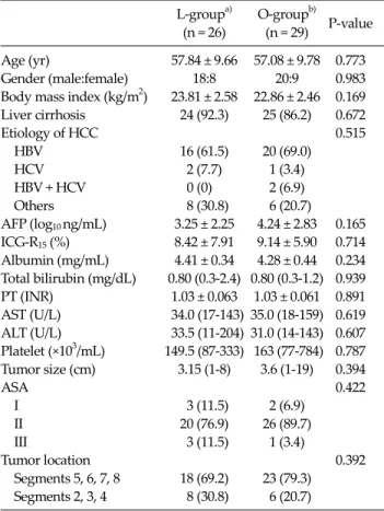
Table 1. Clinicopathological characteristics of patients with hepatocellular carcinoma (HCC)
L-groupa) O-groupb)
P-value (n = 26) (n = 29)
Age (yr) 57.84 ± 9.66 57.08 ± 9.78 0.773
Gender (male:female) 18:8 20:9 0.983
Body mass index (kg/m2) 23.81 ± 2.58 22.86 ± 2.46 0.169 Liver cirrhosis 24 (92.3) 25 (86.2) 0.672
Etiology of HCC 0.515
HBV 16 (61.5) 20 (69.0)
HCV 2 (7.7) 1 (3.4)
HBV + HCV 0 (0) 2 (6.9)
Others 8 (30.8) 6 (20.7)
AFP (log10 ng/mL) 3.25 ± 2.25 4.24 ± 2.83 0.165 ICG-R15 (%) 8.42 ± 7.91 9.14 ± 5.90 0.714 Albumin (mg/mL) 4.41 ± 0.34 4.28 ± 0.44 0.234 Total bilirubin (mg/dL) 0.80 (0.3-2.4) 0.80 (0.3-1.2) 0.939 PT (INR) 1.03 ± 0.063 1.03 ± 0.061 0.891 AST (U/L) 34.0 (17-143) 35.0 (18-159) 0.619 ALT (U/L) 33.5 (11-204) 31.0 (14-143) 0.607 Platelet (×103/mL) 149.5 (87-333) 163 (77-784) 0.787 Tumor size (cm) 3.15 (1-8) 3.6 (1-19) 0.394
ASA 0.422
I 3 (11.5) 2 (6.9)
II 20 (76.9) 26 (89.7)
III 3 (11.5) 1 (3.4)
Tumor location 0.392
Segments 5, 6, 7, 8 18 (69.2) 23 (79.3) Segments 2, 3, 4 8 (30.8) 6 (20.7)
Values are presented as mean± SD, median (range), or number (%).
HBV, hepatitis-B virus; HCV, hepatitis-C virus; AFP, alpha fetoprotein; ICG-R15, indocyanine green-retention rate at 15 minutes; PT, prothrombin time; INR, international normalized ratio; AST, aspartate aminotransferase; ALT, alanine amino- transferase; ASA, American Society of Anesthesiologists physical status score.
a)Laparoscopic liver resection-group. b)Open liver resection-group.
Outcome measures
The following operative variables were collected for each patient: demographics, tumor location and histology, size of hepatic lesions, operative details, morbidity, most recent follow-up data, disease-free status (e.g., recurrence vs. non-recurrence), and date of recurrence. Morbidity and postoperative hospital stay were evaluated according to the Clavien complication grading system [13,14].
Patients were monitored for the development of post- operative hemorrhage, pleural effusion, and postopera- tive fluid collection. Patterns of recurrence were classified as unifocal intrahepatic, multifocal hepatic, and others (e.g., lumbar spine metastasis). Survival status was de- termined by review of the medical records and through use of the Chonnam Regional Cancer Center death index.
Liver resections were defined according to Brisbane 2000 terminology [15]: left lateral sectionectomy (for a seg- mentectomy of segments 2 to 3), segmentectomy (for a re- section of one segment), right hemihepatectomy (for a seg- mentectomy of segments 5 to 8), and left hemihepatec- tomy (for a segmentectomy of segments 2 to 4).
Discharge criteria were the ability to tolerate a soft or regular hospital diet and pain control with oral analgesics.
After discharge, all patients were followed with a surveil- lance protocol that included a multislice computed to- mography scan, liver function tests, and serum alpha-feto- protein level every 3 months after resection for 2 years, then every 4 to 6 months.
Disease-free survival was calculated from the date of the operation to the date of recurrence or last follow-up.
Statistical analyses
Summary statistics are reported using mean or median values, where appropriate. The Student’s t-test and Mann- Whitney U-test were used for the mean comparison of continuous variables and for ordinal data, respectively, whereas the chi-squared and Fisher exact tests were used to compare frequencies of categorical variables between groups. Survival rates were calculated using the Kaplan- Meier method, and survival curves were compared using the log-rank test. Significance was defined as P ≤ 0.05. All statistical analyses were performed using the SPSS ver.
14.0 (SPSS Inc., Chicago, IL, USA).
RESULTS
Clinicopathological features and tumor characteristics are summarized in Table 1. Additional parameters, such as transaminase levels, total bilirubin, and prothrombin time, were similar between the L- and O-groups.
Intraoperative clinical outcomes
The intraoperative clinical outcomes are summarized in Table 2. In the L-group, there were four right hemi-
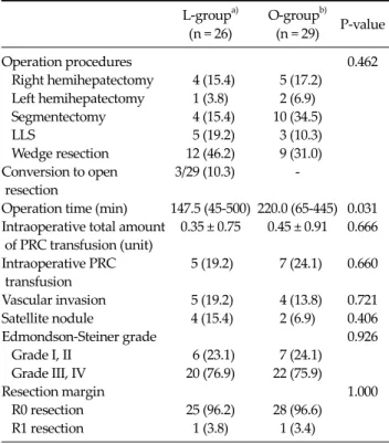
Table 2. Intraoperative clinical outcomes
L-groupa) O-groupb)
P-value (n = 26) (n = 29)
Operation procedures 0.462
Right hemihepatectomy 4 (15.4) 5 (17.2) Left hemihepatectomy 1 (3.8) 2 (6.9) Segmentectomy 4 (15.4) 10 (34.5)
LLS 5 (19.2) 3 (10.3)
Wedge resection 12 (46.2) 9 (31.0) Conversion to open 3/29 (10.3) - resection
Operation time (min) 147.5 (45-500) 220.0 (65-445) 0.031 Intraoperative total amount 0.35 ± 0.75 0.45 ± 0.91 0.666 of PRC transfusion (unit)
Intraoperative PRC 5 (19.2) 7 (24.1) 0.660 transfusion
Vascular invasion 5 (19.2) 4 (13.8) 0.721 Satellite nodule 4 (15.4) 2 (6.9) 0.406
Edmondson-Steiner grade 0.926
Grade I, II 6 (23.1) 7 (24.1) Grade III, IV 20 (76.9) 22 (75.9)
Resection margin 1.000
R0 resection 25 (96.2) 28 (96.6)
R1 resection 1 (3.8) 1 (3.4)
Values are presented as number (%) or median (range).
LLS, left lateral sectionectomy; PRC, packed red blood cell.
a)Laparoscopic liver resection group. b)Open liver resection group.
Table 3. Postoperative clinical course
L-groupa) O-groupb)
P-value (n = 26) (n = 29)
Postoperative mortality 0 (0) 0 (0) NS
Postoperative morbidity 1 (3.8) 7 (24.1) 0.054
Hemorrhage 0 (0) 2 (6.9)
Pleural effusion 1 (3.8) 0 (0)
Abscess 0 (0) 2 (6.9)
Others (ileus, fluid 0 (0) 3 (10.3) collection, etc.)
POD1 bilirubin (mg/dL) 1.4 (0.7-5.3) 1.6 (0.6-3.7) 0.230 POD1 PT (INR) 1.26 ± 0.14 1.38 ± 0.17 0.003 POD1 AST (IU/L) 115.5 (51.0-495.0) 133.0 (63.0-516.0) 0.169 POD1 ALT (IU/L) 114.5 (18.0-294.0) 114.0 (31.0-619.0) 0.853 Postoperative hospital 11.08 ± 4.96 16.07 ± 10.697 0.034 stay (day)
Values are presented as number (%), median (range), or mean ± SD.
POD, postoperative day; PT, prothrombin time; INR, international normalized ratio; AST, aspartate aminotransferase; ALT, alanine aminotransferase; NS, non- specific.
a)Laparoscopic liver resection group. b)Open liver resection group.
hepatectomies, one left hemihepatectomy, four segmen- tectomies, four left lateral sectionectomies, and 13 wedge resections. In the O-group, there were five right hemi- hepatectomies, two left hemihepatectomies, 10 segmen- tectomies, three left lateral sectionectomies, and nine wedge resections. No difference was found between the two groups for the type of resection (P = 0.467). Five pa- tients (19.2%) in the L-group and seven patients (24.1%) in the O-group underwent a major (≥three segments) liver resection (P = 0.660), with a 10.3% conversion rate. The me- dian operation time was shorter in the L-group than that in the O-group (147.5 vs. 220.0 minutes, P = 0.031). The total amount of intraoperative packed red blood cell trans- fusion was 0.35 units in the L-group and 0.45 units in the O-group (P = 0.666). A transfusion was required in five pa- tients (19.2%) in the L-group and seven patients (24.1%) in the O-group (P = 0.660). In both groups, one case had tu- mor exposure at the surgical specimen surface (R1 re- section; P = 1.000). The two groups did not differ sig- nificantly in terms of the presence of vascular invasion,
satellite nodules, or Edmondson-Steiner grade.
The size of the skin incision in the L-group was 6 to 7 cm for the major (≥3 segments) liver resection hepatectomies and <5 cm for the minor (<3 segments) resections, which was shorter than that in the O-group (about 10 to 15 cm).
Postoperative clinical outcomes
We compared the postoperative clinical outcomes be- tween the two groups (Table 3). No perioperative death was recorded in either group. Postoperative complications occurred in one patient in the L-group (3.8%) and in seven patients in the O-group (24.1%; P = 0.054). Based on the surgical complications classification [13,14], complica- tions in the L-group included grade II pleural effusion, which resolved successfully with diuretic treatment. In the O-group, postoperative complications included one case of grade II hemorrhage, one case of grade III hemor- rhage (treated with a re-exploration for bleeding from a re- section margin), two cases of grade II abscess, and one case each of ileus (grade II), ascites (grade II), and fluid collec- tion (grade I).
The peak postoperative values of total bilirubin, aspar- tate aminotransferase, and alanine aminotransferase did not differ statistically between the two groups. However,
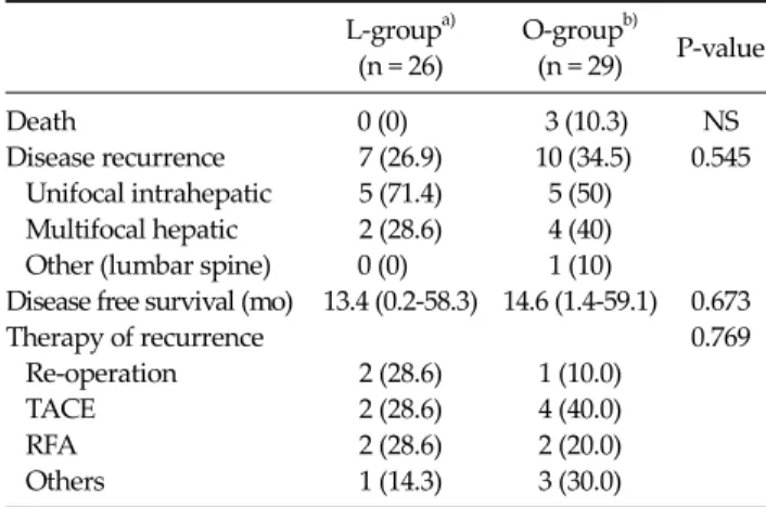
Table 4. Follow-up
L-groupa) O-groupb)
P-value (n = 26) (n = 29)
Death 0 (0) 3 (10.3) NS
Disease recurrence 7 (26.9) 10 (34.5) 0.545 Unifocal intrahepatic 5 (71.4) 5 (50)
Multifocal hepatic 2 (28.6) 4 (40) Other (lumbar spine) 0 (0) 1 (10)
Disease free survival (mo) 13.4 (0.2-58.3) 14.6 (1.4-59.1) 0.673
Therapy of recurrence 0.769
Re-operation 2 (28.6) 1 (10.0)
TACE 2 (28.6) 4 (40.0)
RFA 2 (28.6) 2 (20.0)
Others 1 (14.3) 3 (30.0)
Values are presented as number (%) or median (range).
TACE, transarterial chemoembolization; RFA, radiofrequency ablation; NS, non-specific.
a)Laparoscopic liver resection group; b)Open liver resection group.
Fig. 1. Disease-free survival curves after laparoscopic and open liver resection for hepatocellular carcinoma (P = 0.073).
the prothrombin time value at postoperative day 1 in the L-group was lower than that in the O-group (1.26 vs. 1.38 international normalized ratio, P = 0.003).
The mean hospital stay after surgery was significantly shorter in the L-group than in the O-group (11.08 vs. 16.07 days; P = 0.034).
Follow-up and disease-free survival
Follow-up and survival are summarized in Table 4. The mean follow-up period was 21.75 months (range, 0.7 to 58.4 months) for the L-group and 24.75 months (range, 3.6 to 59.4 months) for the O-group. Seven of the 26 patients in the L-group developed disease recurrence (unifocal intra- hepatic recurrence, n = 5; multifocal hepatic recurrence, n
= 2). No patient developed tumor recurrence at the site of the resection margin, peritoneal dissemination, or port- site metastases. Ten of the 29 patients in the O-group de- veloped tumor recurrence (unifocal intrahepatic recur- rence, n = 5; multifocal hepatic recurrence, n = 4; lum- bar-spine metastasis, n = 1). The 1-year disease-free surviv- al rate was 84.6% for the L-group and 82.8% for the O-group (P = 1.000) (Fig. 1).
In the L-group, recurrence were treated by further sur- gery (2 patients), transarterial chemoembolization (trans- arterial chemoembolization [TACE]; 2), radio-frequency ablation (radiofrequency ablation [RFA]; 2), and refusal of any form of therapy [1], while patients with a recurrence
were treated by re-resection [1], TACE [4], RFA [2], radio- therapy [1], and refusal of any form of therapy [2] in the O-group.
No patients died in the L-group, whereas three patients died in the O-group, and median overall survival was 25.6 months (range, 3.6 to 59.4 months).
DISCUSSION
Since the first report of laparoscopic liver resection by Gagner et al. [16] in 1992, an increasing number of small prospective studies have been published. These studies have reported encouraging results for the feasibility and safety of the procedure. Laparoscopic resection has been more frequently proposed as a curative treatment for HCC [3,6-8,11,17-19] or as a preliminary treatment before trans- plantation [20].
However, laparoscopic liver resection for HCC is still challenging for both surgeons and patients, because most HCCs are associated with underlying liver disease, such as chronic hepatitis and liver cirrhosis. Moreover, apply- ing laparoscopic resection to HCC has been limited by tu- mor location. Most reported cases had peripheral lesions located in the anterolateral segments (segments 2, 3, 4b, 5, and 6) [3,7,17-19,21]. Additionally, major liver resections (i.e., right or left hemihepatectomies) are feasible, but re- main difficult procedures that should be reserved for ex- perienced surgeons [12]. In our study, tumor location in
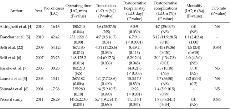
Table 5. Published series of matched comparative studies of laparoscopic and open hepatic resection for hepatocellular carcinoma
Author Year No. of cases (L:O)
Operating time (L:O, min)
(P-value)
Transfusion (L:O, n (%))
(P-value)
Postoperative hospital stay
(L:O, day) (P-value)
Postoperative complications (L:O, n (%))
(P-value)
Mortality (L:O, n (%))
(P-value)
DFS rate (P-value)
Aldrighetti et al. [4] 2010 16:16 150:240 4:6 (25:37.5) 6.3:9 4:7 (25:43.7) 0:0 NS
(0.044) (NS) (0.039) (NS) (NS)
Tranchart et al. [5] 2010 42:42 233.1:221.8 4:7 (9.5:16.7) 6.7:9.6 5:12 (11.9:28.5) 1:1 (2.4:2.4) -
(0.90) (0.51) (<0.0001) (0.10) (1.00)
Belli et al. [22] 2009 54:125 167:185 6:31 (11:25.6) 8.4:9.2 10:45 (19:36) 1:5 (2:4) 0.864
(0.012) (0.030) (0.113) (0.020) (0.615)
Belli et al. [6] 2007 23:23 148:125.2 0:4 (0:17.3) 8.2:12.04 3:11 (13:47.8) 1:0 (4.3:0) -
(0.016) (0.036) (0.048) (0.01) (NS)
Kaneko et. al. [7] 2005 30:28 182:210 - 14.9:21.6 3:5 (10:18) 0 : 0 NS
(NS) (<0.005) (NS) (NS)
Laurent et al. [3] 2003 13:14 267:182 1:4 (7.7:28.6) 15.3:17.3 4:7 (36:50) 0:2 (0:14) NS
(0.006) (0.490) (0.830) (NS) (0.2)
Shimada et al. [8] 2001 17:38 325:280 1:4 (5.9:10.5) 12:22 1:4 (5.9:10.5) - NS
(0.18) (0.990) (<0.001) (0.99)
Present study 2011 26:29 147.5:220.0 5:7 (19.2:24.1) 11.1:16.1 1:7 (3.8:24.1) 0:0 0.673
(0.031) (0.660) (0.034) (0.054) (NS)
L, Laparoscopic resection; O, Open resection; DFS, disease-free survival, NS, non-specific.
laparoscopic liver resections included three cases (11.5%) in segment 7 and two cases (7.7%) in segment 8. Five pa- tients (19.2%) in the L-group underwent major (≥3 seg- ments) liver resection. This result suggests that the limi- tations of laparoscopic resection based on tumor location and the extent of resection will be overcome with further experience and technical advances.
One of the advantages of laparoscopic liver resection is a shorter operation time than for the open method.
Aldrighetti et al. [4] reported that the laparoscopic ap- proach resulted in a shorter operating time (150 vs. 240 mi- nutes, P = 0.044) in a case-matched analysis of laparoscopic (n = 16) and open liver resection (n = 16) of HCC.
Additionally, Belli et al. [22] reported a retrospective anal- ysis of a prospectively maintained database of 179 liver re- sections including major hepatectomies. They showed a shorter operating time with laparoscopic versus open liver resection (167 vs. 185 minutes, P = 0.012). These results are comparable with the results of the present study.
Sarpel et al. [9] matched 20 laparoscopic liver resections for HCC to 56 open resections for HCC. The adjusted odds ratio for a length of stay ≥6 days was significantly lower in patients who underwent a laparoscopic resection. These results are comparable with the results of the present
study showing a shorter postoperative hospital stay for patients who underwent a laparoscopic resection (11.1 vs.
16.1 days, P = 0.034).
The main concern with using the laparoscopic techni- que for malignancies is the risk of inadequate tumor resection. However, no difference has been observed in margin-free resections between laparoscopic and open liv- er resection in many comparative studies [3,5-6,9,12, 23-26]. In our series, one case in each group had tumor ex- posure at the surgical specimen surface (R1 resection); this was not significant (P = 1.000).
Another concern about laparoscopic resection of malig- nancies is the risk for a port-site tumor recurrence, which was not recorded in our patients. With more than 3,000 cases of minimally invasive hepatic resection in the liter- ature, no incidence of port-site recurrence or tumor seed- ing has been reported [3-8,22]. Thus, this concern should not prevent surgeons from conducting a laparoscopic approach.
Both groups underwent surgery performed by the same surgeon, confirming the feasibility and safety of the lapa- roscopic approach when performed by a surgeon with ex- perience in both open and laparoscopic liver surgery. In fact, the mortality and the morbidity rates for the laparo-
scopic resection group were 0% and 3.8%, respectively.
No prospective, randomized controlled trial has been reported comparing laparoscopic with open liver resec- tion. However, several studies have provided outcomes of matched comparisons between laparoscopic and open hepatic resection for HCC (Table 5). Belli et al. [22] pro- vided the largest matched comparison of laparoscopy (n = 54) with open liver resection (n = 125) of HCC in patients with cirrhosis. Mortalities at 30 days were similar between the two groups; however, morbidity was significantly lower in the laparoscopic group (19 vs. 36%, P = 0.020). The 3-year overall survival (67 vs. 62%, P = 0.347) and disease- free survival (52 vs. 59%, P = 0.864) were not significantly different between the laparoscopic and open groups.
These results are comparable with the results of other studies showing an overall 3-year survival of 60 to 93%, and 3-year disease-free survival of 52 to 64% after laparo- scopic liver resection for HCC [11,18,19]. In our study, three patients died in the O-group, and the 2-year overall survival rate was 93.1%. No patients died in the L-group during the follow-up period. Thus, overall survival in the L-group could not be calculated and compared with that of the O-group. Nevertheless, our study confirmed the fea- sibility, safety, and benefits of laparoscopic liver resection for selected patients, including HCCs located in segments 7 and 8 and major hepatectomies.
The results of our study are limited by the non-random- ized design, small number of cases, and the selection bias related to the choice of approach based merely on tumor characteristics. Although the potential historical bias was reduced by the study design, resulting in an open re- section group that was well matched with a laparoscopic resection group for age, gender, ASA class, tumor location and size, type of liver resection, and degree of liver cir- rhosis, our disease-free survival results after laparoscopic liver resection for HCC are short-term results. Thus, a larg- er group of patients and further examinations (longer- term follow-up) are necessary to analyze the role of lapa- roscopic liver resection.
In conclusion, the present study showed that laparo- scopic liver resection for HCC is feasible and safe in se- lected patients and can lead to good surgical results with a shorter postoperative hospital stay, shorter operating
time, less intraoperative bleeding, and similar outcomes in terms of disease-free survival when compared with open surgery.
CONFLICTS OF INTEREST
No potential conflict of interest relevant to this article was reported.
REFERENCES
1. Nguyen KT, Gamblin TC, Geller DA. World review of lap- aroscopic liver resection-2,804 patients. Ann Surg 2009;
250:831-41.
2. Mala T, Edwin B. Role and limitations of laparoscopic liver resection of colorectal metastases. Dig Dis 2005;23:142-50.
3. Laurent A, Cherqui D, Lesurtel M, Brunetti F, Tayar C, Fagniez PL. Laparoscopic liver resection for subcapsular hepatocellular carcinoma complicating chronic liver disease. Arch Surg 2003;138:763-9.
4. Aldrighetti L, Guzzetti E, Pulitanò C, Cipriani F, Catena M, Paganelli M, et al. Case-matched analysis of totally laparo- scopic versus open liver resection for HCC: short and mid- dle term results. J Surg Oncol 2010;102:82-6.
5. Tranchart H, Di Giuro G, Lainas P, Roudie J, Agostini H, Franco D, et al. Laparoscopic resection for hepatocellular carcinoma: a matched-pair comparative study. Surg Endosc 2010;24:1170-6.
6. Belli G, Fantini C, D'Agostino A, Cioffi L, Langella S, Russolillo N, et al. Laparoscopic versus open liver re- section for hepatocellular carcinoma in patients with histo- logically proven cirrhosis: short- and middle-term results.
Surg Endosc 2007;21:2004-11.
7. Kaneko H, Takagi S, Otsuka Y, Tsuchiya M, Tamura A, Katagiri T, et al. Laparoscopic liver resection of hep- atocellular carcinoma. Am J Surg 2005;189:190-4.
8. Shimada M, Hashizume M, Maehara S, Tsujita E, Rikimaru T, Yamashita Y, et al. Laparoscopic hepatectomy for hep- atocellular carcinoma. Surg Endosc 2001;15:541-4.
9. Sarpel U, Hefti MM, Wisnievsky JP, Roayaie S, Schwartz ME, Labow DM. Outcome for patients treated with laparo- scopic versus open resection of hepatocellular carcinoma:
case-matched analysis. Ann Surg Oncol 2009;16:1572-7.
10. Vittimberga FJ Jr, Foley DP, Meyers WC, Callery MP.
Laparoscopic surgery and the systemic immune response.
Ann Surg 1998;227:326-34.
11. Lai EC, Tang CN, Ha JP, Li MK. Laparoscopic liver re- section for hepatocellular carcinoma: ten-year experience in a single center. Arch Surg 2009;144:143-7.
12. Buell JF, Cherqui D, Geller DA, O'Rourke N, Iannitti D, Dagher I, et al. The international position on laparoscopic
liver surgery: The Louisville Statement, 2008. Ann Surg 2009;250:825-30.
13. Dindo D, Demartines N, Clavien PA. Classification of sur- gical complications: a new proposal with evaluation in a cohort of 6336 patients and results of a survey. Ann Surg 2004;240:205-13.
14. Clavien PA, Barkun J, de Oliveira ML, Vauthey JN, Dindo D, Schulick RD, et al. The Clavien-Dindo classification of surgical complications: five-year experience. Ann Surg 2009;250:187-96.
15. Belghiti J, Clavien P, Gadzijev E, Garden J, Lau W, Makuuchi M, et al. The Brisbane 2000 terminology of liver anatomy and resections terminology committee of the in- ternational hepato-pancreato-biliary association: Chair- man, SM Strasberg (USA). HPB (Oxford) 2000;2:333-9.
16. Gagner M, Rheault M, Dubuc J. Laparoscopic partial hep- atectomy for liver tumor [abstract]. Surg Endosc 1992;6:99.
17. Chen HY, Juan CC, Ker CG. Laparoscopic liver surgery for patients with hepatocellular carcinoma. Ann Surg Oncol 2008;15:800-6.
18. Dagher I, Lainas P, Carloni A, Caillard C, Champault A, Smadja C, et al. Laparoscopic liver resection for hep- atocellular carcinoma. Surg Endosc 2008;22:372-8.
19. Cherqui D, Laurent A, Tayar C, Chang S, Van Nhieu JT, Loriau J, et al. Laparoscopic liver resection for peripheral hepatocellular carcinoma in patients with chronic liver dis- ease: midterm results and perspectives. Ann Surg 2006;243:
499-506.
20. Laurent A, Tayar C, Andréoletti M, Lauzet JY, Merle JC, Cherqui D. Laparoscopic liver resection facilitates salvage liver transplantation for hepatocellular carcinoma. J Hepatobiliary Pancreat Surg 2009;16:310-4.
21. Belli G, Fantini C, D'Agostino A, Belli A, Russolillo N.
Laparoscopic liver resections for hepatocellular carcinoma (HCC) in cirrhotic patients. HPB (Oxford) 2004;6:236-46.
22. Belli G, Limongelli P, Fantini C, D'Agostino A, Cioffi L, Belli A, et al. Laparoscopic and open treatment of hep- atocellular carcinoma in patients with cirrhosis. Br J Surg 2009;96:1041-8.
23. Ito K, Ito H, Are C, Allen PJ, Fong Y, DeMatteo RP, et al.
Laparoscopic versus open liver resection: a matched-pair case control study. J Gastrointest Surg 2009;13:2276-83.
24. Cai XJ, Yang J, Yu H, Liang X, Wang YF, Zhu ZY, et al.
Clinical study of laparoscopic versus open hepatectomy for malignant liver tumors. Surg Endosc 2008;22:2350-6.
25. Endo Y, Ohta M, Sasaki A, Kai S, Eguchi H, Iwaki K, et al. A comparative study of the long-term outcomes after laparo- scopy-assisted and open left lateral hepatectomy for hep- atocellular carcinoma. Surg Laparosc Endosc Percutan Tech 2009;19:e171-4.
26. Topal B, Fieuws S, Aerts R, Vandeweyer H, Penninckx F.
Laparoscopic versus open liver resection of hepatic neo- plasms: comparative analysis of short-term results. Surg Endosc 2008;22:2208-13.