INTRODUCTION
Natural killer (NK) cells comprise 5% to 20% of human peripheral blood lymphocytes and are derived from CD34+
hematopoietic progenitor cells [1]. The precise physiologic sites where NK cells mature and the mechanisms that drive the development of their functional characteristics have not yet been fully clarified but recent studies indicate that these occur in the bone marrow and the lymph nodes [2-4].
NK cells have the morphology of large granular lympho- cytes, and are phenotypically defined by the expression of
CD56 and the lack of CD3 and T-cell receptor molecules [3, 4]. Approximately 10% of NK cells express very high levels of CD56 and also have dim expression of FcγRIII (CD16), a receptor that binds the Fc portion of IgG [5]. These cells mostly are believed to have primarily an immunoregula- tory role exerted through the secretion of cytokines and chemokines [5]. Although less common in blood, bone mar- row and spleen, this cell subset predominates in the sec- ondary lymphoid tissues [6]. The remaining 90% of NK cells in blood express lower levels of CD56 and high levels of CD16 [5]. These cells appear to function predominantly in direct cytotoxicity and antibody-dependent cellular cytotoxicity (ADCC) [5, 7]. NK cells can directly induce apoptosis via the perforin-granzyme pathway, or by expressing death-recep- tor ligands on their cell surface. Such ligands include tumor necrosis factor (TNF)-related apoptosis-inducing ligand (TRAIL), or the Fas ligand, which directly trigger cell death via their respective receptors [8].
NK cells can kill target cells without the need for prior
89 89
Expansion and Activation of Natural Killer Cells for Cancer Immunotherapy
Duck Cho, M.D.
1and Dario Campana, M.D.
1,2Departments of Oncology1and Pathology2, St. Jude Children’s Research Hospital, Memphis, TN, USA
89 89
Natural killer (NK) cells can kill a wide range of cancer cells and are a promising tool for cell ther- apy of cancer. NK cells cytotoxicity is regulated by a balance between stimulatory and inhibitory signals. Interleukin-2 is known to increase NK cell cytotoxicity. Although many cytokines have been studied in efforts to induce durable NK cell expansions, most reports indicate a rather modest effect and the requirement for additional stimuli. We found that contact with the K562 myeloid leukemia cell line, genetically modified to express a membrane-bound form of interleukin-15 and the ligand for the costimulatory molecule 4-1BB, induced vigorous expansion of NK cells from peripheral blood.
Based on these findings, we developed a method for large-scale clinical-grade expansion of NK cells. This method is currently used to expand allogeneic NK cells for infusion in patients with leukemia and solid tumors. We here summarize methods for expansion and activation of NK cells from human peripheral blood mononuclear cells as well as clinical-scale methods to produce NK cells for immu- notherapy under Current Good Manufacturing Practices (cGMP) conditions. (Korean J Lab Med 2009;
29:89-96)
Key Words : NK cells, Cell therapy, Acute myeloid leukemia, Acute lymphoblastic leukemia, Chimeric receptors
Received :February 18, 2009 Manuscript No :KJLM09-026 Revision received :March 25, 2009
Accepted :March 27, 2009
Corresponding author :Dario Campana, M.D., Ph.D.
Department of Oncology, St. Jude Children’s Research Hospital, 262 Danny Thomas Place, Memphis, TN 38105, USA Tel : +901-4595 2528, Fax : +901-4595 5947 E-mail : [email protected]
*This work was supported by grants CA113482 and CA21765 from the National Cancer Institute, grants from the Assisi Foundation and from the Fondation de Gouverneurs de l’Espoir, and by the American Lebanese Syri- an Associated Charities (ALSAC).
sensitization, an effect that is regulated by the balance of stimulatory and inhibitory signals [4, 9, 10]. Many of the signaling receptors on the surface of NK cells engage major histocompatibility complex (MHC) class I and MHC class I-like molecules. A well-described mechanism is the inhi- bition of NK activity by increasing expression of MHC or human leukocyte antigen (HLA) class I in target cells [11].
NK cells express killer immunoglobulin-like receptors (KIRs), most of which recognize specific corresponding HLA class I molecules on target cells, and deliver inhibitory signals [4, 9, 10]. These inhibitory signals from HLA can override activating signals and suppress the function of NK cells. A concept that has recently emerged is that interaction bet- ween NK cells and HLA molecules might also be important for their functional maturation and the generation of NK cells that are tolerant towards self molecules, a process which has been termed ‘‘licensing’’[12].
This review summarizes methods for expansion and acti- vation of NK cells from human peripheral blood mononu- clear cells as well as clinical-scale methods to produce NK cells for immunotherapy under Current Good Manufacturing Practices (cGMP) conditions.
METHODS TO EXPAND NK CELLS 1. Cytokines and stimulants
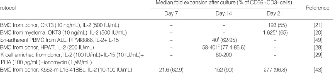
Several protocols for NK-cell expansion have been re- ported (summarize in Table 1; see also www.nkcells.info/
wiki/index.php/NK_cell_expansion). Many cytokines have
been studied in efforts to induce durable NK cell expansion as well as to increase their cytotoxicity [13]. Interleukin (IL)- 2 can enhance the cytotoxicity of NK cells within 24 hr of incubation [14, 15]. IL-2 can also stimulate their prolifer- ation but only a minority of NK cells can maintain prolifer- ation after the initial response [14, 16, 17]. IL-4, IL-7 and IL-12 also induced some proliferative stimuli but are over- all less potent than IL-2 [18]. Likewise, IL-15 alone or in combination with IL-2 typically results in minimal NK cell expansion [8]. We found that IL-2 (1,000 IU/mL) or IL-15 (10 ng/mL) did not induce significant expansion of NK cells [19]. It appears that cytokines may be necessary but not sufficient for optimal proliferation of NK cells.
Recently, Alici et al. [20] used culture conditions using IL-2 (500 IU/mL) with an anti-CD3 antibody (Orthoclone OKT-3, 10 ng/mL) and reported that the number of NK cells from the peripheral blood of 7 newly diagnosed, untreated patients with multiple myeloma had expanded on average 1,600-fold after 20 days of culture. This is in striking con- trast with the 190-fold expansion obtained using NK cells from healthy individuals [21], implying that NK cells from myeloma patients may somehow be more susceptible to stimulation than NK cells from healthy individuals. It is also unclear how CD3 stimulation contributed to the expansion of CD3-NK cells in these studies.
It should be noted that the cell populations that results from the stimulation of peripheral blood mononuclear cells with IL-2 termed lymphokine activated killer (LAK) cells are comprised of mostly of polyclonal T-cells and only a small fraction is CD56+NK cells [22].
*Measured on day 20; �day 10; and �day 10-21.
Abbreviations: PBMC, peripheral blood mononuclear cells; ALL, acute lymphoblastic leukemia.
Protocol Median fold expansion after culture (% of CD56+CD3- cells)
Day 7 Day 14 Day 21
Reference
PBMC from donor, OKT3 (10 ng/mL), IL-2 (500 IU/mL) - - 193 (55) [21]
PBMC from myeloma, OKT3 (10 ng/mL), IL-2 (500 IU/mL) - - 1,625* (65) [20]
Non-adherent PBMC from ALL, RPMI8866, IL-2+IL-15 - 40�(62-95) - [49]
PBMC from donor, HFWT, IL-2 (200 IU/mL) - 58-401�(77.4-85.6) - [28]
NK cell enriched from donor, IL-2 (100 IU/mL)+IL-15 (10 IU/mL)+ - 80-200 - [29]
PHA (100 μg/mL)+ionomycin (1 μM/mL)
PBMC from donor, K562-mIL15-41BBL, IL-2 (10-100 IU/mL) 21.6 (62.9) 152 (90) 277 (96.8) [43]
Table 1. Selected protocols for expansion and activation of NK cells
2. Expansion of NK cells with accessory cells
Most investigators believe that sustained proliferations of NK cells require additional signals [23, 24], such as the presence of monocytes [23] or B-lymphoblastoid cells [16, 25, 26]. Miller et al. [23] reported an approximate 30-fold expansion of NK cells after 18 days of culture with 1,000 IU/mL IL-2 and monocytes. Perussia et al. [27] found that contact with irradiated B-lymphoblastoid cells induced as high as a 25-fold expansion of NK cells after 2 weeks of stimulation. Harada et al. [28] reported that HFWT, a Wilms tumor-derived cell line, stimulates up to 400-fold NK ex- pansion after 2 weeks. Other investigators have used allo- geneic mononuclear cells [29], autologous lymphocytes [30], mitogen activated lymphocytes [25], and umbilical cord mes- enchymal cells [31].
Contact with K562 cells, a cell line derived from a patient with myeloid blast crisis of chronic myelogenous leukemia and bearing the BCR-ABL1translocation, is known to in- duce modest proliferations of NK cells [28, 32], and aug- ment NK cell proliferation in response to IL-15 [24]. We genetically modified K562 cells to express two NK stimu- latory molecules. One, the ligand for 4-1BB (4-1BBL), in- duces activation signals through 4-1BB (CD137), a costim- ulatory molecule expressed on the surface of NK cells [33].
The other, IL-15, is known to support NK-cell maturation and survival [34-37]. IL-15 has greater activity when bound to the cell membrane of stimulatory cells, rather than in its soluble form [38-42]; hence, we made a construct con- taining the human IL-15 gene fused to the gene encoding the human CD8αtransmembrane domain, and used it to transduce K562 cells. The resulting cell line (K562-mb15- 41BBL) induced 21.6-fold (range, 5.1 to 86.6-fold; n=50) ex- pansion of CD56+CD3-NK cells from peripheral blood after 7 days, which was considerably superior to that produced by stimulation with IL-2, IL-12, IL-15 and/or IL-21 [19, 43]. These cultures were performed using irradiated K562- mb15-41BBL cells and 10 IU/mL IL-2. We observed mini- mal or no proliferation of CD3+lymphocytes. NK cells could be further expanded with K562-mb15-41BBL cell stimula- tion by prolonging the cultures and adding 100 IU/mL IL-
2 after day 7. Thus, median NK cell recovery increased to 152-fold after 14 days, and 277-fold after 21 days of cul- ture [19, 43]
NK cells expanded by K562-mb15-41BBL cells stimula- tion were significantly more potent than purified unstim- ulated or IL-2-stimulated NK cells against acute myeloid leukemia (AML) cells in vitro and could eradicate AML in murine models [43]. Preliminary studies indicate that these NK cells are also cytotoxic against cell lines derived from patients with Ewing sarcoma, rhabdomyosarcoma and neu- roblastoma (D. Cho, D. Shook, D. Campana, unpublished results).
Although expanded NK cells acquired powerful cytotox- icity against a variety of cancer cell types, their capacity to kill acute lymphoblastic leukemia cells (ALL) remained overall limited. To overcome this resistance we transduced expanded NK cells with artificial receptors directed against CD19, a molecule expressed by ALL cells and cells of other B-cell malignancies. Anti-CD19 receptors linked to CD3ζ markedly enhanced NK-cell mediated killing of ALL cells, a result that was further improved by adding the 4-1BB costimulatory molecule to the chimeric anti-CD19-CD3ζ receptor [19]. Addition of 4-1BB was also associated with increased production of IFN-γand GM-CSF [19].
CLINICAL LARGE-SCALE NK CELL ACTIVATION AND EXPANSION 1. Methodologic considerations
It is now feasible to obtain clinical-grade purified func- tional NK cells for infusion [44]. NK cells can be obtained from different sources: from the patient (autologous), the patient’s human leukocyte antigen (HLA)-matched siblings or haploidentical family members, or unrelated donors. It might be convenient if NK cells could be collected in advance, cryopreserved and thawed before infusion. Unstimulated cryopreserved and thawed NK cells have phenotype and cytotoxicity that resembles those of fresh cells [45]. Whether this is also the case for activated and expanded NK cells re- mains to be established.
For clinical-grade NK cell activation and expansion, cells need to be cultured for a period of time that varies between less than 24 hr to several weeks [20, 46]. Several tissue cul- ture media have proven effective including stem cell growth medium (SCGM) (CellGenix, Freiburg, Germany) [20, 43]
and X-VIVO serum-free media (BioWhittaker, Verviers, Belgium) [47]. The media can be supplemented with fetal bovine serum (from certified sources) or human serum from AB blood donor [47] is used for clinical applications. NK cells can be cultured in flasks or in bags such as Teflon (FEP) bags [29, 48] and Baxter Lifecell bags [8]. If stimulatory cells are used, it is important to prevent their overgrowth and to ensure that no viable cells are infused with the cul- tured NK cells. Irradiation, at doses of 30 Gy [49], 50 Gy [28], 70 Gy [29] or 100 Gy [19] is a safe and effective method.
Our clinical protocol used cGMP guidelines to process apheresis products and obtain expanded activated NK cells [43] (Fig. 1). Mononuclear cells are Ficoll-separated and placed in culture in SCGM medium supplemented with 10%
FBS, gentamicin sulfate (50 mg/L), and 10 IU/mL human IL-2 at a concentration of 0.5×10⁵CD56+CD3-cells/mL.
Irradiated (100 Gy) K562-mb15-41BBL cells (from a Mas- ter Cell Bank) are added at a ratio of 1 CD56+CD3-cell: 10 K562-mb15-41BBL cells. Cultures are performed in a closed VueLife bags system (American Fluoroseal, Gaithersburg, MD, USA). Cells are fed after 2 and 5 days, and harvested after 7 days of culture. The cell product is then depleted of residual T cells using the CliniMACS System (Miltenyi).
Finally, cells are washed and resuspended in PlasmaLyte- 148 (Baxter, Deerfield, IL, USA) with 0.5% human serum albumin. Under these conditions, we obtained a median 90.5-fold expansion of CD56+CD3-NK cells (n=12) after 7 days of culture [43]. The expansion in these large-scale cultures was higher than that observed in small-scale ex- periments, most likely because of the use of SCGM tissue culture medium instead of RPMI-1640 (SCGM appears to be well suited to support NK cell growth) [20]. We there- fore estimate that it would be feasible to obtain the num- ber of NK cells planned for infusion in our protocol (maxi- mum dose, 5×10⁷NK cells/kg) and more from a leukaphe- resis product. For example, considering that an average apheresis from a normal adult donor gives about 1×10¹⁰ nucleated cells and the average percentage of NK cells is 7.0%, a 90-fold expansion would result in 6.3×10¹⁰NK cells.
If necessary, larger numbers could be obtained by prolong- ing the cultures beyond 7 days.
An alternative to expand NK cells is represented by the continuously growing NK cell lines NK-92, derived from a patient with non-Hodgkin lymphoma, that is cytotoxic against a wide range of malignant cancer cells [50]. These cells have practical appeal, but irradiation is mandatory before infusion in patients, which may limit their efficacy in vivo.
2. Therapeutic applications of NK cells
In the setting of hematopoietic stem-cell transplantation, donor NK cells may exert an anti-leukemia effect if they do not express KIRs reacting with the HLA class I epitope expressed by the patient’s leukemia cells. In animal mod- Fig. 1. Schematic representation of protocols using expanded NK
cells at St Jude Children’s Research Hospital. The leukapheresis product obtained from a haploidentical donor is mixed with irradi- ated K562-mb15-41BBL cells. After 7 days of culture, most cells recovered are activated NK cells. After T-cell depletion using the CliniMACS system, NK cells are infused in patients with NK-sen- sitive malignancies such as acute myeloid leukemia (AML), Ewing sarcoma or rhabdomyosarcoma. For patients whose neoplasia is less sensitive to NK cytotoxicity, such as B-lineage acute lym- phoblastic leukemia (ALL) or B-cell non-Hodgkin lymphoma (B- NHL), expanded NK cells are transduced with an anti-CD19 chi- meric receptor before infusion.
Monocyte
NK cell
7 days
T cell depletion
AML Sarcoma
Irradiated K562-mb15-41BBL
and 10 IU/mL IL2
ALL B-NHL
Genetic modifi- cation Anti-CD19 chimeric
receptor T cell
els, donor NK cells killed host leukemic cells and lympho- hematopoietic cells without affecting non-hematopoietic tissues [51], suggesting the possibility of an NK-mediated graft-versus-leukemia (GVL) effect without systemic dis- ease. Therefore, it is now a common practice at some clin- ical centers to select donors with an HLA and KIR type that facilitates NK cell activation [52-54].
In addition to their use in the context of allogeneic stem cell transplantation, allogeneic NK cells can be directly in- fused in non-myeloablated patients. Miller et al. [46] first demonstrated the potential utility of this approach. These investigators treated 19 adult patients with high risk AML with cyclophosphamide (60 mg/kg for 1 or 2 doses), fluda- rabine (25 mg/m2daily for 5 doses), IL-2 (10 million units per dose for 6 to 9 doses), and an infusion of 2×10⁷/kg CD3- depleted NK cell product (NK cells enriched by approximate- ly 40%) that was activated for 18 hr with 1,000 IU/mL IL- 2. Eight of 15 AML patients showed at least 1% engraftment at day 7 or later after the infusion, and 5 patients achieved a complete remission. Interestingly, lymphodepletion in- duced higher levels of IL-15 which in turn might have been important in prolonging the survival of the infused NK cells [46]. Our current protocol using NK cells expanded by stim- ulation with the K562-mb15-41BBL cell line uses a frame- work identical to that described by Miller et al. [46]. Thus, patients are treated for 7 days with cyclophosphamide and fludarabine and receive subcutaneous IL-2 after infusion of the expanded NK cells.
If cancer cells present a tumor-specific antigen in the HLA context they can be recognized and lysed by cytotoxic T lymphocytes (CTL) specific for the antigen. For example, expanded T lymphocytes specific for Epstein-Barr virus (EBV)-associated molecules have been applied for the treat- ment and prophylaxis of EBV-associated lymphoprolifer- ative disease and lymphoma [55]. Other EBV-associated tumors may also be amenable to this form of therapy [56, 57]. However, most cancers lack identifiable virus-associ- ated antigens [58], although other molecules, such as WT1 and Pr3, are overexpressed in some cancer cells and are pos- sible targets for adoptive T-cell therapy [59, 60]. NK cells offer some potential advantage over CTL therapy. First, a
wide range of cancer cells appear to be sensitive to NK cell cytoxicity. In addition to AML, NK-sensitive malignancies include soft-tissue sarcomas (D. Cho, D. Shook, D. Cam- pana, unpublished), neuroblastoma [59, 60] and malignant glioma [59]. Second, they can be used in an allogeneic set- ting without the risk of graft-versus-host disease [44].
Thirdly, with the method described in this review, large numbers of cytotoxic NK cells can be reliably obtained in a relatively short period of time.
For malignancies that are relatively resistant to NK cells, strategies that can be explored include genetic modification with artificial receptors, such as the one that we reported using anti-CD19 receptors for the treatment of B-cell neo- plasias [19]. To this end, the overall strategy that we have described is not limited to CD19+leukemia and lymphoma cells but is also applicable to many other molecules expressed by cancer cells and can be implemented by replacing the anti-CDc19 scFv with the scFv of another antibody [61, 62].
REFERENCES
1. Galy A, Travis M, Cen D, Chen B. Human T, B, natural killer, and dendritic cells arise from a common bone marrow progenitor cell subset. Immunity 1995;3:459-73.
2. Farag SS and Caligiuri MA. Human natural killer cell development and biology. Blood Rev 2006;20:123-37.
3. Lanier LL. NK cell recognition. Annu Rev Immunol 2005;23:225-74.
4. Caligiuri MA. Human natural killer cells. Blood 2008;112:461-9.
5. Cooper MA, Fehniger TA, Caligiuri MA. The biology of human natural killer-cell subsets. Trends Immunol 2001;22:633-40.
6. Fehniger TA, Cooper MA, Nuovo GJ, Cella M, Facchetti F, Colon- na M, et al. CD56 bright natural killer cells are present in human lymph nodes and are activated by T cell-derived IL-2: a potential new link between adaptive and innate immunity. Blood 2003;101:
3052-7.
7. Lundqvist A, Abrams SI, Schrump DS, Alvarez G, Suffredini D, Berg M, et al. Bortezomib and depsipeptide sensitize tumors to tumor necrosis factor-related apoptosis-inducing ligand: a novel method to potentiate natural killer cell tumor cytotoxicity. Cancer Res 2006;
66:7317-25.
8. Srivastava S, Lundqvist A, Childs RW. Natural killer cell immuno-
therapy for cancer: a new hope. Cytotherapy 2008;10:775-83.
9. Moretta L and Moretta A. Unravelling natural killer cell function:
triggering and inhibitory human NK receptors. EMBO J 2004;23:
255-9.
10. Lanier LL. Up on the tightrope: natural killer cell activation and inhibition. Nat Immunol 2008;9:495-502.
11. Ljunggren HG and Karre K. In search of the ‘missing self’: MHC molecules and NK cell recognition. Immunol Today 1990;11:237-44.
12. Yokoyama WM and Kim S. Licensing of natural killer cells by self- major histocompatibility complex class I. Immunol Rev 2006;214:
143-54.
13. Terme M, Ullrich E, Delahaye NF, Chaput N, Zitvogel L. Natural killer cell-directed therapies: moving from unexpected results to successful strategies. Nat Immunol 2008;9:486-94.
14. Trinchieri G, Matsumoto-Kobayashi M, Clark SC, Seehra J, London L, Perussia B. Response of resting human peripheral blood natural killer cells to interleukin 2. J Exp Med 1984;160:1147-69.
15. Phillips JH and Lanier LL. Dissection of the lymphokine-activated killer phenomenon. Relative contribution of peripheral blood natu- ral killer cells and T lymphocytes to cytolysis. J Exp Med 1986;164:
814-25.
16. London L, Perussia B, Trinchieri G. Induction of proliferation in vitro of resting human natural killer cells: IL 2 induces into cell cycle most peripheral blood NK cells, but only a minor subset of low density T cells. J Immunol 1986;137:3845-54.
17. Lanier LL, Buck DW, Rhodes L, Ding A, Evans E, Barney C, et al.
Interleukin 2 activation of natural killer cells rapidly induces the expression and phosphorylation of the Leu-23 activation antigen. J Exp Med 1988;167:1572-85.
18. Robertson MJ, Manley TJ, Donahue C, Levine H, Ritz J. Costimula- tory signals are required for optimal proliferation of human natu- ral killer cells. J Immunol 1993;150:1705-14.
19. Imai C, Iwamoto S, Campana D. Genetic modification of primary natural killer cells overcomes inhibitory signals and induces specif- ic killing of leukemic cells. Blood 2005;106:376-83.
20. Alici E, Sutlu T, Bjorkstrand B, Gilljam M, Stellan B, Nahi H, et al.
Autologous antitumor activity by NK cells expanded from myelo- ma patients using GMP-compliant components. Blood 2008;111:
3155-62.
21. Carlens S, Gilljam M, Chambers BJ, Aschan J, Guven H, Ljunggren HG, et al. A new method for in vitro expansion of cytotoxic human
CD3-CD56+ natural killer cells. Hum Immunol 2001;62:1092-8.
22. Rosenberg SA, Lotze MT, Muul LM, Leitman S, Chang AE, Etting- hausen SE, et al. Observations on the systemic administration of autologous lymphokine-activated killer cells and recombinant inter- leukin-2 to patients with metastatic cancer. N Engl J Med 1985;313:
1485-92.
23. Miller JS, Oelkers S, Verfaillie C, McGlave P. Role of monocytes in the expansion of human activated natural killer cells. Blood 1992;80:
2221-9.
24. Robertson MJ, Cameron C, Lazo S, Cochran KJ, Voss SD, Ritz J. Cos- timulation of human natural killer cell proliferation: role of acces- sory cytokines and cell contact-dependent signals. Nat Immun 1996- 1997;15:213-26.
25. Rabinowich H, Sedlmayr P, Herberman RB, Whiteside TL. Increased proliferation, lytic activity, and purity of human natural killer cells cocultured with mitogen-activated feeder cells. Cell Immunol 1991;
135:454-70.
26. Igarashi T, Wynberg J, Srinivasan R, Becknell B, McCoy JP Jr, Taka- hashi Y, et al. Enhanced cytotoxicity of allogeneic NK cells with killer immunoglobulin-like receptor ligand incompatibility against melanoma and renal cell carcinoma cells. Blood 2004;104:170-7.
27. Perussia B, Ramoni C, Anegon I, Cuturi MC, Faust J, Trinchieri G.
Preferential proliferation of natural killer cells among peripheral blood mononuclear cells cocultured with B lymphoblastoid cell lines. Nat Immun Cell Growth Regul 1987;6:171-88.
28. Harada H, Saijo K, Watanabe S, Tsuboi K, Nose T, Ishiwata I, et al.
Selective expansion of human natural killer cells from peripheral blood mononuclear cells by the cell line, HFWT. Jpn J Cancer Res 2002;93:313-9.
29. Luhm J, Brand JM, Koritke P, Hoppner M, Kirchner H, Frohn C.
Large-scale generation of natural killer lymphocytes for clinical application. J Hematother Stem Cell Res 2002;11:651-7.
30. Condiotti R, Zakai YB, Barak V, Nagler A. Ex vivo expansion of CD56+ cytotoxic cells from human umbilical cord blood. Exp Hema- tol 2001;29:104-13.
31. Boissel L, Tuncer HH, Betancur M, Wolfberg A, Klingemann H.
Umbilical cord mesenchymal stem cells increase expansion of cord blood natural killer cells. Biol Blood Marrow Transplant 2008;14:
1031-8.
32. Phillips JH and Lanier LL. A model for the differentiation of human natural killer cells. Studies on the in vitro activation of Leu-11+ gran-
ular lymphocytes with a natural killer-sensitive tumor cell, K562. J Exp Med 1985;161:1464-82.
33. Melero I, Johnston JV, Shufford WW, Mittler RS, Chen L. NK1.1 cells express 4-1BB (CDw137) costimulatory molecule and are required for tumor immunity elicited by anti-4-1BB monoclonal antibodies.
Cell Immunol 1998;190:167-72.
34. Carson WE, Fehniger TA, Haldar S, Eckhert K, Lindemann MJ, Lai CF, et al. A potential role for interleukin-15 in the regulation of human natural killer cell survival. J Clin Invest 1997;99:937-43.
35. Cooper MA, Bush JE, Fehniger TA, VanDeusen JB, Waite RE, Liu Y, et al. In vivo evidence for a dependence on interleukin 15 for sur- vival of natural killer cells. Blood 2002;100:3633-8.
36. Fehniger TA and Caligiuri MA. Ontogeny and expansion of human natural killer cells: clinical implications. Int Rev Immunol 2001;20:
503-34.
37. Wu J and Lanier LL. Natural killer cells and cancer. Adv Cancer Res 2003;90:127-56.
38. Musso T, Calosso L, Zucca M, Millesimo M, Ravarino D, Giovarelli M, et al. Human monocytes constitutively express membrane-bound, biologically active, and interferon-gamma-upregulated interleukin- 15. Blood 1999;93:3531-9.
39. Dubois S, Mariner J, Waldmann TA, Tagaya Y. IL-15Ralpha recycles and presents IL-15 In trans to neighboring cells. Immunity 2002;17:
537-47.
40. Koka R, Burkett P, Chien M, Chai S, Boone DL, Ma A. Cutting edge:
murine dendritic cells require IL-15R alpha to prime NK cells. J Im- munol 2004;173:3594-8.
41. Burkett PR, Koka R, Chien M, Chai S, Boone DL, Ma A. Coordinate expression and trans presentation of interleukin (IL)-15Ralpha and IL-15 supports natural killer cell and memory CD8+ T cell home- ostasis. J Exp Med 2004;200:825-34.
42. Kobayashi H, Dubois S, Sato N, Sabzevari H, Sakai Y, Waldmann TA, et al. Role of trans-cellular IL-15 presentation in the activation of NK cell-mediated killing, which leads to enhanced tumor immu- nosurveillance. Blood 2005;105:721-7.
43. Fujisaki H, Kakuda H, Shimasaki N, Imai C, Ma J, Lockey T, et al.
Expansion of highly cytotoxic human natural killer cells for cancer cell therapy. Cancer Res 2009; In press.
44. Leung W, Iyengar R, Leimig T, Holladay MS, Houston J, Hand- gretinger R. Phenotype and function of human natural killer cells purified by using a clinical-scale immunomagnetic method. Cancer
Immunol Immunother 2005;54:389-94.
45. Meehan KR, Wu J, Webber SM, Barber A, Szczepiorkowski ZM, Sentman C. Development of a clinical model for ex vivo expansion of multiple populations of effector cells for adoptive cellular thera- py. Cytotherapy 2008;10:30-7.
46. Miller JS, Soignier Y, Panoskaltsis-Mortari A, McNearney SA, Yun GH, Fautsch SK, et al. Successful adoptive transfer and in vivo ex- pansion of human haploidentical NK cells in patients with cancer.
Blood 2005;105:3051-7.
47. Klingemann HG and Martinson J. Ex vivo expansion of natural killer cells for clinical applications. Cytotherapy 2004;6:15-22.
48. McKenna DH Jr, Sumstad D, Bostrom N, Kadidlo DM, Fautsch S, McNearney S, et al. Good manufacturing practices production of natural killer cells for immunotherapy: a six-year single-institution experience. Transfusion 2007;47:520-8.
49. Torelli GF, Guarini A, Maggio R, Alfieri C, Vitale A, Foa R. Expan- sion of natural killer cells with lytic activity against autologous blasts from adult and pediatric acute lymphoid leukemia patients in com- plete hematologic remission. Haematologica 2005;90:785-92.
50. Tam YK, Martinson JA, Doligosa K, Klingemann HG. Ex vivo ex- pansion of the highly cytotoxic human natural killer-92 cell-line under current good manufacturing practice conditions for clinical adoptive cellular immunotherapy. Cytotherapy 2003;5:259-72.
51. Ruggeri L, Capanni M, Urbani E, Perruccio K, Shlomchik WD, Tosti A, et al. Effectiveness of donor natural killer cell alloreactivity in mis- matched hematopoietic transplants. Science 2002;295:2097-100.
52. Ruggeri L, Capanni M, Casucci M, Volpi I, Tosti A, Perruccio K, et al. Role of natural killer cell alloreactivity in HLA-mismatched he- matopoietic stem cell transplantation. Blood 1999;94:333-9.
53. Giebel S, Locatelli F, Lamparelli T, Velardi A, Davies S, Frumento G, et al. Survival advantage with KIR ligand incompatibility in he- matopoietic stem cell transplantation from unrelated donors. Blood 2003;102:814-9.
54. Leung W, Iyengar R, Turner V, Lang P, Bader P, Conn P, et al. Deter- minants of antileukemia effects of allogeneic NK cells. J Immunol 2004;172:644-50.
55. Rooney CM, Smith CA, Ng CY, Loftin SK, Sixbey JW, Gan Y, et al.
Infusion of cytotoxic T cells for the prevention and treatment of Ep- stein-Barr virus-induced lymphoma in allogeneic transplant recipi- ents. Blood 1998;92:1549-55.
56. Wagner HJ, Bollard CM, Vigouroux S, Huls MH, Anderson R, Pren-
tice HG, et al. A strategy for treatment of Epstein-Barr virus-posi- tive Hodgkin’s disease by targeting interleukin 12 to the tumor envi- ronment using tumor antigen-specific T cells. Cancer Gene Ther 2004;11:81-91.
57. Comoli P, De Palma R, Siena S, Nocera A, Basso S, Del Galdo F, et al. Adoptive transfer of allogeneic Epstein-Barr virus (EBV)-specific cytotoxic T cells with in vitro antitumor activity boosts LMP2-spe- cific immune response in a patient with EBV-related nasopharyn- geal carcinoma. Ann Oncol 2004;15:113-7.
58. Klein G and Klein E. Surveillance against tumors--is it mainly im- munological? Immunol Lett 2005;100:29-33.
59. Main EK, Lampson LA, Hart MK, Kornbluth J, Wilson DB. Human neuroblastoma cell lines are susceptible to lysis by natural killer cells but not by cytotoxic T lymphocytes. J Immunol 1985;135:242-6.
60. Raffaghello L, Prigione I, Airoldi I, Camoriano M, Morandi F, Bocca P, et al. Mechanisms of immune evasion of human neuroblastoma.
Cancer Letters 2005;228:155-61.
61. Sadelain M, Riviere I, Brentjens R. Targeting tumours with geneti- cally enhanced T lymphocytes. Nat Rev Cancer 2003;3:35-45.
62. Imai C and Campana D. Genetic modification of T cells for cancer therapy. J Biol Regul Homeost Agents 2004;18:62-71.