Evaluation of the Wound-healing Activity of Rice Cell Extracts in Vitro
Z-Hun Kim
1†, Sun-Mi Kim
1†, Jin Ho Park
1, Chan-Mi Park
1, Hong-Yeol Choi
2, Hoomin Lee
2, Jae Kweon Park
3, Soonjo Kwon
2, Dong-Il Kim
2, Kyu-Ho Chang
2, Yong-Soo Choi
1*, and Sang-Min Lim
2*
1
Department of Biotechnology, CHA University, Seongnam 13488, Republic of Korea
2
Department of Biological Engineering, Inha University, Incheon 22212, Republic of Korea
3
Department of Life Sciences, Gachon University, Seongnam 13120, Republic of Korea
Received: May 30, 2016 / Revised: August 3, 2016 / Accepted: August 4, 2016
Introduction
Wounds are generated by injuries such as mechanical trauma, burn, surgical procedures, etc. In general, wounds can be restored to a normal structure and func- tion through the wound healing process, the normal physiological response to injury [3]. This process involves a series of cellular and biochemical events, and is divided into inflammation, proliferation and remodeling
phases [8, 22]. These stages are accomplished by compli- cated and overlapping actions of numerous cells, growth factors and cytokines, which accelerate the wound heal- ing process [9, 23]. However, additional treatments (e.g., wound dressing and medicines) are necessary to facili- tate regeneration and prevent inflammation in the pres- ence of severe or extensive wounds. Thus development of materials that facilitate wound healing by stimulating those cells and factors is important.
Plants have long been used for wound healing [17, 28, 30], and active components have been identified by in vitro and in vivo studies. Medicinal plants also exert effects on inflammation, collagenation, epithelialization and antioxidant activity. In addition, they influence pro- duction of extracellular matrix (ECM), cell proliferation, antioxidant activity and production of wound healing- related factors [1]. As a representative plant-based com- In the present study, we evaluated the in vitro wound-healing properties of two types of rice cell extracts (RCEs; prepared using ethanol and pressurized hot water extraction methods), using human dermal fibro- blasts and keratinocytes. The effects of the RCEs (at 25–100 µg/ml) on cytotoxicity and cell migration were assessed. Both RCEs were not cytotoxic to the two cell types, instead increasing their proliferation by up to 25% in a dose-dependent manner compared with the controls. Furthermore, both RCEs significantly enhanced the migratory ability of the two cell types (fibroblast, 230–450%; keratinocyte, 170–350%). Addi- tionally, we examined the effect of the RCEs on type I collagen synthesis, which is important in the wound reconstruction process. The RCEs significantly increased collagen type I mRNA and protein levels to a degree comparable to that induced by vitamin C. These results suggest the RCEs to be candidate materials for use in promoting wound healing, through their actions of increasing cell migration and accelerating wound re-epithelialization.
Keywords: Rice cell extract, wound healing, fibroblast, keratinocyte, collagen type I
*Corresponding authors Y.-S. C.
Tel: +82-31-881-7125, Fax: +82-31-881-7231 E-mail: [email protected]
S.-M. L.
Tel: +82-32-860-9229, Fax: +82-32-873-7518 E-mail: [email protected]
†
These authors contributed equally to this work.
© 2016, The Korean Society for Microbiology and Biotechnology
mercialized medicine, Madecassol
®, which contains extract of Centella asiatica, has been widely used for pro- moting wound repair [32]. It has significant wound heal- ing activity by promoting fibroblast proliferation and collagen synthesis [21]. It also inhibits proliferative activity related to formation of keloid and hypertrophic scars, which are abnormal healing responses to cutaneous injuries caused by hyper-proliferation of dense fibrous tissue, excessive fibrosis, and decomposition of extracellular matrix [11, 29, 35]. Research into plant substances for wound healing is ongoing.
Rice is a cereal grain that provides the daily calories consumed by more than half of the world’s population. It contains substantial bioactive compounds such as ste- rols, waxes, and organic acids including fatty acids, amino acids, etc. [34]. These compounds have been reported to reduce the risk of several chronic diseases, such as cancer, cardiovascular disease, diabetes, and obesity [27]. In particular, phenolic compounds in rice have marked antioxidant activities. Moreover, rice reduces the risk of oxidative damage, which facilitates wound healing [10]. An ongoing inflammatory cascade results in increased tissue destruction and necrosis rather than healing, so compounds with antioxidant properties will be useful not only against oxidative dam- age but also contribute to wound healing [2]. Thus, rice may have potential as a wound-healing agent. In addi- tion, the main advantage of this is that a large amount of rice callus can be produced from a few seeds within a short time starting from callus induction [7, 16].
Discovery and commercialization of drugs from natural compounds has been limited by building up and maintaining a high-quality product but seasonal or environmental variations in production such as production area, source, and limited harvest time cause problems [18].
No study of rice treatment for wound healing is extant. The present study, therefore, aimed to evaluate the wound-healing properties of hot water and ethanolic extraction of rice callus cells (Oryza sativa L.) using an in vitro assay. We carried out cytotoxicity and cell migra- tion assays to evaluate the wound healing potential of RCEs using two human skin cell types (dermal fibro- blasts and keratinocytes). In addition, the effects of RCEs on collagen type I mRNA and protein levels in human dermal fibroblasts were investigated.
Materials and Methods
Rice cell culture
Oryza sativa L. was provided by the Rural Develop- ment Administration (Korea) and used for generation of callus. The rice callus was suspended in N6 medium comprising 2 mg/l of 2,4-dichlorophenoxyacetic acid, 30 g/l of sucrose, 0.02 mg/l of kinetin, 300 mg/l of casamino acids, 500 mg/l of proline, 500 mg/l of glutamine, 500 mg/
l of 2-(N-morpholino) ethanesulfonic acid and 10 mg/l of myo-inositol. Medium pH was adjusted to 5.8 and auto- claved prior to use. The suspension cells were cultured at 120 rpm and 28°C in a shaking incubator and the cul- ture medium was replaced with fresh medium every 10 days. Rice cells were harvested using Whatman No. 1 filter paper. The filtered cells were washed three times with phosphate-buffered saline (PBS; pH 7.4) and then stored in a deep freezer at -80°C until used for extraction.
Preparation of rice cell extracts
For pressurized hot water extraction of rice cells (PHWE-RC), 100 g of freeze-dried rice cells were added to a 1 L Pyrex glass bottle containing 0.3 L of distilled water. The bottle was tightly closed with a stopper and heated in an autoclave at 125°C and 1.5 bar for 30 min.
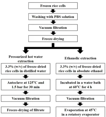
Then, the extracted mixture was filtered through What- man No.1 filter paper and the filtrate was freeze-dried and stored at -20°C until use. For ethanolic extraction of rice cells (E-RC), freeze-dried calli were added to a 0.25 L glass bottle containing 0.15 L of ethanol (99%) and incubated in a water bath at 60°C for 4 h. The extracted mixture was separated by filtration and the filtrate was evaporated at 45°C in a rotatory evaporator (N-1001S- W, Rikakikai Co., Ltd., Japan) (Fig. 1).
Determination of total phenolic and flavonoid contents Total phenolic contents of two rice extracts were ana- lyzed using the Folin-Ciocalteu method [23]. Briefly, each extract sample (100 µg) was resolved in 1 ml dis- tilled water. Then, 0.2 ml of the sample solution was mixed with 5.6 ml of distilled water and 0.2 ml of 50%
Folin-Ciocalteu reagent and reacted for 3 min at room
temperature. Then, after 4 ml of 2% Na
2CO
3was added
in the solution, it was incubated for 1 h at 37 ℃. Absor-
bance of the sample was determined at 725 nm using a
microplate reader (BioTek Inc., USA). Gallic was used as a standard.
The total flavonoid contents of two rice extracts were determined according to the previous report [31].
Briefly, after 200 µl of 10-fold diluted extracts, 200 µl of 1 N NaOH, and 2 ml of diethyl glycol were mixed each other, the solution was incubated for 1 h at 37 ℃. Absor- bance of the sample was determined at 510 nm. Querce- tin was chosen as a standard.
Human dermal fibroblast and keratinocyte cultures Human primary dermal fibroblasts (PCS-201-012, American Type Culture Collection, USA) and the spon- taneously immortalized Human adult low Calcium high Temperature (HaCaT) cells, established by Professor N.E. Fusenig (German Cancer Research Center, Ger- many) [4], were used for in vitro studies. Dermal fibro- blasts and HaCaT cells were cultured in high-glucose Dulbecco’s Modified Eagle’s Medium (DMEM; Invitro- gen, USA) supplemented with a fibroblast growth kit (PCS-201-041, ATCC) and 1% penicillin-streptomycin (P/S; Hyclone, USA) and in high-glucose DMEM supple- mented with a keratinocyte growth kit (PCS-200-040, ATCC) and 1% P/S at 37 ℃ in a 5% CO
2incubator,
respectively. The cell lines were seeded at 5 × 10
5and 1 × 10
6cells in a T-175 flask (SPL Life Sciences, Korea), respectively, and then grown to 80 −90% confluence. Der- mal fibroblasts at passages 6 −7 and HaCaT cells at pas- sages 4 −5 were used in the experiments.
Cytotoxicity test of rice cell extract
The effect of 25 −100 µg/ml RCE on dermal fibroblast and HaCaT cell viability was evaluated by WST-1 assay.
Fibroblasts and HaCaT cells were seeded at 3 × 10
3and 1 × 10
4/well in 96-well plates, respectively. Both cell lines were maintained in DMEM (Hyclone) supple- mented with 10% fetal bovine serum (FBS; Hyclone) at 37°C in a 5% CO
2incubator for 24 h. The cells were then treated with 10% v/v WST-1 reagent (LPS Solution, Korea) and incubated at 37°C in a 5% CO
2incubator for 2 h. The absorbance at 450 nm of the samples was deter- mined using a microplate reader (BioTek Inc.).
Scratch migration assay
Dermal fibroblasts and HaCaT cells were seeded at 2 × 10
4and 4 × 10
4per well in six-well plates, respec- tively. The cells were allowed to form a confluent mono- layer at 37°C in a 5% CO
2incubator. Cells were then changed to serum-free medium for 24 h. Subsequently, vertical wounds were created using a sterile pipette tip.
The cells were extensively rinsed with PBS to remove cellular debris prior to treatment with E-RC and PHWE- RC at 25 −100 µg/ml. As positive and negative controls, 10% FBS (Hyclone) and non-treated cells were used, respectively. After 24 and 48 h, images of migrated cells were obtained using a digital camera (TCC-5.01CE, Tucsen Imaging Technology, China) connected to an inverted microscope (Eclipse E600, Nikon Corp., Japan) to observe wound closure.
Quantification of collagen type I mRNA by RT-PCR Dermal fibroblasts were seeded at 2 × 10
4cells per well in six-well plates. The cells were changed to serum- free medium for 24 h. The culture medium was replaced with fresh serum-free medium containing PHWE-RC or E-RC at 25 −100 µg/ml, and then incubated at 37°C in a 5% CO
2incubator for 24 h. Cells were harvested and the total RNA was extracted using the Easy-spin
TM[DNA free] Total RNA Extraction Kit (iNtRON Biotechnology, Korea) according to the manufacturer’s protocols.
Fig. 1. The procedure for extraction from rice cells.
Extracted RNAs were diluted in 20 µl of elution buffer.
The quantity and purity of RNAs were determined using an ND-1000 spectrophotometer (NanoDrop Technolo- gies, USA). cDNA synthesis was performed from 1 µg of pure total RNA using Reverse Transcription Master Pre- mix (iNtRON Biotechnology). Amplification was per- formed under the following conditions: pre-denaturation at 94°C for 5 min; 25 cycles of denaturation at 94°C for 30 s, annealing at 60°C for 30 s, extension at 72°C for 30 s, and a final extension at 72°C for 10 min. PCR prod- ucts were visualized by electrophoresis in a 1.5% (w/v) agarose gel. Primer sequences were as follows: COL1A2 (forward: 5'-ACC TGG TCA AAC TGG TCC TG-3', reverse: 5'-CGT CCT CTC TCA CCA GGA AG-3' and GAPDH (forward: 5'-TGA AGG TCG GTG TGA ACG-3', anti-sense: 5'-TGA TGG CAA CAA TGT CCA C-3'). The amounts of PCR products were normalized to that of the housekeeping gene, GAPDH, to determine the relative expression ratios of each mRNA in relation to the control. Media without RCEs and containing 500 µM vitamin C were used as negative and positive controls, respectively. mRNA was quantified in triplicate inde- pendently.
Measurement of collagen type I in human dermal fibro- blasts by ELISA
Collagen type I protein was quantified using an enzyme-linked immunosorbent assay (ELISA) kit (Human Pro-collagen I a1 DY6220-05, R&D Systems, USA) according to the manufacturer’s instructions.
Briefly, human dermal fibroblasts (5 × 10
3/well) were grown to confluence in 24-well tissue culture plates.
Cells were switched to serum-free medium for 24 h. The serum-free medium was then replaced with that con- taining 25–100 µg/ml of PHWE-RC or E-RC. Media without RCEs and containing 500 µM vitamin C were used as negative and positive controls, respectively.
After 24 h of treatment, culture supernatant was col- lected and stored at -80°C until use. The assay was performed in three independent experiments.
Statistical analysis
The Student’s t-test or one-way ANOVA followed by Dunnett’s test was used for comparison of multiple means with the SigmaPlot 12.0 software (Systat Soft- ware, USA). A p-value of 0.05 represents a significant
difference between groups.
Results and Discussion
In this study, rice cells were extracted by pressurized hot water and ethanolic extraction methods. Several methods can be available for extraction of natural com- pounds. Among them, we used the above methods because they are able to extract roughly active constitu- ents by polarity [12]. In addition, these tests are suitable for studies of human wound healing. Although other sol- vents such as acetone, methanol, and chloroform can also be used, residues may affect the results [20].
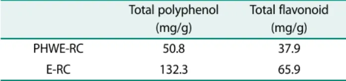
Total polyphenol and flavonoid contents of rice cell extracts were evaluated. As shown in Table 1, total poly- phenol contents of PHWE-RC and E-RC were 50.8 mg/g and 132.3 mg/g, respectively. Total flavonoid contents of E-RC (65.9 mg/g) were also higher than that of PHWE- RC (37.9 mg/g). Ethanolic extraction was more effective method due to polarity of the compounds and the result was corresponded to previous report [31]. The result indicated rice cell extracts contained comparable level of those compounds to other plant extracts [25]. It has been well studied that phenolic compounds showed immune and inflammatory cell function as well as antioxidant activity [24]. Thus, we expect that existence of the com- pounds in rice extracts could contribute to promoting wound healing by treatment of rice extracts.
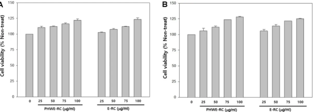
As a preliminary experiment, we evaluated the cyto- toxicity of 25 −100 µg/ml of both RCEs against dermal fibroblasts and HaCaT cells by WST-1 assay. As shown in Fig. 2A and B, treatment of both RCEs did not result in significant cytotoxicity compared with the non-treated control. Furthermore, both RCEs promoted growth of both cell lines in a dose-dependent manner. Treatment with 100 µg/ml RCEs increased the proliferation of both cell types by 25% compared with the control.
We quantified fibroblast and HaCaT cell migration
Table 1. Total polyphenol and flavonoid content of two rice extracts.
Total polyphenol (mg/g)
Total flavonoid (mg/g)
PHWE-RC 50.8 37.9
E-RC 132.3 65.9
Fig. 2. Cytotoxic effects of RCE on the viability of human dermal fibroblasts (A) and HaCaT cells (B). The cells were treated in DMEM serum-free medium with 25, 50, 75 and 100 µg/ml RCE for 24 h. Results are expressed as percentage viability compared to non-treated controls.
Fig. 3. Effect of the RCEs on scratch wound closure in fibroblast and HaCaT cell layers. Phase contrast micrographs of dermal fibroblasts and HaCaT cells taken at 0, 24 and 48 h after treatment with various concentration and type of RCEs. Scale bar 200 µm,
×40 (A). For quantifications, the area covered by dermal fibroblasts (B) and HaCaT cells (C) after incubation for 24 and 48 h was
measured using ImageJ and normalized to control cells. The effect of REC was statistically significant in comparison to the control
sample, as evaluated by the Student's t-test; *p < 0.05, **p < 0.01, **p < 0.001. The error bars indicate S.D. (n = 3).
using an in vitro scratch wound assay at 24 and 48 h post-wounding. Both RCEs at 25 −100 µg/ml were applied to wounded monolayer cultures of both cell types. As shown in Fig. 3B, significant migration of dermal fibro- blast cells into the scratch wound area was observed in the presence of both RCEs. The cell migration was pro- portional to the RCE concentration. In particular, E-RC treatment resulted in 130 −350% greater cell migration compared with the control (p < 0.001). Notably, the effect of 25 µg/ml E-RC was comparable to that of 100 µg/ml PHWE-RC. Similar results were obtained using HaCaT cells (Fig. 3C). Treatment with E-RC prompted significant migration of HaCaT cells in a dose- dependent manner (p < 0.001). Superior migratory abili- ties of HaCaT cell migration was accelerated 70 −250%
by E-RC compared with PHWE-RC. Healing involves complex processes, but re-epithelialization by prolifera- tion and migration of these cells is essential [5, 19], because those cells migrate into wound area to cover damaged tissue surface as well as to promote secretion of various factors involved in wound healing such as growth factors, cytokines, and extracellular matrix [33].
Therefore, cell-to-cell signaling by growth factors and cytokines occurs in the wound area. Thus, these results indicate that E-RC actively recruited cells adjacent to the wound and accelerated cell proliferation.
The effects of the RCEs on collagen type I in dermal fibroblasts were examined compared with that of 500 µM vitamin C, which induces collagen production, as a positive control [26]. Indeed, wound dressings con- taining vitamin C enhance release of growth factors involved in wound healing. Collagens play a critical role in wound healing by activating integrins, which initiate signaling cascades that produce matrix metalloprotein- ases (MMPs) [6, 14, 15]. The enzymes degrade the colla- gen matrix and facilitate keratinocyte migration to the wound area. This results in rapid re-epithelialization and accelerated wound healing. As shown in Fig. 4(A), treatment with >50 µg/ml PHWE-RCs significantly increased collagen type I mRNA levels (p < 0.05); how- ever, this increase was less than that induced by vitamin C. In contrast to PHWE-RC, E-RC treatment enhanced mRNA levels in a dose-dependent manner. In addition treatment with >75 µg/ml E-RC had an effect compara- ble to that of 500 µM vitamin C.
We also quantified collagen type 1 protein levels fol-
lowing treatment with 25 −100 µg/ml RCEs. Dermal
fibroblast cultures were collected at 24 h after the treat-
ments. As shown in Fig. 4(B), both RCEs significantly
promoted collagen production in a dose-dependent man-
ner. PHWE-RC enhanced production of collagen type I
Fig. 4. Effect of the RCEs on collagen type I mRNA (A) and
protein (B) levels in human dermal fibroblasts. (A) mRNA
levels were determined by RT-PCR at 24 h after treatment with
various concentrations of RCEs. Upper panel, representative RT-
PCR analysis. Lower panel, quantification of collagen type I
mRNA expression. (B) Production of collagen type I was evalu-
ated by ELISA of fibroblast culture supernatant at treatment
after 24 h (*p < 0.05, **p < 0.01 vs. the non-treated group). Each
value and error bar represent the mean of triplicate samples
and its S.D.
by up to 47% compared with the non-treated control, but the magnitude of the increase was less than that of vita- min C, even at 100 mg/ml (p < 0.05). In contrast, E-RC at 25 µg/ml stimulated production of collagen type I by 1.8-fold compared to the non-treated control (p < 0.01), comparable to the effect of vitamin C. E-RC at 100 µg/ml resulted in a 2.2-fold increase in collagen production.
Therefore, both RCEs promoted collagen type 1 synthe- sis in human dermal fibroblasts. The effect of E-RC on collagen production was greater than that of PHWE-RC.
As mentioned above, collagen type I gives not only ECM for cell migration and adhesion but also strength of tis- sues. This stimulation by RCEs might contribute to wound healing.
This study is first report on the wound healing prop- erty of RCEs in vitro. PHWE-RC and E-RC exhibited no cytotoxicity to human dermal cells. In addition, both RCEs promoted the proliferation of human fibroblasts and keratinocytes. They increased collagen type I mRNA and protein levels, and thus may play a critical role in the wound healing process. However, further studies such as identification and separation of effective materi- als in RCEs should be performed. In addition, efficacy should be evaluated in an animal study. These prelimi- nary data suggest RCEs to be potential wound healing materials.
Acknowledgments
This research was supported by the National Research Foundation of Korea funded by the Ministry of Education, Korea (Project No.: NRF- 2013R1A1A2061960), for which the authors are grateful.
References
1. Agnihotri S, Wakode S, Agnihotri A. 2010. An overview on anti- inflammatory properties and chemo-profiles of plants used in traditional medicine. Indian J. Nat. Prod. Resour. 1: 150-167.
2. Atiyeh BS, Hayek SN. 2004. An update on management of acute and chronic open wounds: the importance of moist environ- ment in optimal wound healing. Med. Chem. Rev. 1: 111-121.
3. Bajaj P, Schweller RM, Khademhosseini A, West JL, Bashir R. 2014.
3D biofabrication strategies for tissue engineering and regenera- tive medicine. Annu. Rev. Biomed. Eng. 16: 247-276.
4. Boukamp P, Petrussevska RT, Breitkreutz D, Hornung J, Markham A, Fusenig NE. 1988. Normal keratinization in a spontaneously immortalized aneuploid human keratinocyte cell line. J. Cell Biol.
106: 761-771.
5. Broughton II G, Janis JE, Attinger CE. 2006. The basic science of wound healing. Plast. Reconstr. Surg. 117: 12S-34S.
6. Caley MP, Martins VL, O'Toole EA. 2015. Metalloproteinases and wound healing. Adv. Wound Care 4: 225-234.
7. Cheon SH, Lee KH, Kwon JY, Choi SH, Song MN, Kim DI. 2009.
Enhanced delivery of siRNA complexes by sonoporation in trans- genic rice cell suspension cultures. J. Microbiol. Biotechnol. 19:
781-786.
8. Choi JC, Uyama H, Lee CH, Sung MH. 2015. Promotion effects of ultra-high molecular weight poly- γ-glutamic acid on wound healing. J. Microbiol. Biotechnol. 25: 941-945.
9. Gurtner GC, Werner S, Barrandon Y, Longaker MT. 2008. Wound repair and regeneration. Nature 453: 314-321.
10. Havsteen BH. 2002. The biochemistry and medical significance of the flavonoids. Pharmacol. Ther. 96: 67-202.
11. Huang C, Akaishi S, Hyakusoku H, Ogawa R. 2014. Are keloid and hypertrophic scar different forms of the same disorder? A fibrop- roliferative skin disorder hypothesis based on keloid findings. Int.
Wound J. 11: 517-522.
12. Kim A, Kim H, Ryu R, Han H, Han Y, Lee M, et al. 2015. A mixture of ethanol extracts of persimmon leaf and Citrus junos Sieb improves blood coagulation parameters and ameliorates lipid metabolism disturbances caused by diet-induced obesity in C57BL/6J mice. J. Microbiol. Biotechnol. 26: 295-308.
13. Kim DG, Kim EY, Kim YR, Kong IS. 2015. Construction of chimeric human epidermal growth factor containing short collagen-bind- ing domain moieties for use as a wound tissue healing agent. J.
Microbiol. Biotechnol. 25: 119-126.
14. Kim KO, Lee Y, Hwang JW, Kim H, Kim SM, Chang SW, et al. 2014.
Wound healing properties of a 3-D scaffold comprising soluble silkworm gland hydrolysate and human collagen. Colloids Surf. B 116: 318-326.
15. Kim ZH, Lee Y, Kim SM, Kim H, Yun CK, Choi YS. 2015. A compos- ite dermal filler comprising cross-linked hyaluronic acid and human collagen for tissue reconstruction. J. Microbiol. Biotechnol.
25: 399-406.
16. Kwon JY, Yang YS, Cheon SH, Nam HJ, Jin GH, Kim DI. 2013. Biore- actor engineering using disposable technology for enhanced production of hCTLA4Ig in transgenic rice cell cultures. Biotech- nol. Bioeng. 110: 2412-2424.
17. Lai PKK, Chan JYW, Wu SB, Cheng L, Ho GKW, Lau CP, et al. 2014.
Anti‐inflammatory activities of an active fraction isolated from the root of Astragalus membranaceus in RAW 264.7 Macro- phages. Phytother. Res. 28: 395-404.
18. Lam KS. 2007. New aspects of natural products in drug discovery.
Trends Microbiol. 15: 279-289.
19. Lee KM, Shim H, Lee GS, Park IH, Lee OS, Lim SC, Kang TJ. 2013.
Chitin from the extract of cuttlebone induces acute inflamma- tion and enhances MMP1 expression. Biomol. Ther. 21: 246-250.
20. Lu Y, Foo LY. 2002. Polyphenolics of Salvia-a review. Phytochemis- try 59: 117-140.
21. Maquart F-X, Bellon G, Gillery P, Wegrowski Y, Borel J-P. 1990.
Stimulation of collagen synthesis in fibroblast cultures by a triter-
pene extracted from Centella asiatica. Connect. Tissue Res. 24:
107-120.
22. Martin P. 1997. Wound healing-aiming for perfect skin regenera- tion. Science 276: 75-81.
23. Meda A, Lamien CE, Romito M, Millogo J, Nacoulma OG. 2005.
Determination of the total phenolic, flavonoid and proline con- tents in Burkina Fasan honey, as well as their radical scavenging activity. Food Chem. 91: 571-577.
24. Middleton Jr E. 1998. Effect of plant flavonoids on immune and inflammatory cell function, pp. 175-182. In Manthey JA, Buslig BS (eds.), Flavonoids in the Living System, Springer US, New York.
25. Miean KH, Mohamed S. 2001. Flavonoid (myricetin, quercetin, kaempferol, luteolin, and apigenin) content of edible tropical plants. J. Agric. Food Chem. 49: 3106-3112.
26. Niiyama H, Kuroyanagi Y. 2014. Development of novel wound dressing composed of hyaluronic acid and collagen sponge con- taining epidermal growth factor and vitamin C derivative. J. Artif.
Organs 17: 81-87.
27. Okarter N, Liu RH. 2010. Health benefits of whole grain phyto- chemicals. Crit. Rev. Food Sci. Nutr. 50: 193-208.
28. Reuter J, Merfort I, Seelinger G, Wölfle U, Schempp CM. 2009.
Botanicals in dermatology and skin health. pp. 33-65. In Cooper
R, Kronenberg F (eds.) Botanical Medicine. From Bench to Beside.
Mary Ann Liebert Inc., New Rochelle.
29. Robles DT, Berg D. 2007. Abnormal wound healing: keloids. Clin.
Dermatol. 25: 26-32.
30. Shi G, Wang B, Wu Q, Wang T, Wang C, Sun X, et al. 2014. Evalua- tion of the wound-healing activity and anti-inflammatory activ- ity of aqueous extracts from Acorus calamus L. Pak. J. Pharm. Sci.
27: 91-95.
31. Shin SL, Lee CH. 2011. Antioxidant activities of ostrich fern by dif- ferent extraction methods and solvents. J. Life Sci. 21: 56-61.
32. Suguna L, Sivakumar P, Chandrakasan G. 1996. Effects of Centella asiatica extract on dermal wound healing in rats. Indian J. Exp.
Biol. 34: 1208-1211.
33. Werner S, Grose R. 2003. Regulation of wound healing by growth factors and cytokines. Physiol. Rev. 83: 835-870.
34. Yanagawa H, Kato T, Kitahara Y, Kato Y. 1972. Chemical compo- nents of callus tissues of rice. Phytochemistry 11: 1893-1897.
35. Xiu J, Qi S, Li T, Huang B, Tang J, Xu Y, et al. 2009. Effect of asiatico- side on hypertrophic scar in the rabbit ear model. J. Cutan.
Pathol. 36: 234-239.
국문초록
In vitro 실험을 통한 벼세포 추출물의 창상 치유 효능 평가
김지훈
1†, 김선미
1†, 박진호
1, 박찬미
1, 최홍열
2, 이후민
2, 박제권
3, 권순조
2, 김동일
2, 장규호
2, 최용수
1*, 임상민
2*
1
차의과학대학교 바이오공학과
2
인하대학교 생명공학과
3