144
144 THE EWHA MEDICAL JOURNALTHE EWHA MEDICAL JOURNAL
Avascular Necrosis of the Femoral Head in Patient Visiting for Abdominal Pain
Jae Hyun Jung, Young Ho Seo, Sung Jae Choi
Division of Rheumatology, Department of Internal Medicine, Korea University Medical College, Seoul, Korea
A 36-year-old man with abdominal pain presented with a 9-month history of low back pain after traffic accident. He was a chronic daily alcohol drinker, but had any other medical problem. He was diagnosed as a herniation of nucleus pulpo- sus previously and had been treated by acupuncture and pain killer. In spite of treatment, wax and wane pattern low back pain continued, and left ankle pain newly developed. Abdominal pain, which was dominant on the right side, also appeared at 5 months after the accident. Abdominal pain occurred abruptly and disappeared by rest, and it was not associated with meal.
We could not find abnormal finding on esophagogastroduode- noscopy, total colonoscopy, abdominal ultrasonography, and
laboratory examination. Erythrocyte sedimentation rate (2 mm/
hr) and C-reactive protein (0.113 mg/dL) level were not el- evated.
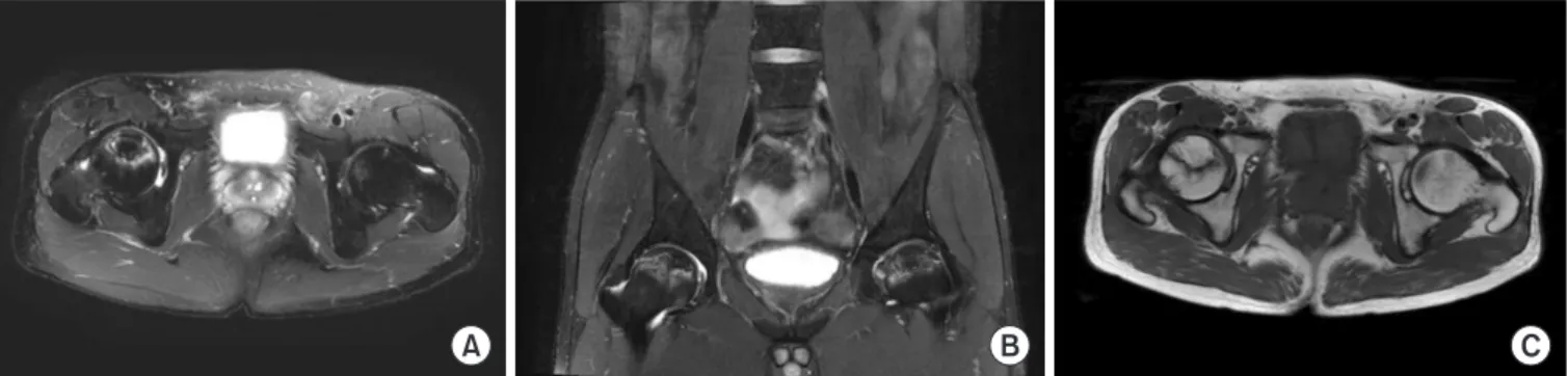
Back pain began when he was younger than 40 years old, aggravated at night, and was not improved with rest. Initially, we thought that he might have inflammatory back pain, and ankle pain was one of extra-axial manifestations of spondylo- arthropathy. Simple radiography (Fig. 1) showed suspicious cam type femoroacetabular impingement at right. Sacroiliac joint magnetic resonance imaging (Fig. 2) showed avascular necrosis (AVN) of the femoral head, Association Research Circulation Osseous (ARCO) stage II [1]. The T1-weighted image revealed
Images and Solution
Ewha Med J 2015;38(3):144-145
http://dx.doi.org/10.12771/emj.2015.38.3.144 pISSN 2234-3180 • eISSN 2234-2591
Received July 8, 2015, Accepted August 20, 2015
Corresponding author Sung Jae Choi, Division of Rheumatology, Department of Internal Medicine, Korea University Ansan Hospital, Korea University College of Medicine, 123 Jeokgeum-ro, Danwon-gu, Ansan 15355, Korea
Tel: 82-31-412-6760, Fax: 82-31-412-5984, E-mail: [email protected]
A B
Fig. 1. Sacro-illiac joint AP, both (A) and right side (B). The prominence of the head-neck junction produces a cam effect as the prominence of the femoral head impinges against the associated region of the acetabulum when the femoral head rotates into the hip joint [4].
145
THE EWHA MEDICAL JOURNAL Avascular Necrosis of the Femoral Head in Patient Visiting for Abdominal Pain
a large osteonecrotic lesion. Low-signal-intensity bands or lines within the femoral head were seen surrounding the area that corresponded to ischemic bone on T1- and T2-weighted im- ages. The appearance on T2-weighted image is known as the
“double-line sign” and was considered highly specific for AVN of the femoral head [2].
Finally, he was diagnosed as AVN of the femoral head. Ab- dominal pain was considered as a referred pain. Referred ab- dominal pain results from the integration of nervous impulses arising in the two main divisions of the nervous system [3].
Clinical suspicion should be raised for not only spondyloar- thropathy but also AVN of the femoral head in cases of low back pain with abdominal pain.
References
1. Orban HB, Cristescu V, Dragusanu M. Avascular necrosis of the femoral head. Maedica (Buchar) 2009;4:26-34.
2. Stoica Z, Dumitrescu D, Popescu M, Gheonea I, Gabor M, Bog- dan N. Imaging of avascular necrosis of femoral head: familiar methods and newer trends. Curr Health Sci J 2009;35:23-28.
3. Gallegos NC, Hobsley M. Abdominal wall pain: an alternative diagnosis. Br J Surg 1990;77:1167-1170.
4. Beall DP, Sweet CF, Martin HD, Lastine CL, Grayson DE, Ly JQ, et al. Imaging findings of femoroacetabular impingement syn- drome. Skeletal Radiol 2005;34:691-701.
5. Kaushik AP, Das A, Cui Q. Osteonecrosis of the femoral head: an update in year 2012. World J Orthop 2012;3:49-57.
Fig. 2. MR images. They show serpiginous high signal intensity areas at both femoral heads are seen on T2 fat saturation images, transverse (A) and coronal (B) section, and on the low sacro-illiac line on T1-weighted image (C) without significant flattening or joint space narrowing. Find- ings are compatible with avascular necrosis, Association Research Circulation Osseous (ARCO) stage II; demarcating sclerosis in femoral head, no collapse [5].
A B C