https://doi.org/10.5624/isd.2019.49.1.53
Introduction
Bisphosphonates, which are inhibitors of osteoclastic bone resorption,
1are useful for the treatment of osteoporo- sis and bone metastases.
2-6However, they are also implicat- ed in the onset of medication-related osteonecrosis of the jaw (MRONJ).
1,7,8Patients with MRONJ are often referred to a larger hospital for the evaluation of bisphosphonate-in- duced changes in the jaws, along with the differential diag- nosis of other diseases of the jaws, such as osteoradionecro-
sis (ORN) or osteomyelitis.
9ORN is a pernicious complication of radiotherapy for head and neck carcinomas.
10The most common cause of ORN is radiation arteritis,
11-13which leads to the onset of a hypocellular, hypovascular, and hypoxic environment. Jaws with MRONJ or ORN must be evaluated before any medi- cal procedure is performed.
9Tc-99m hydroxymethylene diphosphonate (HMDP) scin- tigraphy is capable of demonstrating physiologic changes in bone, and it has been shown that scintigraphy is effective for detecting MRONJ.
14Furthermore, multiple imaging modalities, such as Tc-99m HMDP scintigraphy, computed tomography (CT), and magnetic resonance (MR) imaging, are useful for detecting MRONJ.
8However, to the best of our knowledge, the imaging features of MRONJ and ORN on scintigraphy, CT, and MR imaging have not been pre-
Tc-99m hydroxymethylene diphosphonate scintigraphy, computed tomography, and magnetic resonance imaging of osteonecrosis in the mandible: Osteoradionecrosis versus medication- related osteonecrosis of the jaw
Ichiro Ogura
1,*, Yoshihiko Sasaki
2, Mikiko Sue
2, Takaaki Oda
2, Ayako Kameta
1, Kazuhide Hayama
11Department of Oral and Maxillofacial Radiology, The Nippon Dental University, School of Life Dentistry at Niigata, Niigata, Japan
2Radiology, The Nippon Dental University, Niigata Hospital, Niigata, Japan
ABSTRACT
Purpose: To present characteristic findings of Tc-99m hydroxymethylene diphosphonate(HMDP) scintigraphy, computed tomography(CT), and magnetic resonance(MR) imaging for osteonecrosis in the mandible, especially osteoradionecrosis(ORN) and medication-related osteonecrosis of the jaw(MRONJ).
Materials and Methods: Thirteen patients with MRONJ and 7 patients with ORN in the mandible underwent Tc-99m HMDP scintigraphy, CT, and MR imaging(T1-weighted images[T1WI], T2-weighted images[T2WI], short inversion time inversion recovery images[STIR]), diffusion-weighted images[DWI], and apparent diffusion coefficient[ADC]
mapping). The associations of scintigraphy, CT, and MR imaging findings with MRONJ and ORN were analyzed using the chi-square test with the Pearson exact test.
Results: Thirteen patients with MRONJ and 7 patients with ORN in the mandible showed low signal intensity on T1WI and ADC mapping, high signal intensity on STIR and DWI, and increased uptake on scintigraphy. Periosteal bone proliferation on CT was observed in 69.2% of patients with MRONJ(9 of 13) versus 14.3% of patients with ORN(1 of 7)(P=0.019).
Conclusion: This study presented characteristic imaging findings of MRONJ and ORN on scintigraphy, CT, and MR imaging. Our results suggest that CT can be effective for detecting MRONJ and ORN.(Imaging Sci Dent 2019; 49: 53-8) KEY WORDS: Osteonecrosis; Tomography, X-Ray Computed; Radionuclide Imaging; Magnetic Resonance Imaging
Copyright ⓒ 2019 by Korean Academy of Oral and Maxillofacial Radiology
This is an Open Access article distributed under the terms of the Creative Commons Attribution Non-Commercial License(http://creativecommons.org/licenses/by-nc/3.0) which permits unrestricted non-commercial use, distribution, and reproduction in any medium, provided the original work is properly cited.
Imaging Science in Dentistry·pISSN 2233-7822 eISSN 2233-7830
*This work was supported by JSPS KAKENHI Grant Number JP 18K09754.
Received October 30, 2018; Revised December 18, 2018; Accepted December 19, 2018 Correspondence to: Prof. Ichiro Ogura
Department of Oral and Maxillofacial Radiology, The Nippon Dental University, School of Life Dentistry at Niigata, 1-8 Hamaura-cho, Chuo-ku, Niigata, Niigata 951- 8580, Japan
Tel) 81-25-267-1500, E-mail) [email protected]
sented in the literature. The aim of this study was to evalu- ate the Tc-99m HMDP scintigraphy, CT, and MR imaging findings of osteonecrosis in the mandible, especially ORN and MRONJ.
Materials and Methods
Thirteen patients with MRONJ and 7 patients with ORN in the mandible underwent Tc-99m HMDP scintigraphy, CT, and MR imaging at Radiology, The Nippon Dental
University Niigata Hospital from July 2013 to December 2017. Table 1 characteristics of the patients with ORN and MRONJ.
The images were acquired using a 16-MDCT apparatus (Aquilion TSX-101A; Canon Medical Systems, Otawara, Japan), a 1.5-T MR imaging system (EXCELART Vantage MRT-2003; Canon Medical Systems, Otawara, Japan), and a SNC-5100R scintigraphy apparatus (Shimadzu, Kyoto, Japan) with Tc-99m HMDP (Clear Bone Injection; Nihon Medi-Physics, Tokyo, Japan), following our institutional
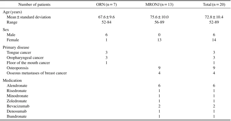
Table 1. Characteristics of the patients with osteoradionecrosis (ORN) and medication-related osteonecrosis of the jaw(MRONJ)
Number of patients ORN(n=7) MRONJ(n=13) Total(n=20)
Age(years)
Mean±standard deviation
Range 67.6±9.6
52-84 75.6±10.0
56-89 72.8±10.4
52-89 SexMale
Female 6
1 0
13 6
14 Primary disease
Tongue cancer Oropharyngeal cancer Floor of the mouth cancer Osteoporosis
Osseous metastases of breast cancer
33
1 9
4
33 19 4 Medication
Alendronate Risedronate Minodronate Zoledronate Bevacizumab Denosumab Ibandronate
61 11 21 1
61 11 21 1
Table 2. Comparison between osteoradionecrosis (ORN) and medication-related osteonecrosis of the jaw(MRONJ) with computed to- mography(CT), magnetic resonance image(MRI) and bone scintigraphy
Imaging features ORN(n=7) MRONJ(n=13) P-value
Bone scintigraphy
Increased uptake 7(100%) 13(100%) -
CT images
Osteolytic changes of the jaws Sequestrum separation Periosteal bone proliferation
6(85.7%) 6(85.7%) 1(14.3%)
12(92.3%) 10(76.9%) 9(69.2%)
0.639 0.639 0.019 MR images
Low-signal intensity on T1WI High-signal intensity on T2WI High-signal intensity on STIR High-signal intensity on DWI Low-signal intensity on ADC map
7(100%) 4(57.1%) 7(100%) 5/5(100%) 5/5(100%)
13(100%) 10(76.9%) 13(100%) 8/8(100%) 8/8(100%)
0.357- - - - T1WI: T1-weighted image, T2WI: T2-weighted image, STIR: short TI inversion recovery image, DWI: diffusion-weighted image, ADC: apparent diffusion coefficient
Fig. 1. Medication-related osteonecrosis of the right side of the mandible in an 86-year- old woman. A. Axial bone tissue algorithm computed tomography shows an osteolytic lesion with sequestrum separation and peri- osteal bone proliferation in the right mandi- ble(arrow). B. On magnetic resonance im- aging, an axial T1-weighted image shows heterogeneous, low-signal intensity(arrow).
C. An axial T2-weighted image shows het- erogeneous, low-signal intensity(arrow). D.
An axial short TI inversion recovery image shows heterogeneous, high-signal intensity (arrow). E. An axial diffusion-weighted image shows heterogeneous, high-signal intensity(arrow). F. An axial apparent diffu- sion coefficient map shows heterogeneous, low-signal intensity(arrow). G. A maxi- mum intensity projection(diffusion-weight- ed image) shows the lesion in an improved way(arrow). H-J. Bone scintigraphy shows increased uptake in the mandible(arrow).
A B
C D
E F G
H I J
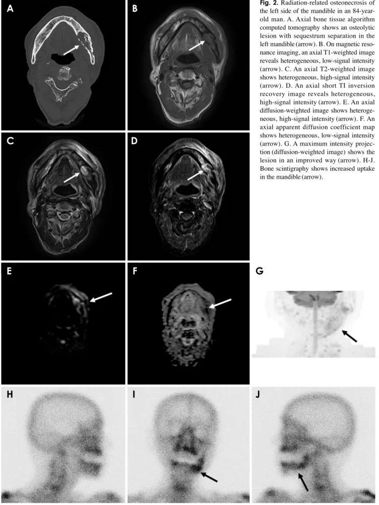
Fig. 2. Radiation-related osteonecrosis of the left side of the mandible in an 84-year- old man. A. Axial bone tissue algorithm computed tomography shows an osteolytic lesion with sequestrum separation in the left mandible(arrow). B. On magnetic reso- nance imaging, an axial T1-weighted image reveals heterogeneous, low-signal intensity (arrow). C. An axial T2-weighted image shows heterogeneous, high-signal intensity (arrow). D. An axial short TI inversion recovery image reveals heterogeneous, high-signal intensity(arrow). E. An axial diffusion-weighted image shows heteroge- neous, high-signal intensity(arrow). F. An axial apparent diffusion coefficient map shows heterogeneous, low-signal intensity (arrow). G. A maximum intensity projec- tion(diffusion-weighted image) shows the lesion in an improved way(arrow). H-J.
Bone scintigraphy shows increased uptake in the mandible(arrow).
A B
C D
E F G
H I J
protocol.
8,14-16The findings of Tc-99m HMDP scintigraphy, CT, and MR imaging of ORN and MRONJ were evaluated by 2 oral radiologists. Scintigraphy was used to analyze areas of in- creased uptake. CT was used to evaluate osteolytic chang- es of the jaws, sequestrum separation, and periosteal bone proliferation. In MR imaging, T1-weighted images (T1WI), T2-weighted images (T2WI), short inversion time inversion recovery images (STIR), diffusion-weighted images (DWI), and apparent diffusion coefficient (ADC) maps were ob- tained.
The associations of scintigraphy, CT, and MR imaging findings with MRONJ and ORN were analyzed using the chi-square test with the Pearson exact test. P values
<0.05 were considered to indicate statistical significance.
Results
The bone scintigraphy, CT, and MR imaging findings are compared between MRONJ and ORN in Table 2. Thir- teen patients with MRONJ and 7 patients with ORN in the mandible showed low signal intensity on T1WI and ADC mapping, high signal intensity on STIR and DWI, and increased uptake on scintigraphy. Periosteal bone pro- liferation on CT was observed in 69.2% of patients with MRONJ (9 of 13) versus 14.3% of patients with ORN (1 of 7) (P=0.019). Figures 1 and 2 show images of MRONJ and ORN, respectively.
Discussion
Imaging findings are unclear both in the early stages of ORN and when the disease is advanced.
11-13Although the ra- diological findings are nonspecific, they do appear to play a role in the management of MRONJ.
17In this study, we have presented the characteristic imaging findings of MRONJ and ORN on bone scintigraphy, CT, and MR imaging.
Tc-99m HMDP scintigraphy is an effective diagnostic tool for detecting bone changes, and has a higher sensitiv- ity than that of radiography.
14Many authors have reported that scintigraphy showed increased uptake at sites affect- ed by MRONJ.
5,17,18In our study, all cases (MRONJ and ORN) showed positive findings on bone scintigraphy. Arce et al.
3reported that the presence of increased uptake in the bone on Tc-99m HMDP depends on osteoblastic activity and skeletal vascularity. We therefore suggest that scintig- raphy shows areas of increased uptake at the sites affected by mandibular diseases.
Bisdas et al.
9reported that CT was useful for detecting
predominantly osteolytic diseases and sclerotic lesions in the jaws, with or without periosteal bone proliferation. In our study periosteal bone proliferation on CT was observed in 69.2% of patients with MRONJ (9 of 13) versus 14.3%
of patients with ORN (1 of 7) (P=0.019). Therefore, we propose that periosteal bone proliferation is a distinctive characteristic of MRONJ on CT.
MR imaging is useful for the diagnosis of bone marrow disease, and necrosis is usually detected as areas of de- creased signal on T1WI.
13Krishnan et al.
2reported that the earliest MR imaging finding of MRONJ was the loss of the normal T1 hyperintensity of fatty marrow in the mandible.
In our study, all cases (ORN and MRONJ) showed low sig- nal intensity on T1WI and high signal intensity on STIR.
Driessen et al.
19reported that DWI demonstrated consis- tently high accuracy and high negative predictive value for head and neck cancer. Bonello et al.
20showed that DWI could provide information on microstructural tumor charac- teristics. Previous studies have reported that DWI and ADC maps were useful for the differential diagnosis of oral and maxillofacial lesions.
15,16In our study, all cases (ORN and MRONJ) showed high signal intensity on DWI, and low signal intensity on ADC maps. We therefore consider that DWI and ADC maps reflect the histopathological differenc- es between malignant tumors and inflammatory diseases in the mandible.
Regarding the reasons underlying the different imaging characteristics of MRONJ and ORN, we propose that peri- osteal bone proliferation on CT in association with ORN is rare because ORN is caused by radiation arteritis.
11-13All cases of MRONJ and ORN in our study showed low signal intensity on T1WI and ADC maps, high signal in- tensity on STIR and DWI, and increased uptake on bone scintigraphy. We suggest that the increased uptake on bone scintigraphy may be correlated with MR imaging findings, especially those on DWI and ADC maps, although the sample was relatively small in this study.
In conclusion, this study presented the characteristic im- aging findings of MRONJ and ORN on bone scintigraphy, CT, and MR imaging. Our results suggest that CT can be effective for detecting MRONJ and ORN.
References
1. Taniguchi T, Ariji Y, Nozawa M, Naitoh M, Kuroiwa Y, Kurita K, et al. Computed tomographic assessment of early changes of the mandible in bisphosphonate-treated patients. Oral Surg Oral Med Oral Pathol Oral Radiol 2016; 122: 362-72.
2. Krishnan A, Arslanoglu A, Yildirm N, Silbergleit R, Aygun N.
Imaging findings of bisphosphonate-related osteonecrosis of the
jaw with emphasis on early magnetic resonance imaging find- ings. J Comput Assist Tomogr 2009; 33: 298-304.
3. Arce K, Assael LA, Weissman JL, Markiewicz MR. Imaging findings in bisphosphonate-related osteonecrosis of jaws. J Oral Maxillofac Surg 2009; 67(5 Suppl): 75-84.
4. Leite AF, Ogata Fdos S, de Melo NS, Figueiredo PT. Imaging findings of bisphosphonate-related osteonecrosis of the jaws:
a critical review of the quantitative studies. Int J Dent 2014;
2014: 784348.
5. Paulo S, Abrantes AM, Laranjo M, Carvalho L, Serra A, Botel- ho MF, et al. Bisphosphonate-related osteonecrosis of the jaw:
specificities. Oncol Rev 2014; 8: 254.
6. Koth VS, Figueiredo MA, Salum FG, Cherubini K. Bisphos- phonate-related osteonecrosis of the jaw: from the sine qua non condition of bone exposure to a non-exposed BRONJ entity.
Dentomaxillofac Radiol 2016; 45: 20160049.
7. Ruggiero SL. Diagnosis and staging of medication-related osteonecrosis of the jaw. Oral Maxillofac Surg Clin North Am 2015; 27: 479-87.
8. Ogura I, Sasaki Y, Kameta A, Sue M, Oda T. Characteristic multimodal imaging of medication-related osteonecrosis of the jaw: comparison between oral and parenteral routes of medica- tion administration. Pol J Radiol 2017; 82: 551-60.
9. Bisdas S, Chambron Pinho N, Smolarz A, Sader R, Vogl TJ, Mack MG. Biphosphonate-induced osteonecrosis of the jaws:
CT and MRI spectrum of findings in 32 patients. Clin Radiol 2008; 63: 71-7.
10. Chronopoulos A, Zarra T, Tröltzsch M, Mahaini S, Ehrenfeld M, Otto S. Osteoradionecrosis of the mandible: a ten year sin- gle-center retrospective study. J Craniomaxillofac Surg 2015;
43: 837-46.
11. Chronopoulos A, Zarra T, Ehrenfeld M, Otto S. Osteoradione- crosis of the jaws: definition, epidemiology, staging and clinical and radiological findings. A concise review. Int Dent J 2018; 68:
22-30.
12. van de Meent MM, Pichardo SE, Rodrigues MF, Verbist BM, van Merkesteyn JP. Radiographic characteristics of chronic dif-
fuse sclerosing osteomyelitis/tendoperiostitis of the mandible: a comparison with chronic suppurative osteomyelitis and osteora- dionecrosis. J Craniomaxillofac Surg 2018; 46: 1631-6.
13. Hatakeyama H, Fujima N, Tsuchiya K, Mizoguchi K, Mizuma- chi T, Sakashita T, et al. Osteoradionecrosis of the hyoid bone after intra-arterial chemoradiotherapy for oropharyngeal cancer:
MR imaging findings. Cancer Imaging 2017; 17: 22.
14. Hayama K, Tsuchimochi M, Yamaguchi H, Oda T, Sue M, Kameta A, et al. Dynamic analysis of technetium-99m HMDP accumulation and its effect on regional bone metabolism and bone blood flow in bisphosphonate-related osteonecrosis of the jaw. Oral Radiol 2013; 29: 135-9.
15. Ogura I, Sasaki Y, Kameta A, Sue M, Oda T. Diffusion-weight- ed imaging in the oral and maxillofacial region: usefulness of apparent diffusion coefficient maps and maximum intensity projection for characterization of normal structures and lesions.
Pol J Radiol 2017; 82: 571-7.
16. Oda T, Sue M, Sasaki Y, Ogura I. Diffusion-weighted magnetic resonance imaging in oral and maxillofacial lesions: prelimi- nary study on diagnostic ability of apparent diffusion coefficient maps. Oral Radiol 2018; 34: 224-8.
17. Morag Y, Morag-Hezroni M, Jamadar DA, Ward BB, Jacobson JA, Zwetchkenbaum SR, et al. Bisphosphonate-related osteo- necrosis of the jaw: a pictorial review. Radiographics 2009; 29:
1971-84.
18. O’Ryan FS, Khoury S, Liao W, Han MM, Hui RL, Baer D, et al. Intravenous bisphosphonate-related osteonecrosis of the jaw:
bone scintigraphy as an early indicator. J Oral Maxillofac Surg 2009; 67: 1363-72.
19. Driessen JP, van Kempen PM, van der Heijden GJ, Philippens ME, Pameijer FA, Stegeman I, et al. Diffusion-weighted imag- ing in head and neck squamous cell carcinomas: a systematic review. Head Neck 2015; 37: 440-8.
20. Bonello L, Preda L, Conte G, Giannitto C, Raimondi S, An- sarin M, et al. Squamous cell carcinoma of the oral cavity and oropharynx: what does the apparent diffusion coefficient tell us about its histology? Acta Radiol 2016; 57: 1344-51.