RESEARCH ARTICLE
Received: August 26, 2019, Revised: September 19, 2019, Accepted: October 1, 2019 eISSN 2233-7679
†Correspondence to: Soo-Jeong Hwang, https://orcid.org/0000-0003-4725-1512
Department of Dental Hygiene, College of Medical Science, Konyang University, 158 Gwanjeodong-ro, Seo-gu, Daejeon 35365, Korea Tel: +82-42-600-8444, Fax: +82-42-600-8408, E-mail: [email protected]
Copyright © The Korean Society of Dental Hygiene Science.
Relationship of Oral Bacterial Load Over One Year of Smoking Cessation
Sunghyun Kim
1, Min-Seock Seo
2, and Soo-Jeong Hwang
3,†1
Department of Clinical Laboratory Science, College of Health Sciences, Catholic University of Pusan, Busan 46252,
2
Department of Conservative Dentistry, Wonkwang University Daejeon Dental Hospital, Daejeon 35233,
3
Department of Dental Hygiene, College of Medical Science, Konyang University, Daejeon 35365, Korea
Background: Smoking exerts an adverse effect on the periodontal tissue by reorganizing the ecosystem of oral microorganisms
and is considered to be an important factor in the development of periodontal disease. Although cross-sectional studies on smokers and non-smokers have been attempted to investigate the microbial differences in periodontal oral cavity, only few studies have been conducted to investigate the changes in oral microorganisms during smoking cessation. The purpose of this study was to investigate the changes of bacteria in saliva and gingival crevicular fluid (GCF) over a period of one year among 11 smokers trying to quit smoking.
Methods: Eleven smokers trying to quit smoking visited the clinic at baseline, two weeks, two months, four months, six months,
and 12 months to give saliva and GCF samples. The amounts of 16S rRNA, Porphyromonas gingivalis , Treponema denticola , Prevotella intermedia , Fusobacterium nucleatum subsp. nucleatum , Streptococcus mutans , and Streptococcus sobrinus in saliva and GCF were quantified using real-time polymerase chain reaction TaqMan probe assay. The results were analyzed by nonparametric statistical analysis using Friedman test and Spearman correlation coefficient.
Results: After cessation of smoking, the amounts of 16S rRNA corresponding to
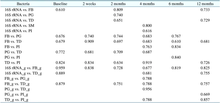
P. gingivalis , F. nucleatum , P. intermedia , and T. denticola in saliva decreased and then again increased significantly. The amount of F. nucleatum 16S rRNA in GCF decreased significantly after smoking cessation. Positive correlations were observed between 16S rRNA and F. nucleatum and between F.
nucleatum and T. denticola in saliva and GCF.
Conclusion:
Even if the number of subjects in this study was small, we suggest that smoking cessation may reduce the total bacterial amount and F. nucleatum in GCF. However, the results regarding changes in the microbial ecosystem due to smoking or smoking cessation were inconsistent. Therefore, further in-depth studies need to be carried out.
Key Words: Bacteria, Periodontal disease, Smoking, Smoking cessation
Introduction
Smoking has an adverse effect on the periodontal tissue and is considered to be an important factor in the progression of periodontal disease. Previous studies have demonstrated the clinical impact of smoking, reporting that scaling and root planing were less effective in smokers than in non-smokers
1), and that periodontal treatment combined with smoking cessation resulted in greater reduction in average periodontal pocket depth
2).
When the amounts of matrix metalloproteinase 9 in gingival crevicular fluid (GCF) were measured in the smoking, non-smoking, and smoking cessation groups, significant differences between the groups were found after adjusting for age and gingival index
3), with the smoking group showing higher concentration of enzymes that destroy periodontal tissues.
In addition, other studies have reported that smoking reorganizes the ecosystem of oral microorganisms.
Smoking was reported to stimulate formation of diverse
and relatively unstable biofilms at gingival margins and subgingival spaces
4), while it was also reported that smokers are more susceptible to Porphyromonas gingivalis infection. In addition, smoking alters the expression of surface components of P. gingivalis to ultimately cause damage to immunoglobulins
5). Shah et al.
6)reported that smoking increases pro-inflammatory and oxidative stress response to virulence-enhanced commensal biofilms.
Assessing oral microorganisms or the health status of periodontal tissues in smokers and non-smokers through a cross-sectional study is less difficult than a longitudinal study. However, the follow-up survey study with smoking cessation as an intervening factor showed a low 12-month success rate in smoking cessation (2.5∼16.9%)
7), and thus, it was not easy to measure any changes. Although there was a study that regularly monitored patients undergone periodontal treatment to investigate the effects of periodontal treatment and smoking cessation
8), there are limited studies that exclusively investigated changes in periodontal tissues during smoking cessation. The purpose of the present study was to observe and analyze changes in oral bacterial load during the smoking cessation process, without involvement of periodontal treatment, in 11 subjects who successfully quit smoking. These subjects were followed up for 12 months in public health center smoking cessation clinics.
Materials and Methods
1. Study subjects and collection of GCF and saliva
The study was performed on a population of 122 men who had signed a written consent to participate in the study and enrolled in public health center smoking cessation clinics. The exclusion criteria of the present study consisted of the following: those with uncontrolled systemic disease; those taking steroids or anti-inflammatory drugs during the study period or within three months prior to participating in the study; those using an oral rinse product during the study period or within three months prior to participating in the study; those receiving dental treatment during the study period; those with ≤20 teeth, excluding the third molars; and those with any area of
periodontal pocket ≥5.5 mm. The subjects were in- structed to visit the public health center at baseline, two weeks, and two, four, six, and 12 months. Due to a very high drop-out rate, only 26 subjects visited the public health center smoking cessation clinics at all time points.
Among them, 15 subjects were unable to quit smoking and only 11 subjects successfully quit smoking for 12 months
9). The success of smoking cessation was determined by verbal reports and using a carbon monoxide meter.
Each time a subject visited the health center, 2 to 4 ml of unstimulated saliva sample was collected, while GCF was collected after removing biofilm and drying. Paper points were used 25 times for 1 minute absorption by intra- crevicular “superficial” method from five interdental spots in maxillary anterior teeth and five interdental spots in mandibular anterior teeth. The samples were frozen immediately after collection at the health center and kept at –20°C until the experiment was performed.
2. Measurement of some oral bacterial load real-time polymerase chain reaction
TaqMan probe assay
Amounts of certain bacteria in saliva and GCF from mandibular anterior region were analyzed using real-time polymerase chain reaction (qPCR) TaqMan probe assay.
Bacteria were received from the Korean Collection for Type Cultures (KCTC) and the following were selected: P.
gingivalis (KCTC 5352, ATCC 33277), Treponema denticola (KCTC 15104, ATCC 35405), Prevotella intermedia (KCTC 5692, KCOM 1107), Fusobacterium nucleatum subsp. nucleatum (KCTC 2640, ACTC25586), Streptococcus mutans (KCTC 3065, ACTC 25175), and Streptococcus sobrinus (KCTC 3308, ACTC 27607).
Molecular biological quantitative bacterial load test was performed at the Department of Clinical Laboratory Science, Catholic University of Pusan, with the primers and TaqMan probe sets prepared as suggested in the previous studies
10-12).
3. Statistics
The cycle threshold (Ct) values of real-time PCR were
used in the analysis without converting them to bacterial
count. This was based on the assumption that analysis
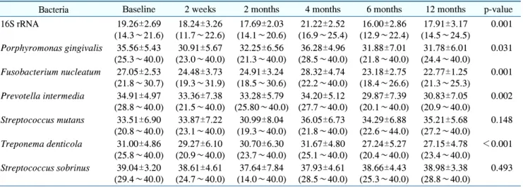
Table 1. Change of Cycle Threshold Values in Real-Time Polymerase Chain Reaction on Each Bacteria in the Saliva of 11 Stop-Smokers
Bacteria Baseline 2 weeks 2 months 4 months 6 months 12 months p-value
16S rRNA 19.26±2.69
(14.3∼21.6)
18.24±3.26 (11.7∼22.6)
17.69±2.03 (14.1∼20.6)
21.22±2.52 (16.9∼25.4)
16.00±2.86 (12.9∼22.4)
17.91±3.17 (14.5∼24.5)
0.001
Porphyromonas gingivalis 35.56±5.43 (25.3∼40.0)
30.91±5.67 (23.0∼40.0)
32.25±6.56 (21.3∼40.0)
36.28±4.96 (28.5∼40.0)
31.88±7.01 (21.8∼40.0)
31.78±6.01 (24.4∼40.0)
0.031
Fusobacterium nucleatum 27.05±2.53 (21.8∼30.7)
24.48±3.73 (19.3∼31.9)
24.91±3.24 (18.5∼30.6)
28.32±4.74 (22.2∼40.0)
23.18±2.75 (18.4∼26.6)
22.77±1.25 (21.3∼25.3)
0.001
Prevotella intermedia 34.91±4.97 (28.8∼40.0)
33.36±7.38 (21.5∼40.0)
33.28±5.79 (25.80∼40.0)
34.20±5.12 (27.7∼40.0)
29.87±7.39 (20.1∼40.0)
30.83±7.05 (20.9∼40.0)
0.002
Streptococcus mutans 33.51±6.90 (20.8∼40.0)
33.87±7.22 (23.1∼40.0)
30.99±8.04 (19.3∼40.0)
36.05±6.73 (21.8∼40.0)
34.29±6.88 (22.6∼44.0)
35.21±5.68 (27.2∼40.0)
0.148
Treponema denticola 31.00±4.86 (25.8∼40.0)
29.27±6.10 (20.9∼40.0)
30.70±6.30 (23.7∼40.0)
31.67±4.80 (25.1∼40.0)
27.24±5.27 (20.4∼40.0)
27.15±4.78 (23.4∼40.0)
<0.001
Streptococcus sobrinus 39.04±3.20 (29.4∼40.0)
38.61±4.61 (24.7∼40.0)
37.64±7.84 (14.0∼40.0)
37.93±4.61 (28.5∼40.0)
38.66±4.43 (25.3∼40.0)
38.98±3.38 (28.8∼40.0)
0.493
Values are presented as mean±standard deviation (minimum∼maximum).
Table 2. Change of Cycle Threshold Values in Real-Time Polymerase Chain Reaction on Each Bacteria in the Gingival Crevicular Fluid of 11 Stop-Smokers
Bacteria Baseline 2 weeks 2 months 4 months 6 months 12 months p-value
16S rRNA 24.72±2.66
(19.8∼28.8)
25.36±2.12 (21.9∼30.2)
25.19±2.15 (22.8∼29.8)
27.14±3.07 (22.6∼31.3)
28.35±1.76 (24.4∼31.1)
28.09±2.18 (24.7∼30.8)
0.001
Porphyromonas gingivalis 38.11±3.29 (32.3∼40.0)
40.00±0.00 (40.0)
39.14±2.86 (30.5∼40.0)
38.26±3.01 (32.9∼40.0)
39.45±1.84 (33.9∼40.0)
38.47±2.63 (33.8∼40.0)
0.358
Fusobacterium nucleatum 26.89±7.16 (21.0∼40.0)
26.35±5.47 (19.6∼40.0)
25.93±5.18 (21.8∼40.0)
32.72±7.22 (22.5∼40.0)
34.64±6.49 (25.6∼40.0)
32.10±7.02 (22.10∼40.0)
<0.001
Prevotella intermedia 35.46±5.44 (26.6∼40.0)
36.84±4.49 (29.0∼40.0)
35.33±4.52 (30.4∼40.0)
39.08±3.05 (29.9∼40.0)
38.98±3.38 (28.8∼40.0)
38.55±3.24 (31.7∼40.0)
0.090
Streptococcus mutans 38.83±2.62 (33.1∼40.0)
39.28±2.38 (32.1∼40.0)
39.14±2.86 (30.5∼40.0)
39.54±1.54 (34.9∼40.0)
39.27±2.41 (32.0∼40.0)
40.00±0.00 (40.0)
0.681
Treponema denticola 34.20±4.94 (27.6∼40.0)
36.93±4.40 (29.3∼40.0)
34.96±4.19 (28.7∼40.0)
37.43±4.52 (28.7∼40.0)
38.94±3.53 (28.3∼40.0)
37.82±3.74 (31.7∼40.0)
0.077
Values are presented as mean±standard deviation (minimum∼maximum).
using Ct values would be appropriate since specific bacteria may not be detected in some samples, and thus, the final variable was presented as Ct value in the table.
Moreover, since the number of subjects was low (n=11) and did not show normal distribution, parametric statistics could have been problematic, and thus, non-parametric statistics were used. For statistical analysis of changes in bacteria at each time point, the Friedman test was used, while the Spearman’s correlation analysis was used for analyzing the correlation between variables. The statistical program IBM SPSS 20.0 (IBM Corp., Armonk, NY, USA) was used and the significance level was set to 0.05.
Results
As shown in Table 1, with respect to the bacteria in saliva, the mean Ct value of 16S rRNA, which reflected the total bacterial count, decreased up to two months;
increased at four months; and decreased again at six and
12 months. Therefore, the total bacterial count showed a
pattern of increasing first, and then decreasing, and the
differences between time point were significant. The Ct
values of P. gingivalis, F. nucleatum, P. intermedia, and
T. denticola also showed a pattern of decreasing at two
weeks and two months, as compared to the baseline;
Table 3. Significant Spearmann Coefficients among Cycle Threshold Values of Oral Bacteria in Saliva and Gingival Crevicular Fluid