416 http://www.jchestsurg.org
JCS
Journal of Chest SurgeryCase Report
Unilateral Giant Bullae: Pulmonary Placental Transmogrification Should Be Kept in Mind: Case Reports
Abdel-Mohsen M. Hamad, M.D.
1, Mona M. Nosseir, M.D.
2, Saleh M. Alorainy, M.D.
1Departments of
1Thoracic Surgery and
2Pathology, King Fahd Specialist Hospital, Buraydah, Saudi Arabia
ARTICLE INFO
Received October 8, 2020 Revised November 11, 2020 Accepted November 29, 2020 Corresponding author Abdel-Mohsen M. Hamad Tel 966-565618155 Fax 966-565618155
E-mail [email protected] ORCID
https://orcid.org/0000-0003-1303-0818
Placental transmogrification is a peculiar clinical entity of the lung of uncertain etiology.
We report 2 cases of pulmonary placental transmogrification in 2 patients of different nationalities. Both of them had no history of smoking or chronic lung disease. The main presentations were dyspnea and chest pain. Radiologic studies showed a unilateral giant bulla in both patients; additional pneumothorax was present in only 1patient. They un- derwent surgical bullectomy. Histopathologic studies revealed the presence of intracystic placenta-like villous structures and a diagnosis of placental transmogrification was made.
Placental transmogrification should be considered in cases of unilateral bullae.
Keywords: Emphysema, Bullae, Placenta, Case report
Copyright
© 2021, The Korean Society for Thoracic and Cardiovascular Surgery
This is an Open Access article distributed under the terms of the Creative Commons Attribution Non-Commercial License (http://creativecommons.org/licenses/
by-nc/4.0) which permits unrestricted non-commercial use, distribution, and reproduction in any medium, provided the original work is properly cited.
Case report
We present 2cases of placental transmogrification (PT) of the lung. Data were collected from patients’ files; the study was approved by the hospital review committee.
Case 1
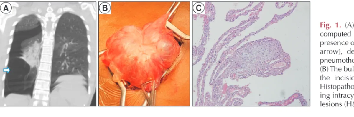
A 25-year-old non-smoking African woman with no his- tory of previous chest troubles presented with acute-onset chest pain and shortness of breath. Chest radiography gave the impression of loculated pneumothorax. Computed to- mography (CT) of the chest verified the presence of right- side pneumothorax, with a large bulla in the right lower lobe (Fig. 1A) and multiple variably-sized bullae involving
most of the outer surface of the upper lobe. A chest drain was inserted and on a later date the patient underwent sur- gical resection of the lower lobe bulla (Fig. 1B) and electro- cautery ablation of the upper lobe bullae, in addition to mechanical pleural abrasions. We started with video-as- sisted thoracoscopic surgery (VATS) and then converted to small muscle-sparing lateral thoracotomy with the assis- tance of a thoracoscopic camera due to marked adhesions of the upper lobe. At her last visit, she was doing well 1 year after surgery.
Case 2
A 28-year-old Jordanian woman presented with right- side vague chest pain and shortness of breath of 2 weeks’
A B C
Fig. 1. (A) Coronal view of chest computed tomography showing the presence of a double air line (white arrow), denoting the presence of pneumothorax and lower lobe bulla.
(B) The bulla was extracted through the incision before resection. (C) Histopathologic examination show- ing intracystic placenta-like villous lesions (H&E stain, ×4).
https://doi.org/10.5090/jcs.20.133
pISSN: 2765-1606 eISSN: 2765-1614
J Chest Surg. 2021;54(5):416-418
417
Abdel-Mohsen M. Hamad, et al. Pulmonary Placental Transmogrification
http://www.jchestsurg.org
JCS
duration. She was non-smoker and had no history of chronic lung diseases. At the time of presentation, she had an average body build and her vital signs and oxygen satu- ration were within normal ranges. The chest X-ray was ini- tially interpreted as showing right-side pneumothorax, but CT of the chest enabled a diagnosis of right-side giant bulla with a compression effect on the lung and shift of the me- diastinum (Fig. 2A, B). The patient underwent right-side VATS bullectomy (Fig. 2C) and mechanical pleural abra- sions. She was doing well 8 months after surgery.
Histopathologic examinations of the resected bullae of both patients showed intracystic proliferation of papillary structures; the papillary cores contained congested blood vessels and were surrounded by hyperplastic alveolar pneumocytes. Therefore, a diagnosis of PT of the lung was made (Figs. 1C, 2D).
This study was conducted in accordance with the amended declaration of Helsinki and the approval of our institutional review board. Both patients provided written informed consent for publication of their clinical details and images.
Discussion
Bullous emphysema is a form of emphysema character- ized by the presence of bullae. The main predisposing fac- tors for its occurrence are tobacco smoking and α
1-anti- trypsin deficiency. In advanced cases, the pathologic process is usually diffuse and affects both lungs. However, bullae may occur in lungs that are otherwise normal [1].
Unilateral bullae occupying most of the hemithorax are seldom seen. In 1979, McChesney [2] described a rare con- genital form of giant bullous emphysema; he coined the term “pulmonary placental transmogrification” because of the morphological resemblance of the intracystic papillary or villous structures to immature placental chorionic villi.
However, in fact, PT has no biological or biochemical rela- tionship to the placenta [2].
Fewer than 40 cases of PT have been reported in the En- glish-language literature. The condition is most common in middle-aged men. In this report, we presented 2 female cases of different nationalities diagnosed within a period of 1 year. Our center serves a multinational community, which may explain this unusual presentation. However, some issues about the actual prevalence of the condition should be raised. It is possible that the diagnosis is missed or ignored in some cases. Furthermore, reluctancy to pub- lish new cases could be another cause of the low reported prevalence of the condition. We encourage good commu- nication between the surgeon and the pathologist when the clinical diagnosis of PT is suspected.
In his first report, McChesney [2] considered PT as an unrecognized hamartoma. Even more than 40 years after that report, the exact etiology of PT remains controversial.
In view of the frequent occurrence of PT with emphysema, some authors consider it as a variant or a complication of bullous emphysema [3]. However, Xu et al. [4] confirmed the presence of PT in 6 cases of pulmonary fibrochondro- matous hamartoma, and argued that PT is a hamartoma- tous lesion. On the contrary, Cavazza et al. [5], using im- munohistochemical, ultrastructural, and molecular studies, identified genotypic alterations in the clear cell component of PT, but not in the adjacent normal tissue;
accordingly, they attributed PT to benign proliferation of immature interstitial “clear cells” with secondary cystic changes, rather than being a variant of emphysema. An- other proposal was introduced by Yang et al. [6], who re- ported PT in a newly developed pulmonary nodule and therefore considered PT to be a pattern of benign morpho- logical changes that may be encountered in both congenital and acquired lesions, rather than being an independent disease. In view of these conflicting reports, we consider
A B C D
R