대한소화기학회지 2001;38:193-202
In t ro d u c t io n1 )
The type species of the genus Helicobacter pylori (H.
pylori) has been established as an etiologic agent of chronic
Received: 10 April 200 1, Accepted: 19 July 200 1.
Correspondence to: Dr . Dong Ki Lee, Department of Internal Medicine, Wonju College of Medicine, Yonsei University, 162 Ilsan-Dong, Wonju, 220-70 1, Republic of Korea.
Tel: (033) 74 1-1222, Fax: (033) 745-6782 E-mail: dklee @wonju.yonsei.ac.kr
active gastritis, gastroduodenal ulcers, and carcinoma of the stomach.1- 8 The rate of H. pylori infection is known to vary in different populations.9 - 14 In 1994, the International Agency for Cancer Reasearch reviewed the available evidence and declared that H. pylori was a carcinogen of humans.3
Since the growth of H. pylori has been shown to be inhibited in human bile in vitro, it is considered not to be associated with hepatobiliary diseases.15 However several Helicobacter spp. have been isolated from the liver and biliary duct in several animals and poultry with hepatobiliary
D et e ct ion of H elicob a ct er S p e cie s fr om K or e an P at ien t s w it h G a lls t on e s U s in g P oly m er a s e Ch ain R e a ct ion s
D o n g K i L e e , M.D ., S o o n Ku B a ik , M .D ., S a n g Ok Kw o n , M.D ., H y u n S o o K i m , M.D ., J o n g B a e Ki m , D .V.M.*, Ge u n H e e K i m *,
H o n g K im *, an d S o o H y u n H a *
Dep artment of Internal Medicine, Wonj u College of Medicine; Department of Medical Laboratory Science*, College of Health Sciences, Yonsei University, Wonj u, Korea
한국 인 담석 환자 로 부터 P o ly m e ra s e C h a i n R e a c t i o n 을 이용한 H e l i c o b a c t e r 속 세균 검출
연세대학교 원주의과대학 원주기독병원 내과 , 연세대학교 보건과학대학 임상병리학과 * 이동기・백순구・권상옥・김현수・김종배 *・김근희 *・김 홍 *・하수현 *
목적 : Helicobacter pylori와 동물에 서식하는 것으로 알려진 담즙 내성 Helicobacter속 세균들이 간담관계 질환 환자로부터 분리되고 있으며, 담석 및 담낭염 발생과 밀접한 관련이 있음이 보고되고 있다. 본 연구에서는 위생검 조직, 위산, 담낭 조직, 담즙, 담석 등의 시료를 이용하여 한국인 담석 환자의 담관내 Helicobacter속 세균 보균율을 조사하였다. 대상 및 방법 : 한국 인 담석증 환자 68명을 대상으로 하여 담석 51예, 담즙 43예, 담낭 조직 29예를 무균적인 방법으로 채취하였으며, 이와 동시 에 위생검 조직 60예 및 위액 42예를 담낭절제술시에 수집하였다. 이들 시료로부터 DNA를 추출한 다음 Helicobacter속 16S rRNA에 특이적인 primer와 Helicobacter종 16S rRNA에 특이적인 primer set를 이용하여 Helicobacter속 세균 검출에 이용하 였으며, 각각의 PCR 산물에 특이적인 probe를 제작하여 PCR 결과를 다시 확인하였다. 결과 : 29예의 담낭 조직 시료 중 4예 에서 H. pylori, 1예에서 H. hepaticus, 2예에서 H. f elis가 검출되었다. 43예의 담즙 시료에서는 12예에서 H. pylori, 1예에서 H. hep aticus, 그리고 2예에서 H. pullorum이 검출되었다. 한편 5 1예의 담석 시료 중에서는 11예의 H. pylori만 검출되었으며 다른 종류의 Helicobacter spp. DNA는 검출되지 않았다. 또한 12예의 위생검 조직, 5예의 위액, 및 1예의 담즙 시료는 H.
pylori와 H. hepaticus가 동시에 검출되었다. 이와 같은 결과는 각각의 PCR 산물에 특이적인 probe를 이용한 Southern blot hybridization 결과와 모두 일치하였다. 결론 : 이상의 결과를 종합할 때 한국인의 담관계 질환에는 H. pylori와 H. hep aticus가 중복 감염되는 것과 관련이 있을 가능성이 높은 것으로 추정된다.
(Korean J Gastroenterol 2001;38 :193-202)
색 인 단어 : 담즙내성 Helicobacter속, PCR, 담석, 담도계 질환
1 9 4 대한소화기학회지 :제 38 권 제 3호, 2001
diseases which were similar to human diseases.16 - 19 Therefore, it is possible that Helicobacter spp. are associated with hepatobiliary diseases in human.2 0 - 2 2
Gallstones can be classified as mixed cholesterol stone and pigmented stone, according to their compositions.
Recently the prevalence of mixed cholesterol gallstone rather than the pigmented gallstone has been steadily increased in Korea with the increased consumption of western food.
The mechanism of cholelithiasis in Korea still needs to be investigated thoroughly. Lee et al2 3 , 2 4 identified the presence of bacteria in the mixed cholesterol gallstone using PCR techniques for the detection of prokaryotic 16S rRNA genes, and they suggested that bio-production of mixed cholesterol gallstone is closely related with abnormal cholesterol metabolism and bacterial infections in the biliary system.
Human bile is sterile under normal conditions. To protect against the invasion of bacteria, the biliary tract has been equipped with several defense mechanisms, which include anatomic barriers (tight junctions between hepatocytes and the sphincter of Oddi), physical mechanisms, chemical factors as bile salts, and immunological defense as Kupffer cells and IgA.2 5 However even the biliary system has several defense mechanisms, it seems that hepatobiliary tract is infected transiently or repeatedly via ascending or descending route by microorganisms.
H. pylori has been shown to be sensitive in vitro to the major free bile acids in human bile.15 O'Connor et al2 6 observed that H. pylori infection was decreased after surgery for stomach diseases. However, under certain pathological conditions, these inhibitory factors for the growth of H.
pylori can be changed. For example, the concentration of different components in the bile can be altered in bile duct obstruction. Furthermore, the inhibitory effect of the bile acids on H. pylori in vivo is uncertain. Since studies by different groups concerning the role of H. pylori in bile reflux gastritis have reached conflicting results. In some reports, the bile reflux (from duodenum to antrum) does not seem to affect the growth of H. pylori in the antrum.2 7 , 2 8 Thus the inhibitory effect of bile acids to H. pylori could be less significant in vivo.
Moreover Fox et al2 9 recently found DNAs of various Helicobacter spp. in bile samples and gallbladder tissue obtained from Chilean patients with chronic cholecystitis.
Helicobacter DNA was detected by PCR and then subj ected to sequence. These results of sequence were represented
strains of H. bilis, H. rapp ini, and H. p ullorum. They concluded that bile-resistant Helicobacter spp. may be associated with gallbladder diseases. However subsequently a similar study was performed in Germany with contrasting results.3 0 The discrepancy in the results of the two studies might be explained by regional differences in the distribution of bile-resistant Helicobacter spp.
Because of the fastidious nature of the Helicobacter spp.
and the requirement of long term culture, rapid and sensitive assay for the detection of the organism has been required.
Recently many PCR-based methods using different gene targets of Helicobacter species have been described to detect the organisms directly in clinical specimens.14 , 3 1- 3 4 Among these targets of PCR, phlyogenetic comparison of rRNA sequences has become a powerful method for the systematic classification and detection of microbial organisms because some segments of rRNA are functionally and evolutionally conserved while others are variable to different degrees.3 5 - 3 8 The 16S rRNA gene of Helicobacter spp. could be a highly specific target for PCR amplification because of the high copy number of rRNA per bacterial cell and has been used previously to help reclassify and detect the organisms.3 9 - 4 3
Although the prevalence has regional differences, H.
pylori and bile-resistant Helicobacter spp. which inhabit in animals were detected in human samples and seem to be associated with the diseases of hepatobiliary tract such as cholelithiasis and cholecystitis. Because the rate of H. pylori infection is above 90% here in Korea, it is important to investigate the prevalence of Helicobacter spp. in biliary tract. The aim of this study was to investigate the prevalence of Helicobacter spp. in gastric biopsy, gastric juice, gallbladder tissue, bile juice, and gallstone samples obtained from cholelithiasis patients in Korea using 16S rRNA gene-specific PCR and Southern blot hybridization.
Ma t e r ia ls a n d Me t h o d s
1. P at ien t s an d s am ples
Gallstones (n=51), bile juice (n=43), and gallbladder tissue (n=29) were obtained by surgical resection employing aseptic technique from 68 Korean patients with symptomatic gallstones. Concomitantly gastric mucosa (n=60) by endoscopy and gastric juice samples (n=42) by Levin tube were obtained before cholecystectomy.
이동기 외 7인, 한국인 담석 환자로부터 Polymerase Chain Reaction을 이용한 Helicobacter속 세균 검출 1 9 5
2 . Met h ods 1) Ba ct e ria l s t ra ins
The Helicobacter spp. type strains used in this study were H. pylori American Type Culture Collection (ATCC) 43504, H. bilis ATCC5 1631, Helicobacter canis ATCC5 140 1, Helicobacter f elis ATCC49479, Helicobacter hep aticus ATCC5 1448, and H. p ullorum ATCC51864. Four other bacterial species were used in the specificity study as control strains. They were Campylobacter j ej uni ATCC33560, Escherichia coli ATCC 25922, Staphylococcus aureus ATCC 13565 and Salmonella typ hi ATCC 19430.
2 ) Cult ure co nd it ions
Six Helicobacter species were inoculated on trypticase soy agar supplemented with 5% bovine blood, 10 ㎍/mL vancomycin, 25 ㎍/mL nalidixic acid, 1 ㎍/mL amphotericin B, and 8 IU/mL polymyxin B, then incubated at 37 under microaerophilic conditions for 5 days. Control strains were inoculated on brain heart infusion agar (Difco, Detroit, MI, U.S.A.) and then incubated at 37 .
3 ) DNA e xt ra ct ion
Gastric mucosa and gallbladder tissue was ground with Stomacher Lab-Blander 80 (Seward Medical, London, England), and DNA was extracted with a QIAamp DNA Mini Kit (QIAGEN Inc., Valencia, CA, U.S.A) according to the manufacturer's instructions. Gastric and bile juice samples were centrifuged for 30 min at 12,000 rpm and the pellet was washed with PBS, and then 10 volumes of 1%
sodium dodecyl sulfate (SDS) were added and incubated for overnight. To this mixture was added lithium chloride to a final concentration of 0.8 M, it was incubated at room temperature for 1 hour. This was centrifuged at 12000 rpm for 30 min and supernatant was placed on new tube. DNA was isolated by extracting with an equal volume of phenol-chloroform-isoamylalchol (25:24:1), precipitated with 0.3 M sodium acetate and 2 volumes of absolute ethanol, and placed on -80 deep freezer for 18 hours. DNA was then pelleted by centrifugation of 14000 rpm, rinsed with 70% ethanol, and vacuum dried. The DNA pellet was dissolved in autoclaved distilled water and frozen at -20 . Gallstone samples were ground with Stomacher Lab-Blander 80 (Seward Medical) and DNA was extracted as described above. And bacterial genomic DNAs from Helicobacter
species and control strains were prepared using boiling lysis4 4 method and stored at -20 for PCR analysis.
4 ) O ligonuc le ot ide prime rs a nd pro be s
Table 1 shows the novel nucleotide sequences of primers and probes and their conditions of PCR. Each Helicobacter species-specific 16S rRNA sequences was selected on the basis of alignments performed with Oligo software (National Biosciences, Polymouth, MN, U.S.A) and BLAST 2 SEQUENCES (web page, www.ncbi.nlm.nih.gov/gorf/bl2.
htm).
5 ) P C R fo r He lico ba cte r ge nus
PCR was performed in a reaction volume of 20 μL consisting of 2 μL of template DNA, 0.2 mM (each) dNTPs (dATP, dGTP, dTTP, dCTP), PCR buffer (50 mM KCl, 10 mM Tris-HCl, pH 8.3), 1.5 mM MgCl2, 40 pmol each primer(Table 1), and 0.5 U of Taq polymerase (Perkin-Elmer Cetus, Norwalk, CT, U.S.A.). The reaction mixture was subjected to 35 cycles of 94 for 1 min, 50 59 for 1 min depending on the primers used, and 72 for 1 min 30s and additional extension step for 15 min at 72 was performed at the end of amplification. The PCR products were analyzed by 1.5% agarose gel electrophoresis with 0.5 ㎍/mL ethidium bromide and visualized under UV light. To avoid the false positivity in PCR due to laboratory contamination, reagent mixing, sample addition and thermocycling were done in separated laboratory, and the precautions, such as using micropipette tips with aerosol barrier, were also taken throughout this study.
Initially, samples were amplified by prokaryotic 16S rRNA-specific primers, POmod-PC5,4 5 to identify the presence of bacterial DNAs. And using Helicobacter genus-specific 16S rRNA primers, C97-C05,2 9 PCR analysis was carried out with the samples showing a positive result in prokaryotic 16S rRNA PCR.
6 ) S pe c ie s - s pe c if ic P C R a na lys is
Samples generating a positive result in Helicobacter genus-specific PCR were subsequently analyzed with an additional six different sets of species-specific primers (Table 1), which were originally designed in this study based on Helicobacter species-specific 16S rRNA sequences.
7 ) S o ut he rn b lot hyb rid iza t io n
For confirmation of the H. pylori, H. hep aticus, and H.
1 9 6 The Korean Journal of Gastroenterology : Vol. 38, No. 3, 200 1
f elis specific PCR-amplified DNA, Southern blot hybridi- zation were performed. Three kinds of probes used were hpp225, hfp 1107 and hhp961, listed in Table 1. The nucleotide sequences of these probes correspond to an internal region of the 642-bp, 366-bp, and 42 1-bp fragment in 16S rRNA gene of H. pylori, H. f elis and H. hepaticus, respectively. Ten ㎕ of each PCR product was electrophoresed through a 1.5% agarose gel and transferred onto nylon membrane using capillary transfer method. The membrane was baked at 80 for 3 hours, hybridized with 3'-end flouresceine-11-dUTP-labeled probe (Amersham, RPN 2130; Buckinghamshire, England), and exposed in the presence of luminol onto Hyperfilm-ECL (Amersham, RPN 2 103; Buckinghamshire, England) according to the manufacture's instructions.
8 ) Nuc le ot ide s e q ue nce a cce s s io n numbe rs The GenBank nucleotide sequence accession numbers for the 16S rRNA sequences, which were deposited previously by other investigators, for H. pylori are U00679, and
U01328 to U0 1332. And the accession numbers of other members of the genus Helicobacter are U5 1873 (H. bilis), L 13464, L 14634 and U04344 (H. canis), M37643 and M57398 (H. f elis), L39122 (H. hepaticus), and AF047850 (H. p ullorum).
R e s u lt s
1. P CR specificit y
The specificity of the Helicobacter genus-specific primer set, C97-C05, was determined with six Helicobacter species and other microorganisms. A product of the 1,200-bp expected size was observed with only Helicobacter species (data not shown). And the specificity of Helicobacter species-specific primer sets was examined with DNA extracted from the six species of Helicobacter strains including H. pylori. PCR amplicons of Helicobacter spp.
used in this study are shown in Fig. 1 with H. bilis-specific primers (plate A), H. canis-specific primers (plate B), and Table 1. The Sequences of PCR Primers and Oligonucleotide Probes Used for the Detection of Helicobacter spp.
Target gene (16s rRNA) specific to
Oligonucleotide primers or probes Annealing Tm
(℃) Reference
Primers and probes Sequence (5' to 3')
Procaryote Pomod
PC5
AGAGTTTGATCMTGG
TACCTTGTTACGACTT 50 45
Helicobacter spp. C97 C05
GCTATGACGGGTATCC
ACTTCACCCCAGTCGCTG 55 29
H. pylori HPU 185
HPL826 hpp225
CCTACGGGGGAAAGATTTAT AGCTGCATTACTGGAGAGACT CTTGTTGGTAAGGTAATGGCTT
52 This study
H. bilis HBU 176
HBL358
GGCTTTCAATAAAGAATTTCTC
AGGCTTTCAATAAAAAATTCG 5 1 This study
H. canis HCU 187
HCL382
TACGGGGGAAAGTAGCAC
GGCTGATCCTTTAGCGAG 50 This study
H. f elis HFU787
HFL 1152 hfp 1107
GTTGGGGGGCTTGTCCTC GTCGTCCTCACCTTCCTCCTG TCTCTAAGAATACTGCCTG
59 This study
H. hep aticus HHPU577 HHPL997 hhp961
AGAACTGCATTTGAAACT TTTCAAGCTCCCCGAAGGG ACTAGAGATAGTGGAGTGC
52 This study
H. pullorum HPRU776
HPRL969
CTTGTCATTGCAGTAATGCAG
GGCAAGCCAGCACTCCG 55 This study
Dong Ki Lee, et al. Detection of Helicobacter Species fr om Korean Patient s with Gallstones Using Polymer ase Ch ain Reactions 1 9 7
H. p ullorum-specific primers (plate C). PCR amplification
with each species-specific primer set successfully amplified only in homologous bacterial DNA sample with the expected size of 642 bp (H. pylori), 183 bp (H. bilis), 366 bp (H. f elis), 42 1 bp (H. hep aticus), and 194 bp (H.
p ullorum). However, H. canis-specific primers produced amplicons, only in H. canis DNA samples, of approximately 380 bp instead of the expected size of 196 bp. H. pylori, H. f elis and H. hep aticus-specific amplicons were confirmed by Southern blot hybridization with internal probe hpp225 (Fig. 2), hfp 1107 (Fig. 3) and hhp961 (Fig. 4), respectively.
2. Det ect ion of H e lic obac te r s pp .
Biliary specimens of gallbladder tissues, bile juice and gallstones along with gastric biopsy and gastric juice were amplified first with prokaryotic 16S rRNA-specific primers, POmod-PC5, followed by Helicobacter genus-specific 16S rRNA primers, C97-C05, in case of prokaryotic 16S rRNA-positive samples to test for the presence of Helicobacter spp. in general.
As shown in Table 2, DNAs extracted from biliary specimens were found to be highly positive in prokaryotic 16S rRNA-specific PCR analysis, and a high level of PCR positivity was found in the gastric biopsy specimens (50 of 60) and gastric juice specimens (35 of 42) from Korean patients with gallstones. And genus-specific PCR positivity was also found in biliary specimens, ranging from 46.5%
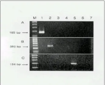
(20 of 43 bile juice specimens) to 34.5% (10 of 29 Fig. 1. PCR amplification of Helicobacter spp. with H.
bilis-specific (A, 183 bp), H. canis-specific (B, 380 bp), and H.
pullorum-specific (C, 194 bp) primer set. Lane M, DNA molecular size standards (100-bp ladder, GenoTech Corp., Taejon); lane 1, H.
bilis ATCC 51631; lane 2, H. canis ATCC 51401; lane 3, H. f elis ATCC 49479; lane 4, H. hepaticus ATCC 51448; lane 5, H.
pullorum ATCC 51864; lane 6, H. pylori ATCC 43504; lane 7, negative control (autoclaved distilled water).
Fig. 2. PCR amplification products of Helicobacter spp. with H.
pylori-specific primer sets (plate A) and corresponding Southern blot hybridization with probes, hpp225 (plate B). Lanes are the same as described in Fig. 1.
Fig. 3. PCR amplification products of Helicobacter spp. with H.
f elis-specific primer sets (plate A) and corresponding Southern blot hybridization with probes, hfp 1107 (plate B). Lanes are the same as described in Fig. 1.
Fig. 4. PCR amplification products of Helicobacter spp. with H.
hepaticus-specific primer sets (plate A) and corresponding Southern blot hybridization with probes, hhp961 (plate B). Lanes are the same as described in Fig. 1.
1 9 8 대한소화기학회지 :제 38 권 제 3호, 2001
gallbladder tissue specimens).
The Helicobacter genus-positive samples were subjected to further PCR analysis using species-specific primer sets.
As a result, H. pylori-specific amplicons were identified from gastric biopsy (50 of 60), gastric juice (28 of 42), gallbladder tissue (4 of 29), bile juice (12 of 43) and gallstones (11 of 5 1). And 12 gastric biopsy, 9 gastric juice, 1 gallbladder tissue, and 1 bile juice samples were positive in PCR with H. hepaticus-specific primers. One of gastric biopsy and two gallbladder tissue were positive for H. f elis.
The positive reaction for H. p ullorum-specific PCR analysis was found in only one gallbaldder tissue and one bile acid sample. However, none of 23 specimens of gallstones, which were positive in the Helicobacter genus-specific PCR, were positive for H. hepaticus-specific, H. f elis-specific, and H. p ullorum-specific PCR.
Twelve gastric biopsy, 5 gastric juice, and 1 bile juice samples which were positive for H. hepaticus-specific PCR were also positive by PCR with H. pylori-specific primers at the same time.
All of the samples that were positive for H. pylori- specific PCR, H. hep aticus-specific PCR and H. f elis- specific PCR were shown to give positive signal in Southern blot hybridization with corresponding internal probes of hpp225, hhp961 and hfp 1107, respectively.
D is c u s s io n
Among Helicobacter spp. that have a strong host specificity, the type strain of H. pylori was detected in gastric biopsy, gastric juice, dental plaque, and fecal samples and recognized as a causative agent of several gastrointestinal diseases.3 3 ,4 6 - 4 8
However, it is not certain that H. pylori are associated with hepatobiliary diseases including colonization in the biliary system of human.
Recently, several additional Helicobacter species have been isolated from liver and biliary duct of mammals.16 , 17 ,2 0
They have been thought to be associated with hepatitis, cholangiofibrosis, and cholecystitis.16 , 17 ,2 0 ,2 1
Moreover, even though the prevalence has regional differences, H. pylori and bile-resistant Helicobacter spp. which inhabit in animals have been detected in human liver4 9 and biliary specimens,
2 8 ,2 9 ,5 0
and seems to be concerned with the diseases of hepatobiliary tract as cholelithiasis and cholecystitis.2 9 The mechanism by which Helicobacter spp. infection leads to liver injury is unclear at present.
The specificity of the 6 Helicobacter species-specific primer sets designed in this study were examined. Not only Helicobacter genus-specific but also the novel species- specific primer sets were proved to have very good reproducible results. Helicobacter species-specific primer sets, except H. canis-specific primers, produced amplicons with expected size only in corresponding bacterial DNA sample, respectively. The reason for the size difference of H. canis-amplicon is unclear at present. However, it should Table 2. The Results of PCR for the Detection of Helicobacter spp. in Clinical Specimens
Primer sets Gastric biopsy (n=60)
Gastric juice (n=42)
Gallbladder tissue (n=29)
Bile juice (n=43)
Gallstone (n=51) POmod - PC5
for Procaryote 53 (88.3%) 37 (88.1%) 16 (61.5%) 23 (53.5%) 30 (58.8%)
C97 - C05
for Helicobacter spp. 50 (83.3%) 35 (83.3%) 10 (34.5%) 20 (46.5%) 23 (45.1%) HPU 185-HPL826
for H. pylori 50 (83.3%) 28 (66.7%) 4 (13.8%) 12 (27.9%) 11 (2 1.6%)
HHPU577-HHPL997
for H. hep aticus 12 (20.0%) 9 (21.4%) 1 ( 3.5%) 1 ( 2.3%) 0 (0%)
HFU787-HFL 1152
for H. f elis 1 ( 1.7%) 0 (0%) 2 ( 6.9%) 0 (0%) 0 (0%)
HPRU776-HPRL969
for H. p ullorum 0 ( 0%) 0 (0%) 0 (0%) 2 (4.7%) 0 (0%)
이동기 외 7인, 한국인 담석 환자로부터 Polymerase Chain Reaction을 이용한 Helicobacter속 세균 검출 1 9 9
be elucidated the size difference of amplicon in H.
canis-specific primers through nucleotide sequence analysis.
Then H. pylori, H. f elis and H. hep aticus-specific PCR amplicons were further verified by Southern blot hybridization with corresponding internal probes of hpp225, hfp 1107 and hhp961, respectively. This result of species- level PCR specificity test prompted us to apply the species-specific primer sets directly to the clinical biliary specimens.
It was not surprising that DNAs extracted from biliary specimens were found to be highly positive (53.5% 61.5%) in prokaryotic 16S rRNA-specific PCR analysis. Then prokaryotic 16S rRNA-positive samples were subjected to the PCR for detection of the genus Helicobacter.
Determined by the same primer set-based PCR, the detection rates of Helicobacter species in biliary specimens from Korean patients with cholelithiasis (34.5% 46.5%) was slightly lower in this study than those (39.1 56.5%) performed previously in Chile by Fox et al.2 9 To identify the Helicobacter spp. in clinical samples on the species level, the Helicobacter genus-positive samples were sub- jected to PCR using each Helicobacter species-specific primers. H. pylori-specific amplicons were identified from gallbladder tissue (4 of 29), bile juice (12 of 43) and gallstones (11 of 5 1), as shown in Table 2. Lin et al2 7 reported 42.9% of H. pylori infection, by PCR targeting for urease A gene of H. pylori, in the bile juice obtained from the patients with obstructive biliary disorders. And among the Korean patients with obstructive biliary disorders such as cancers in the bile duct and cholelithiaisis, the infection rate of H. pylori in the biliary tract is reported to be 39.3% in case of ure A PCR and 36.7% in cag A PCR.5 0
H. hep aticus-specific PCR products were detected from one gallbladder tissue and one bile juice samples, compared to the positivity of 12 among 60 gastric biopsy specimens and 9 among 42 gastric juice specimens. Whereas two gallbladder tissue specimens and one gastric biopsy specimen were positive in H. f elis-specific PCR, only 2 bile juice specimens were positive in H. p ullorum-specific PCR.
The PCR specificity of these positive results for H.
pylori, H. hep aticus and H. f elis was further verified by Southern blot hybridization with corresponding internal probes of hpp225, hfp 1107 and hhp961, respectively.
However, we could not perform Southern blot hybridization for H. p ullorum-specific amplicons because we could not
select the appropriate oligonucleotide probe due to the lack of internal sequences specific only for H. p ullorum.
Twelve gastric biopsy, 5 gastric juice, and 1 bile juice specimens which were positive for H. hep aticus-specific PCR were also positive by PCR with H. pylori-specific primers. These results might imply that Helicobacter species can be co-infected in patients with biliary diseases. This co-infectivity of Helicobacter species was not found in 11 gallstone samples which were positive for H. pylori-specific PCR.
To our knowledge, these results are the first report in Korea on the PCR detection of bile-resistant Helicobacter species to the species level and co-infectivity with H. pylori in human biliary specimens. The bile-resistant Helicobacter species of H. hep aticus, H. f elis, and H. p ullorum detected in this study were reported that their presence has been associated with hepatobiliary diseases,2 9 although the patho-etiological mechanism has not been clear until now.
Fox et al2 9 reported that 56.5% of bile samples and 39.1%
of gallbladder tissue were positive for Helicobacter by PCR in patients with chronic cholecystitis. Because the chronic cholecystitis with gallstone is a risk factor for gallbladder cancer, and Chile has the highest gallbladder cancer mortality rate in the world, they suggested Helicobacter sp.
play a causative role in the development of gallbladder cancer. However, in Korea, even though the H. pylori infection is highly prevelent, the frequency of gallbladder cancer is relatively low. Our study showed low colonization rate of bile-resistant Helicobacter spp. compared to that of Chile study. Not only the regional differences in the distribution of bile-resistant Helicobacter spp., but also the discrepancy in the result of colonizing the gallbladder may be responsible for differences in the biliary disease pattern.
It is possible that these bacteria are transmitted to humans via ingestion of undercooked meat and poultry products which are colonized with Helicobacter spp. And this study have indicated that bile-resistant Helicobacter spp.
could be co-infected with H. pylori. The transmitted bacteria, in this way, are colonized gastric mucosa and duodenum. Thus it appears that bacteria may pass from gastrointestinal tract into the hepatobiliary duct either by an ascending route, as in the case of the disorder of Sphincter of Oddi, or by the hepatobiliary route via the portal blood.
In summary, bile resistant Helicobacter spp., which is known to be inhabited in other animals, were detected in
2 0 0 The Korean Journal of Gastroenterology : Vol. 38, No. 3, 200 1
gastrointestinal and hepatobiliary tract in humans.
Futhermore, it is possible that H. pylori might be co-infected with other Helicobacter spp. However, the results from molecular microbiologic studies without recovery of Helicobacter spp. have major limit for the explanation of development of the disease. Further studies are needed to ascertain the roles and precise mechanisms of Helicobacter spp. in hepatobiliary diseases.
S u m m a ry
Background/Aims: Helicobacter pylori and bile-resistant Helicobacter spp. which inhabit in animals were reported to be detected in human samples and to be associated with the hepatobiliary tract diseases of such as cholelithiasis and cholecystitis. Because the rate of H. pylori infection is above 90% in Korea, it is important to investigate the prevalence of Helicobacter spp. in biliary tract. We investigated the prevalence of Helicobacter spp. in gastric biopsy, gastric juice, gallbladder tissue, bile juice, and gallstone samples from Korean patients with cholelithiasis.
Methods: Clinical biliary specimens of gallstone (n=5 1), bile juice (n=43), and gallbladder tissue (n=29) were collected from 68 Korean patients with symptomatic gallstone, using aseptic technique during cholecystectomy.
Concomitantly, gastric mucosa (n=60) and gastric juice samples (n=42) were obtained before cholecystectomy.
DNA from these specimens was extracted and amplified with the Helicobacter genus-specific and species-specific novel primers for 16S rRNA gene. For confirmation of the H. pylori, H. hep aticus, and H. f elis specific PCR-amplified DNA, Southern blot hybridization was performed. Results:
The gallbladder tissue specimens were positive for H. pylori in 4, for H. hepaticus in 1, and for H. f elis in 2 samples.
The bile juice specimens were positive for H. pylori in 12, for H. hep aticus in 1, and for H. p ullorum in 2 samples.
The gallstone specimens were positive only for H. pylori in 11 samples. Interestingly, 12 gastric biopsy, 5 gastric juice, and 1 bile juice sample which was positive in H.
hep aticus-specific PCR were also positive in PCR with H.
pylori-specific primers. This result was completely correlated with the result of Southern blot hybridization using corresponding internal probes of hpp225 and hhp961, respectively. Conclusions: The results of this study may imply that H. pylori and H. hepaticus can be co-infected in
patients with biliary diseases.
Key Words: Bile-resistant Helicobacter spp., PCR, Gallstones, Biliary diseases
A c k n o w le d g e m e n t
This work was supported in part by Yonsei University Research Fund of 1999.
R e f e r e n c e s
1. Suerbaum S. Helicobacter pylori-microbiology, virulence factors, and clinical manifestations. Biotest Bulletin
1995;5:115-126.
2. Hazell SL, Borody TJ, Gal A, et al. Campylobacter pyloridis gastritis: detection of urease as a marker of bacterial colonization and gastritis. Am J Gastroenterol
1987;82:292-296.
3. WHO. Infection with Helicobacter pylori. IARC Monogr Eval Carcinog Risk Hum 1994;61:177-240.
4. Graham DY. Helicobacter pylori : its epidemiology and its role in duodenal ulcer disease. J Gastroenterol Hepatol
1991;6:105-113.
5. Marshall BJ, Warren JR. Unidentified curved bacilli in the stomach of patients with gastritis and peptic ulceration.
Lancet 1984;1:1311-1315.
6. Moss S, Calam J. Helicobacter pylori and peptic ulcers: the present position. Gut 1992;33:289-292.
7. Parsonnet J, Friedman GD, Vandersteen DP, et al.
Helicobacter pylori infection and the risk of gastric carcinoma. N Engl J Med 199 1;325:1127-1131.
8. Wotherspoon AC, Ortiz-Hidalgo C, Falzon MR, et al.
Helicobacter pylori-associated gastritis and primary B-cell gastric lymphoma. Lancet 1991;338:1175-1176.
9. Baik SC, Kim JB, Cho MJ, et al. Helicobacter pylori infection among normal Korean adults. J Korean Soc Microbiol 1990;25:455-462.
10. Rhee KH, Cho MJ, Kim JB. Prevalence of Campylobacter pylori in normal Korean persons. J Korean Soc Microbiol
1988;23:242.
11. Rhee KH, Youn HS, Baik SC, et al. Prevalence of Helicobacter pylori infection in Korean. J Korean Soc Microbiol 1990;25:475-490.
12. Marshall BJ, Warren JR, Francis G, Langton SR, Goodwin CS, Blincow ED. Rapid urease test in the management of Campylobacter pyloridis-asscociated gastritis. Am J
Dong Ki Lee, et al. Detection of Helicobacter Species fr om Korean Patient s with Gallstones Using Polymer ase Ch ain Reactions 2 0 1
Gastroenterol 1987;82:200-210.
13. Megraud F, Brassens-Rabbe MP, Denis F, Bellbouri JA, Hoa DQ. Seroloepidemiology of Campylobacter pylori infection in various populations. J Clin Microbiol 1989;27:1870-1873.
14. O'Toole PW, Logan SM, Kostrzynska M, Wadstrom T, Trust TJ. Isolation and biochemical and molecular analysis of a species-specific protein antigen from the gastric pathogen Helicobacter pylori. J Bacteriol 199 1;173:505- 5 13.
15. Hanninen ML. Sensitivity of Helicobacter pylori to different bile salts. Eur J Clin Microbiol Infect Dis 1991;10:5 15- 5 18.
16. Fox JG, Dewhirst FE, Tully JG, et al. Helicobacter hepaticus sp. nov., a microaerophilic bacterium isolated from livers and intestinal mucosal scrapings from mice. J Clin Microbiol 1994;32:1238-1245.
17. Fox JG, Yan LL, Dewhirst FE, et al. Helicobacter bilis sp.
nov., a novel Helicobacter species isolated from bile, livers, and intestines of aged, inbred mice. J Clin Microbiol 1995;33:445-454.
18. Franklin CL, Beckwith CS, Livingston RS, et al. Isolation of a novel Helicobacter species, Helicobacter cholecystus sp. nov., from the gallbladders of syrian hamsters with cholangiofibrosis and centrilobular pancreatitis. J Clin Microbiol 1996;34:2952-2458.
19. Stanley J, Linton D, Burnens AP, et al. Helicobacter p ullorum sp. nov.- genotype and phenotype of a new species isolated from poultry and from human patients with gastroenteritis. Microbiology 1994;140:3441-3449.
20. Fox JG, Drolet R, Higgins R, et al. Helicobacter canis isolated from a dog liver with multifocal necrotizing hepatitis. J Clin Microbiol 1996;34:2479-2482.
2 1. Fox JG, Li X, Yan L, et al. Chronic proliferative hepatitis in A/JCr associated with persistent Helicobacter hep aticus infection: a model of Helicobacter-induced carcinogenesis.
Infect Immun 1996;64:1548-1558.
22. Ward JM, Anver MR, Haines DC, Benveniste RE. Chronic active hepatitis in mice cause by Helicobacter hep aticus.
Am J Pathol 1994;145:959-968.
23. Lee DK, Tarr PI, Haigh WG, Lee SP. Bacterial DNA in mixed cholesterol gallstones. Am J Gastroenterol 1999;94:
3502-3506.
24. Lee DK, Tarr PI, Haigh WG, Lee SP. Bacterial genes in mixed cholesterol gallstones and associated sludge.
Gastroenterology 1998;114:A528.
25. Sung JY, Costerton JW, Shaffer EA. Defense system in the
biliary tract against bacterial infection. Dig Dis Sci 1992;37:689-696.
26. O'Connor HJ, Wyatt JI, Dixon MF, Axon AT. Campylo- bacter like organisms and reflux gastritis. J Clin Pathol 1986;39:531-534.
27. Lin TT, Yeh CT, Wu CS, Liaw YF. Detection and partial sequence analysis of Helicobacter pylori DNA in the bile samples. Dig Dis Sci 1995;40:22 14-22 19.
28. Roe IH, Kim JT, Lee HS, Lee JH. Detection of Helicobacter DNA in bile from bile duct diseases. J Korean Med Sci 1999;14:182-186.
29. Fox JG, Dewhirst FE, Shen Z, et al. Hepatic Helicobacter species identified in bile and gallbladder tissue from Chileans with chronic cholecystitis. Gastroenterology 1998;
114:755-763.
30. Rudy J, Rudy A, Maiwald M, Stremmel W. Helicobacter sp. are not detectable in bile from German patients with biliary disease. Gastroenterology 1999;116:10 16-1017.
31. Bickley J, Owen RJ, Fraser AG, et al. Evaluation of the polymerase chain reaction for detecting the urease C gene of Helicobacter pylori in gastric biopsy samples and dental plaque. J Med Microbiol 1993;39:338-344.
32. Clayton CL, Kleanthous H, Coates PJ, Morgan DD, S.
Tabaqchali S. Sensitive detection of Helicobacter pylori by using polymerase chain reaction. J Clin Microbiol 1992;
30:192-200.
33. Hammer M, Tyszkiewicz T, Wadstrom T, O'Toole. PW.
Rapid detection of Helicobacter pylori in gastric biopsy material by polymerase chain reaction. J Clin Microbiol
1992;30:54-58.
34. Valentine JL, Arthur RR, Mobley HL, Dick JD. Detection of Helicobacter pylori by using the polymerase chain reaction. J Clin Microbiol 199 1;29:689-695.
35. Gray MW, Sankoff D, Cedergren RJ. On the evolutionary descent of organisms and organelles: a global phylogency based on a highly conserved structural core in small subunit ribosomal RNA. Nucleic Acids Res 1984;12:5837-5852.
36. Lane DJ, Pace B, Olsen GJ, Stahl DA, Sogin ML, Pace NR. Rapid determination of 16S ribosomal RNA sequences for phylogenetic analyses. Proc Natl Acad Sci USA
1985;82:6955-6959.
37. Olsen GJ, Lane DJ, Giovannoni SJ, Pace NR, Stahl DA.
Microbial ecology and evolution: a ribosomal RNA approach. Annu Rev Micrbiol 1986;40:337-365.
38. Pace NR, Olsen GJ, Woese CR. Ribosomal RNA phylogeny and the primary lines of evolutionary descent. Cell
1986;45:325-326.
2 0 2 대한소화기학회지 :제 38 권 제 3호, 2001
39. Edwards U, Rogall T, Blocker H, Emde M, Bottger EC.
Isolation and direct complete nucleotide determination of entire genes. Characterization of a gene coding for 16S ribosomal RNA. Nucleic Acids Res 1989;17:7843-7853.
40. Gramley WA, Asghar A, Frierson HF Jr, Powell. SM.
Detection of Helicobacter pylori DNA in fecal samples from infected individuals. J Clin Microbiol 1999;37:
2236-2240.
4 1. Riley LK, Franklin CL, Hook RR Jr, Besch-Williford C.
Identification of murine Helicobacters by PCR and restriction enzyme analyses. J Clin Microbiol 1996;34:
942-946.
42. Shames B, Fox JG, Dewhirst F, Yan L, Shen Z, Taylor NS.
Identification of widespread Helicobacter hep aticus infection in feces in commercial mouse colonies by culture and PCR assay. J Clin Microbiol 1995;33:2986-2992.
43. Weisburg WG, Barns SM, Pelletier DA, Lane DJ. 16S ribiosomal DNA amplification for phylogenetic study. J Bacteriol 1991;173:697-703.
44. Holmes DS, Quigley M. A rapid boiling method for the preparation of bacterial plasmids. Anal Biochem 1981;114:
193-197.
45. Wilson KH, Blitchington RB, Greene RC. Amplification of bacterial 16S ribosomal DNA with polymerase chain reaction. J Clin Microbiol 1990;28:1942-1946.
46. Birac C, Tall F, Albenque M, et al. PCR to detect Helicobacter pylori in the mouth. Irish J Med Sci
1992;161(suppl 10):28.
47. Ho SA, Hoyle JA, Lewis FA, et al. Direct polymerase chain reaction test for detection of Helicobacter pylori in humans and animals. J Clin Microbiol 199 1;29:2543-2549.
48. Mapstone NP, Lynch D, Lewis FA. The polymerase chain reaction in the diagnosis of Helicobacter infection. Ital J Gastroenterol 199 1;23(suppl 2):4.
49. Nilsson H-O, Taneera J, Castedal M, Glatz E, Olsson R, Wadström T. Identification of Helicobacter pylori and other Helicobacter species by PCR, hybridization, and partial DNA sequencing in human liver samples from patients with primary sclerosing cholangitis or primary biliary cirrhosis. J Clin Microbiol 2000;38:1072-1076.
50. Roe IH, Lee MS, Chin YJ. Detection of Helicobacter DNA in the bile from the obstructed bile duct. Korean J Med
1998;55:310-316.