대한소화기학회지 2004;44:206~211
1)
접수: 2004년 월5 24 ,일 승인: 2004년 월9 10일 연락처 김세환: , 700-721, 대구광역시 중구 삼덕 가2 50
경북대학병원 소화기내과
Tel: (053) 940-7220, Fax: (053) 954-7417 E-mail: durumi88@yahoo.co.kr
2)
Correspondence to: Se Hwan Kim, M.D.
Department of Internal Medicine, Kyungpook National University Hospital
50 Samduk-2ga, Jung-gu, Daegu 700-721, Korea Tel: +82-53-940-7220, Fax: +82-53-954-7417 E-mail: durumi88@yahoo.co.kr
위와 대장 관상선종의 크기와 부위에 따른 발현 Cyclooxygenase-2 (COX-2)
경북대학교 의과대학 내과학교실 병리학교실, *
김세환이종협강경희박지현박창근조창민권영오김성국최용환배한익* 김미성*
Cyclooxygenase-2 Expression according to Size and Location of Gastric and Cyclooxygenase-2 Expression according to Size and Location of Gastric and Cyclooxygenase-2 Expression according to Size and Location of Gastric and Cyclooxygenase-2 Expression according to Size and Location of Gastric and
Colorectal Tubular Adenomas Colorectal Tubular Adenomas Colorectal Tubular Adenomas Colorectal Tubular Adenomas
Se Hwan Kim,Se Hwan Kim, Se Hwan Kim,
Se Hwan Kim, M.D., Jong Hyup Lee,Jong Hyup Lee,Jong Hyup Lee,Jong Hyup Lee, M.D., Kyung Hee Kang,Kyung Hee Kang,Kyung Hee Kang, M.D., Jee Hyun Park,Kyung Hee Kang, Jee Hyun Park,Jee Hyun Park, M.D.,Jee Hyun Park, Chang Keun Park,
Chang Keun Park, Chang Keun Park,
Chang Keun Park, M.D., Chang Min Cho,Chang Min Cho,Chang Min Cho,Chang Min Cho, M.D., Young Oh Kweon,Young Oh Kweon,Young Oh Kweon,Young Oh Kweon, M.D. Sung Kook Kim,Sung Kook Kim,Sung Kook Kim,Sung Kook Kim, M.D., Yong Hwan Choi,
Yong Hwan Choi,Yong Hwan Choi,
Yong Hwan Choi, M.D., Han Ik Bae,Han Ik Bae,Han Ik Bae,Han Ik Bae, M.D.*, and Mi Sung KimMi Sung KimMi Sung KimMi Sung Kim*
Departments of Internal Medicine and Pathology *, Kyungpook National University College of Medicine, Daegu, Korea Background/Aims:
Background/Aims:
Background/Aims:
Background/Aims: Recent studies have shown that cyclooxygenase-2 (COX-2) may be involved in the process of invasion, growth and apoptosis in colorectal carcinoma and in the growth and tumorigenesis in familial adenomatous polyposis. This study was conducted to determine the significance of the expression of COX-2 in gastric and colorectal adenomas. Methods: Methods: Methods: Methods: Forty-nine samples of gastric adenoma and fifty-seven samples of colorectal adenoma were obtained by endoscopic mucosal resection or polypectomy from 106 patients from January 2000 to July 2003. COX-2 expression was determined by immunohistochemistry. Correlation between COX-2 expression and several clinical factors were compared in each gastric and colorectal adenomas. Results: Results: Results: Results:
The expression of COX-2 in epithelial cells was significantly higher in the group with large adenoma ( >1 cm) compared with the group with small adenoma ( 1 cm) in gastric (76.5% vs. 46.7%, p=0.04) and colorectal adenomas (75% vs. 41.5%, p=0.023). Moreover, increased COX-2 expression was shown in distal compared to proximal colorectal adenoma (64.3% vs. 37.9%, p=0.047). Conclusions: Conclusions: Conclusions: COX-2 was expressed in a size-dependent Conclusions:
manner in gastric and colorectal tubular adenomas. The expression of COX-2 was different according to the location of colorectal adenoma. The association of COX-2 expression with the size of adenoma may suggest that the role of COX-2 is not related to the early development of adenoma, but related to the progression of adenoma.
(Korean J Gastro (Korean J Gastro (Korean J Gastro
(Korean J Gastroenterol 2004;44:206-211) enterol 2004;44:206-211) enterol 2004;44:206-211) enterol 2004;44:206-211) Key
Key Key
Key Words: Words: Words: Cyclooxygenase-2; Gastric adenoma; Colorectal adenoma Words:
김세환 외10 .인 위와 대장 관상선종의 크기와 부위에 따른 Cyclooxygenase-2 (COX-2)발현
207
서 론
는 프로스타글란딘 합성에 관여하 Cyclooxygenase (COX)
는 조절효소로서COX-1과COX-2의 가지 아형이 존재한2 다. COX-1은 외부 자극과 관계없이 여러 장기에서 존재하 면서 주로 위점막상피의 보호 신장의 혈류 유지 그리고, , 혈소판 응집을 조절하는 데 관여한다 이에 반해. COX-2는 정상 조직에서는 거의 발현되지 않다가 사이토카인 내독, 소 인터루킨 호르몬 등의 외부 자극에 의해서 발현된다, , .1,2 특히 여러 암종에서COX-2의 발현이 증가하며3 암의 침윤 이나 성장에도 관여할 뿐만 아니라4혈관 성장 인자 형성과 미세혈관 형성에도 직접 관여한다.5또한COX-2가 염증 반 응과 발암 과정에서 중요한 역할을 담당한다는 사실이 밝 혀짐에 따라6,7 최근에는 관련 질환의 치료에 활용할 수 있 는 방안이 모색되고 있다.
대장선종과 대장암에서 선종과 암의 크기에 비례하여 발현이 증가하며
COX-2 8,9위선종과 위암에서도COX-2발 현이 보고되었다.10,11또한COX의 활성을 억제하는 아스피 린 등의 비스테로이드성 소염제가 유방암 대장암 위암 등, , 의 발병률을 감소시키고 가족성 선종성 대장용종증에서, 선종의 크기와 수를 현저히 줄인다는 연구 결과를 기반으 로 비선택적COX또는 선택적COX-2 억제제를 이용한 연 구가 활발히 진행되고 있다.12,13
저자들은 아직까지 위선종의 진행과 COX-2 발현에 대 한 연구가 충분히 되어 있지 않기에 위와 대장 관상선종 환 자에서 선종의 크기와COX-2 발현을 비교함으로써 임상적 으로 가질 수 있는 선종의 진행과 COX-2 발현의 관계를 알아보고자 하였다.
대상 및 방법
대상1.
년 월부터 년 월까지 소화기내과에 내원하여 2000 1 2003 7
용종절제술 또는 내시경 점막절제술을 시행받은 관상선종 환자106예 중 위선종49 ,예 대장선종57예를 대상으로 하 였다 대상 환자들의 평균 연령은 위선종 환자에서. 63세였 고 대장선종 환자에서는60세로 두 군의 차이가 크게 없었 으며 성별은 위선종과 대장선종 환자에서 각각 71%, 67%
로 남자가 조금 많았다 선종의 진행을 보기 위해 선종의. 크기를1 cm를 기준으로 할 때 위선종 환자에서는 선종이 이상인 경우가 예 로 많았고 대장선종 환자 1 cm 34 (69.4%)
에서는1 cm 미만이41 (71.9%)예 였다 또한 위선종 환자에. 서 선종이 위각부 이하 부위에 많았고(71%)대장선종 환자 에서는 비장만곡부를 경계로 근 원위부에 비슷한 분포로·
선종이 있었다.
방법 2.
연구 대상 환자들의 임상 기록과 내시경 사진 병리 보고, 서 등을 재검토하여 관상선종을 다시 확인하고 선종의 크 기를 분류하였다 선종의 크기는 용종절제술 또는 내시경. 점막절제술 후의 표본 크기를 사용하였고 크기를 정확히 알 수 없는 경우에는 내시경 사진에서의 크기로 선종의 크 기를 정하였다 관상선종의 점막상피에서. COX-2 발현을 보기 위하여106예의 검체를 대상으로 조직미세배열 방법 을 이용하여 면역조직화학염색을 시행하였다.
조직미세배열 방법 1)
조직미세배열 방법은 먼저 수납 블록으로 한 파라핀 블 록에 직경2 mm의 구멍30개를 만들어 각 증례의 파라핀 블록에서 필요한 부위를 조직 채취 기구를 이용하여 얻은 직경 2 mm의 조직을 수납 블록에 심어 미세배열 블록을 만들었다 이 후. 5 µm로 연속 절편을 내어60 정도로2-4 시간동안 보온기에서 건조시키고 그 이후에 면역조직화학 염색을 시행하였다.
면역조직화학염색 2)
파라핀 포매 조직에서 자이렌으로 파라핀을 제거하였고 알코올로 순차적으로 각각 분간 처리 후
100%, 95%, 75% 3
증류수로 함수시켰다. Microwave오븐을 이용하여 끊는10
인산완충액 에 분간 처리 후 상온에서
mM (PBS, pH 6.0) 10
식혔고 내인성 페록시다아제의 활동을 억제하기 위해 3%
과산화수소를 투여 후methanol에 분간 수세하였다 블록10 . 용액(Zymed, San Francisco, California, USA)으로 분간 처30 리하여 비특이적 결합을 억제한 후4 에서 하룻밤 동안 단 클론 항체인 항COX-2항체(1:100, Transduction Laboratory, 를 반응시켰다 다시 로 수 Lexington, Kentucky, USA) . PBS 세한 후 차 항체2 (Zymed, San Francisco)와 분간 반응시30 키고PBS로 차례 수세하였다 여기에3 . streptoavidin과 과 산화효소가 결합된 용액에30분간 반응시키고10-20분간 으로 발색시킨 다음 헤마토 diaminobenzidine (DAB) Mayer 실린으로 대조염색 후 알코올에 순차적으로 수세시켰다.
그 후 마지막으로 malinol로 표본 처리하였다. 염색 결과의 판정
3)
발현은 점막상피의 핵 주위 염색의 강도와 염색 COX-2
된 세포 수의 면적을 기준으로 염색의 강도는 약 양성 중, 등도 양성 강 양성으로 세분화하였고 면적은 광학현미경, 을 이용하여400배의 배율에서 전체1,000개의 세포 중 염 색된 세포 수를 백분율로 표시하여 양성 세포가 없는 경
The Korean Journal of Gastroenterology: Vol. 44, No. 4, 2004
208
우에 점 선종의0 , 10% 이하 부위에서 약 양성 또는 중등도 의 염색 강도를 보이거나 선종의1% 이하 부위에서 강 양 성의 염색 강도를 보이는 경우에 점 선종의1 , 10-50% 부위 에서 약 양성 또는 중등도의 염색 강도를 보이거나 선종의 부위에서 강 양성의 염색 강도를 보이는 경우에 점
1-10% 2 ,
선종의50%이상 부위에서 약 양성 또는 중등도의 염색 강 도를 보이거나 선종의 10%이상 부위에서 강 양성의 염색 강도를 보이는 경우에 점으로 각각 분류한 후3 0, 1점을 음 성군, 2, 3점을 양성군으로 나누어 통계 처리를 하였다.14,15 또한 항COX-2 항체 대신 증류수로 처리하여 음성 대조군 으로 사용하였으며 강하게 핵 주위에 염색된 위암 또는 대 장암을 양성 대조군으로 하였다.
통계 분석 3.
통계 처리는Window용SPSS 11.0 (SPSS Inc. Chicago, 프로그램을 사용하여 각 군을
IL, USA) 2 test, logistic 를 시행하였다 통 regression test, Pearson's correlation test . 계적 유의성은 값이p 0.05 이하인 경우로 하였다.
결 과
위선종과 대장선종에서의 발현
1. COX-2
전체 대상의 관상선종 106예 중에서 위선종 49예 중 발현의 정도가 점이 예 점이 예 점이 예 COX-2 0 1 , 1 15 , 2 32 , 점이 예였다 선종의 크기에 따라서는 미만의 위
3 1 . 1 cm
선종에서는 점이 예1 8 (53%)였고 1 cm 이상에서는 점이2 예 로 가장 많은 빈도를 보였다 대장선종 예 중
26 (76%) . 57
에서는 점이 예 점이0 4 , 1 24 , 2예 점이25 , 3예 점이 예였고4 마찬가지로 선종의 크기에 따라1 cm미만에서는 점이1 20
예(49%), 1 cm이상에서는 점이2 11 (69%)예 로 크기에 따 라 COX-2 발현의 강도가 증가하였다(Table 1).
위선종과 대장선종에서의 발현 양상
2. COX-2
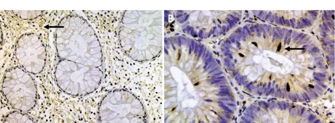
대체적으로 관상선종에서의COX-2발현은 정상 점막에 서도 볼 수 있는 세포간질의 섬유아세포와 림프구에 갈색 으로 염색이 될 뿐만 아니라(Fig. 1A) 정상 점막에서는 볼 수 없는 상피세포의 세포질과 핵 주위에 갈색으로 염색이 되는 것을 볼 수 있었다(Fig. 1B).
3. 위선종과 대장선종의 크기와 부위에 따른 COX-2 발현
위선종의 크기와 부위에 따른COX-2발현의 관계를 보 면 위선종의 크기가1 cm이상의 경우에서1 cm미만보다 의미 있게COX-2발현이 증가하였고(p
=
0.040),위선종의 크기와 COX-2 발현은 유의한 상관성을 보였다(p=0.007, 그러나 위선종을 위각부를 경계로 하여 r=0.305, Table 2).Fig.Fig.Fig.
Fig. 1.1.1. Microscopic findings of COX-2 expression in normal mucosa and tubular adenoma. (A) It shows only brownish staining in1.
lymphocytes, fibroblasts of stroma (arrow) and non-staining in epithelial cells (immunohistochemistry, ×200). (B) It shows brownish staining in cytoplasm and perinucleus of epithelial cells (arrow), lymphocyte and fibroblast of stroma (immunohistochemistry, ×400).
Table 1.
Table 1.
Table 1.
Table 1. COX-2 Expressions in Gastric and Colorectal Adenoma
Histopathology
& size n COX-2
0 1 2 3
Gastric adenoma 1 cm 1 cm
15 34
0 (0) 1 (3)
8 (53) 7 (21)
6 (40) 26 (76)
1 (7) 0 (0) Colorectal adenoma
1 cm 1 cm
41 16
4 (10) 0 (0)
20 (49) 4 (25)
14 (34) 11 (69)
3 (7) 1 (6) Parenthesis: %. COX-2, cyclooxygenase-2.
Kim SH, et al. Cyclooxygenase-2 Expression according to Size and Location of Gastric and Colorectal Tubular Adenomas
209
근위부와 원위부로 구분하였을 때 위각부를 포함한 원위부 에서 위선종이 훨씬 더 많았고 부위에 따른COX-2발현은 유의한 차이가 없었다(p
=
0.136).대장선종에서도 선종의 크 기와 부위에 따른COX-2발현의 차이를 보였으며 위 선종 과 같이 선종의 크기가1 cm이상 시1 cm미만보다 의미 있게COX-2발현이 증가하였으며(p=
0.023)크기를5 mm 미만, 5-10 mm, 10 mm이상으로 세분화하여 비교 시에도 로 의미 있게 발현이 높았다 20%, 48.4%, 75% COX-2또한 로 대장
(p=0.022, Table 2). Pearson's correlation test 선종의 크기와 COX-2 발현의 상관관계를 보았을 때 유의 한 상관성을 보였다(p=0.016, r=0.331).대장선종도 비만곡부 를 경계로 근위부와 원위부에 비슷한 분포를 보이면서 위선 종과는 다르게 근위부보다 원위부에서 의미 있게COX-2발 현이 증가하였고(p
=
0.047) multivariate logistic regression 분석에서도 마찬가지로 원위부에서 의미 있게COX-2발현 이 증가하였다(p=
0.028).관상선종의 크기와 발현
4. COX-2
위선종과 대장선종에서 각각 크기가 증가함에 따라서 발현이 증가하였다 위선종과 대장선종의 구분 없
COX-2 .
이1 cm를 기준으로 크기가 클수록COX-2발현이 증가하 였고(p
=
0.001) 아울러 크기가 1 cm 이상인 관상선종에서 가 발현될 가능성이 배 증가함을 알 수 있었다COX-2 4.22
(Table 2).
고 찰
프로스타글란딘은arachidonic acid의 대사 산물로 여러 가지 기계적 또는 화학적 자극에 의하여 유도되어 염증 발,
열 그리고 통증을 일으키기도 하고 자궁근층의 주기적 수 축 배란 뼈의 재형성에도 관여한다 또한 프로스타글란딘, , . 은 여러 약제들에 의한 위점막 손상에 대해 위점막상피를 보호하는 데 중요한 역할을 한다. COX-2는 유사분열 세포, 접합 면역체계의 감시와 세포사멸에 영향을 주어 암의 발, 생 기전에도 중요한 역할을 한다.
아직까지COX-2의 암의 발생 과정에서의 역할에 대해 정확히 알려져 있지 않다 하지만 최근의 연구들에 의하면.
가 세포사멸의 억제와 관련된
COX-2 Bcl-2, antiapoptotic 과 비례하여 증가하였고
protein 4 침습에 관여하는 matrix 의 활성 metalloproteinase-2, membrane metalloproteinase-1 과 관련되어 있으며16vascular endothelial growth factor
와 같은 혈관 형성 인자의 생성에도 관여한다
(VEGF) .5 그
리고COX-2에 의해 생성된 프로스타글란딘E2가 세포막의 상피세포 수용체에 결합하여 세포성장을 촉진하고 그 외에 면역체계의 감시에도 관여한다 한편 대장암 유방암 식도. , , 암 폐암과 피부암 등에서 프로스타글란딘의 수치가 정상, 조직에 비하여 증가되어 있으며COX억제제가 위암세포의 세포사멸을 촉진하고 세포성장을 억제17하면서 몇 가지 암 의 발생 빈도를 억제한다.13
와 세포 진행에 대한 관계를 보면 먼저 대장선종과 COX-2
대장암에서는 크기에 비례하여 COX-2 발현이 증가하고3,8 가족성 선종성 대장용종증에서는 선종의 크기가 최소한
이상인 경우에 프로스타글란딘이 증가하며
6-7 mm 18대장
의 정상 점막보다는 산발성 선종에서 크기가6 mm이상일 때COX-2 발현이 증가한다.9또한 선종의 부위와 COX-2 발현에 대한 연구는 아직까지 부족하지만 부위에 따라
나 경로에
microsatellite instability chromosomal instability 따른 암 발생 과정의 차이에 기인하여 대장의 근위부보다 Table 2.
Table 2.Table 2.
Table 2. Relationship between COX-2 Expression and Size/Location of Gastric and Colorectal Adenoma COX-2
p-value Odds ratio [95% CI]
Gastric adenoma (n=49)
Size 1 cm
1 cm
7 (46.7) 26 (76.5)
8 (53.3) 8 (23.5)
0.040 3.7 [1.0, 13.4]
Location Proximal Distal
3 (42.9) 30 (71.4)
4 (57.1) 12 (28.6)
0.136 3.3 [0.6, 17.2]
Colorectal adenoma (n=57)
Size 1 cm
1 cm
17 (41.5) 12 (75)
24 (58.5) 4 (25)
0.023 4.2 [1.1, 15.4]
Location Proximal Distal
11 (37.9) 18 (64.3)
18 (62.1) 10 (35.7)
0.047 2.9 [1.0, 8.6]
Overall (n=106)
Size 1 cm
1 cm
24 (42.9) 38 (76.0)
32 (57.1) 12 (24.0)
0.001 4.2 [1.8, 9.7]
Parenthesis: %. COX-2, cyclooxygenase-2; CI, confidence interval.
대한소화기학회지 제 권 제 호: 44 4 , 2004
210
원위부에서COX-2발현이 증가한다.19-21이에 반하여 위선 종과 위암에서는 과형성 위용종에서 용종의 크기가 150 mm2이상일 때 의미 있게COX-2 발현이 증가하고22 37명 의 위암 환자들에서 크기가3.8±2.7 cm보다도6.5±4.6 cm 인 경우에서 의미 있게COX-2 mRNA발현이 증가한다.23 하지만 아직까지 위선종에서의 크기와COX-2 발현에 대한 연구는 미흡한 실정이다.
따라서 본 연구에서는 COX-2 발현이 위선종과 대장선 종의 진행에 미치는 영향을 보기 위해 위 및 대장의 관상선 종 환자에서 선종의 크기와COX-2 발현을 비교하였다 그. 결과 위 및 대장선종 환자에서 크기가 1 cm 이상이면 발현이 의미 있게 증가하였고 대장선종에서는 크기 COX-2
를 세분화하여5 mm미만, 5-10 mm, 10 mm이상으로 구 분하여도 크기에 비례하여 의미 있게COX-2 발현이 증가 하였다 그러나 위선종에서는 대장선종과 같이 크기를 세. 분화하였으나 크기에 따른 각 군의 표본 수 차이로 인해 발현이 의미있게 증가하지는 않았다 한편 선종의
COX-2 .
부위에 따른COX-2 발현을 보았을 때 대장선종에서는 비 만곡부를 경계로 구분하여 위선종과는 다르게 근위부보다 원위부에서 의미 있게 COX-2 발현이 증가하였다 이것은. 아마도 부위에 따른 각 세포의 유전적 변화 차이와 COX-2 조절 유전자의 발현 차이에 의해서 발생한다고 생각된다.19 결론적으로 본 연구에서와 같이 대장선종뿐만 아니라 위 선종에서도COX-2 발현이 크기와 관련성이 있으며 위 선 종의 초기 발생보다는 선종의 진행과 관련이 있음을 알 수 있었다 앞으로 예방적 화학요법이 임상에서 가지는 의미. 가 커지는 만큼 위선종과 위암에서도 예방적 화학요법에 대한 연구가 활발히 진행되어야 할 것으로 생각한다.
요 약
목적: 최근cyclooxygenase-2 (COX-2)는 암의 침윤과 성 장 세포소멸에 관여하며 대장선종과 가족성 선종성 대장, 용종증에서 성장과 발암과정에 관여한다 이에 본 연구에. 서는 위선종과 대장선종의 진행에 있어서 COX-2가 어떤 차이를 나타내는지 알아보고자 했다. 대상 및 방법: 2000 년 월부터1 2003년 월까지 소화기내과에 내원하여 용종절7 제술 또는 내시경 점막절제술을 시행받은 관상선종 환자 명 중 위선종 명 대장선종 명을 대상으로 하였다
106 49 , 57 .
발현은 면역조직화학염색을 이용하였으며 장기에 COX-2
따른 선종의 발생 연령 성별 크기 장기내 위치 등을 인, , , , 자로 COX-2발현을 비교하였다. 결과: 위선종에서는 크기 가 1 cm이상 시1 cm미만보다76.5%, 46.7%로COX-2 발현이 의미 있게 높았다(p=0.04).대장선종에서도1 cm이 상 시1 cm미만보다75.0%, 41.5%로COX-2발현이 높았다
또한 대장선종에서 원위부가 근위부보다
(p=0.023). 64.3%,
로 발현이 높았다
37.9% COX-2 (p=0.047).결론:COX-2발 현은 대장선종 환자에 있어서 크기에 비례하여 증가하였고 부위에 따른 차이도 있었다 위선종에서도 대장선종과 같. 이 크기에 따른 차이가 있었으며 크기가 작을수록 COX-2 의 발현율은 현저히 낮았다 그러므로. COX-2는 선종의 초 기 발생보다는 선종의 진행에 관여할 것으로 생각되며 이 에 대한 많은 연구가 앞으로 필요할 것이다.
색인단어: COX-2, 위선종 대장선종,
참고문헌
1. Herschman HR. Prostaglandin synthase 2. Biochim Biophys Acta 1996;1299:125-140.
2. Jones DA, Carlton DP, McIntyre TM, Zimmerman GA, Prescott SM. Molecular cloning of human prostaglandin endoperoxide synthase type II and demonstration of expression in response to cytokines. J Biol Chem 1993;
268:9049-9054.
3. Eberhart CE, Coffey RJ, Radhika A, Giardiello FM, Ferrenbach S, DuBois RN. Up-regulation of cyclooxygenase 2 gene expression in human colorectal adenomas and adenocarcinomas. Gastroenterology 1994;107:1183-1188.
4. Tsujii M, DuBois RN. Alterations in cellular adhesion and apoptosis in epithelial cells overexpressing prostaglandin endoperoxide synthase 2. Cell 1995;83:493-501.
5. Tsujii M, Kawano S, Tsuji S, Sawaoka H, Hori M, DuBois RN. Cyclooxygenase regulates angiogenesis induced by colon cancer cells. Cell 1998;93:705-716.
6. Lichtenstein DR, Syngal S, Wolfe MM. Nonsteroidal antiinflammatory drugs and the gastrointestinal tract. the double-edged sword. Arthritis Rheum 1995;38:5-18.
7. Prescott SM, Fitzpatrick FA. Cyclooxygenase-2 and carcinogenesis. Biochim Biophys Acta 2000;1470:69-78.
8. Fujita T, Matsui M, Takaku K, et al. Size- and invasion- dependent increase in cyclooxygenase 2 levels in human colorectal carcinomas. Cancer Res 1998;58:4823-4826.
9. Hasegawa K, Ichikawa W, Fujita T, et al. Expression of cyclooxygenase-2 (COX-2) mRNA in human colorectal adenomas. Eur J Cancer 2001;37:1469-1474.
10. Ristimaki A, Honkanen N, Jankala H, Sipponen P, Harkonen M. Expression of cyclooxygenase-2 in human gastric carcinoma. Cancer Res 1997;57:1276-1280.
11. Rajnakova A, Moochhala S, Goh PM, Ngoi S. Expression of nitric oxide synthase, cyclooxygenase, and p53 in different stages of human gastric cancer. Cancer Lett
김세환 외10 .인 위와 대장 관상선종의 크기와 부위에 따른 Cyclooxygenase-2 (COX-2)발현
211
2001;172:177-185.
12. Harris RE, Kasbari S, Farrar WB. Prospective study of nonsteroidal anti-inflammatory drugs and breast cancer.
Oncol Rep 1999;6:71-73.
13. Schreinemachers DM, Everson RB. Aspirin use and lung, colon, and breast cancer incidence in a prospective study.
Epidemiology 1994;5:138-146.
14. Chae SW, Sohn JH, Shin HS, Park YE. Expression of cyclooxygenase-2 protein in gastric carcinogenesis. Cancer Res Treat 2002;34:252-257.
15. Azumaya M, Kobayashi M, Ajioka Y, et al. Size-dependent expression of cyclooxygenase-2 in sporadic colorectal adenomas relative to adenomas in patients with familial adenomatous polyposis. Pathol Int 2002;52:272-276.
16. Tsujii M, Kawano S, DuBois RN. Cyclooxygenase-2 expression in human colon cancer cells increases metastatic potential. Proc Natl Acad Sci USA 1997;94:3336-3340.
17. Kim JH, Kim RH, Yoo K. Induction of apoptosis and inhibition of cellular proliferation in aspirin-treated SNU- 668 human gastric adenocarcinoma cell lines. J Korean
Cancer Assoc 2001;33:71-76.
18. Yang VW, Shields JM, Hamilton SR, et al. Size-dependent increase in prostanoid levels in adenomas of patients with familial adenomatous polyposis. Cancer Res 1998;58:1750- 1753.
19. Dimberg J, Samuelsson A, Hugander A, Söderkvist P.
Differential expression of cyclooxygenase 2 in human colorectal cancer. Gut 1999;45:730-732.
20. Einspahr JG, Krouse RS, Yochim JM, et al. Association between cyclooxygenase expression and colorectal adenoma characteristics. Cancer Res 2003;63:3891-3893.
21. Lindblom A. Different mechanism in the tumorigenesis of proximal and distal colon cancers. Curr Opin Oncol 2001;
13:63-69.
22. Kawada M, Seno H, Wada M, et al. Cyclooxygenase-2 expression and angiogenesis in gastric hyperplastic polyp- association with polyp size. Digestion 2003;67:20-24.
23. Uefuji K, Ichikura T, Mochizuki H. Expression of cyclo- oxygenase-2 in human gastric adenomas and adenocarcinomas.
J Surg Oncol 2001;76:26-30.