Birt-Hogg-Dube′(BHD) syndrome is an autosomal dominant inherited disorder that is characterized by skin fibrofolliculomas, renal tumors and multiple lung cysts with or without spontaneous pneumothorax. The disease is caused by germline mutations in the BHD or FLCN gene located in 17p11.2, and this gene codes for a protein of unknown function called folliculin (1-3). We report here on two cases of BHD syndrome that present- ed with spontaneous pneumothorax.
Case Reports
Case 1
A 47-year-old woman visited our emergency room for an acute onset of dyspnea. Her chest radiograph (Fig.
1A) showed large pneumothorax in the right lung and a radiolucent area in the left lower lung field. The chest CT scan taken after closed thoracostomy showed multi- ple cysts of various sizes and shapes in both lungs (Figs.
1B-C). The pulmonary cysts were sharply demarcated with thin walls and they measured 0.3-5.5 cm in diame- ter. The intervening lung parenchyma between the cysts appeared normal. The initial radiologic impression after the CT scan was lymphangioleiomyomatosis (LAM). The patient had no smoking history. She had a history of thoracoscopic bulla resection for a left pneu- mothorax 15 years ago. The patient reported that her
Spontaneous Pneumothorax in Birt-Hogg-Dube′ Syndrome: Two Case Reports1
Hyoung-ju Bae, M.D., Ok Hee Woo, M.D., Hwan Seok Yong, M.D., Eun-Young Kang, M.D., Hyun Koo Kim, M.D.2, Young Ho Choi, M.D.2, Bong Kyung Shin, M.D.3, Yoon Kyung Kim, M.D.
1Department of Radiology, Korea University School of Medicine, Korea University Guro Hospital
2Department of Thoracic Surgery, Korea University School of Medicine, Korea University Guro Hospital
3Department of Pathology, Korea University School of Medicine, Korea University Guro Hospital
Received August 18, 2010 ; Accepted October 5, 2010
Address reprint requests to : Yoon Kyung Kim, M.D., Department of Radiology, Korea University Guro Hospital, 97 Guro-dong, Guro-gu,
Birt-Hogg-Dube′(BHD) syndrome is a rare autosomal dominant inherited disorder that is characterized by skin fibrofolliculomas, renal tumors and multiple lung cysts with or without spontaneous pneumothorax. The disease is caused by germline muta- tions in the FLCN gene that codes for a protein of unknown function called folliculin.
Patients with BHD syndrome do not always have all three manifestations of the skin, kidney and lung. To the best of our knowledge, there has been no case report of the ra- diologic findings of the lung manifestation in a patient with BHD syndrome in Korea.
We report here on two cases of BHD syndrome that presented with spontaneous pneumothorax. The pulmonary abnormalities consisted of multiple thin-walled cysts of various sizes and shapes in both lungs.
Index words :Lung Cysts
Pneumothorax
tient then underwent wedge resection in the right lung, and this revealed bulla and blebs without evidence of LAM (Fig. 1D).
Eight months after the patient’s surgery, her daughter visited our hospital and presented with recurrent pneu- mothorax. The chest CT scan of the daughter after closed thoracostomy also revealed multiple thin-walled cysts in both lungs (Figs. 1E, F). The daughter had a pre- vious history of wedge resection at our hospital a year previously, and her previous lung specimen revealed bulla only (Fig. 1G).
Case 2
A 54-year-old woman presented with dyspnea, cough and chest pain. Her chest radiograph showed pneu- mothorax in the left lung. The chest CT scan after closed thoracostomy revealed multiple thin-walled cysts and bullae in both lungs (Figs. 2A-C). They were round or
oval shaped and some of them showed a lobulating con- tour. The intervening lung parenchyma between the cysts was normal without any evidence of pulmonary nodule or fibrosis. She had a history of closed thoracos- tomy due to right pneumothorax 35 years ago. She had a 20 pack-year smoking history and had stopped smoking 5 years ago. She had multiple whitish papules measur- ing 2-3 mm on her cheek, and a punch biopsy was per- formed. The pathologic result was perifollicular fibrosis, which was consistent with the histologic diagnosis of fi- brofolliculoma (Fig. 2D). Ultrasonography of the kid- neys did not show any abnormality.
Discussion
Birt-Hogg-Dube′(BHD) syndrome is a rare autosomal dominant inherited disorder that is characterized by skin fibrofolliculomas, renal tumors and multiple lung
A B C
D
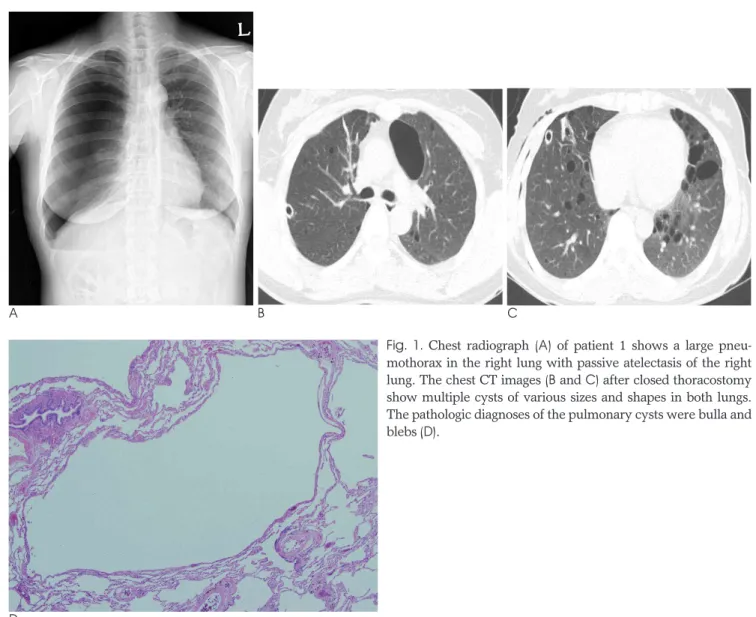
Fig. 1. Chest radiograph (A) of patient 1 shows a large pneu- mothorax in the right lung with passive atelectasis of the right lung. The chest CT images (B and C) after closed thoracostomy show multiple cysts of various sizes and shapes in both lungs.
The pathologic diagnoses of the pulmonary cysts were bulla and blebs (D).
cysts with or without spontaneous pneumothorax (1-3).
Skin fibrofolliculoma is a benign hamartoma of the hair follicle and this is the hallmark of BHD’s cutaneous manifestation. Fibrofolliculomas usually appear as mul- tiple, dome-shaped, whitish papules in the face (4, 5).
The renal tumors associated with BHD syndrome show various histologic types, ranging from benign oncocy- tomas to malignant renal carcinomas. Patients with BHD syndrome have a 7-fold increased risk of develop- ing renal neoplasm and the most life-threatening com- plication of BHD syndrome is renal cancer (4, 6). More than 80% of adult patients with BHD syndrome have multiple lung cysts. Despite the presence of multiple lung cysts, the lung function is usually unaffected (7).
The histology of the pleuropulmonary lesions include intraparenchymal collections of air surrounded by nor- mal parenchyma or a thin fibrous wall, and blebs within the pleura (8). Patients with BHD syndrome have a 50-
fulfillment of one major or two minor criteria. The ma- jor criteria include: 1) at least five fibrofolliculomas with at least one histologically confirmed and 2) pathogenic FLCN germline mutation. The minor criteria include: 1) multiple lung cysts that are bilateral basally located, with or without spontaneous pneumothorax, 2) renal cancer with an early onset (< 50 years old) or multifocal or bilateral renal cancer, and 3) a first-degree relative with BHD syndrome (5). Based on these criteria, our pa- tients could be diagnosed as having BHD syndrome (multiple lung cysts and a familial history of sponta- neous pneumothorax in patient 1, and multiple lung cysts and fibrofolliculomas in patient 2).
Patients with BHD syndrome do not always have all three manifestations, including skin, kidney and lung manifestations. Toro et al. reported that skin fibrofol- liculomas, pulmonary cysts and renal tumors were found in 98%, 89% and 23% of 198 patients, respective- ly (7). According to a report of Tobino and colleagues, pulmonary cysts may develop earlier than the skin and renal lesions; therefore, the detection of pulmonary cysts on chest CT scan may be useful for making an ear- ly diagnosis of this syndrome. On the analysis of the thin-section CT findings of pulmonary cysts in 12 pa- tients with genetically diagnosed BHD syndrome, multi- ple, irregular-shaped cysts of various sizes with a pre- dominance in the lower medial lung zone were charac- teristic for BHD syndrome. In addition, cysts abutting or including the proximal portions of the lower pulmonary arteries or veins were found in all the patients (9). These findings can be helpful for making the radiologic differ- ential diagnosis for diseases associated with multiple pulmonary cysts such as lymphangioleiomyomatosis, Fig. 1. The chest CT images of her daughter also showed multiple thin- walled cysts in both lungs (E and F).
The pathologic diagnosis of the pul- monary cysts was bulla only (G).
G
E F
In summary, multiple lung cysts with or without spontaneous pneumothorax may be the first manifesta- tion of BHD syndrome. Therefore, clinicians must be aware of this syndrome and they should recommend that the affected patient and family members undergo proper screening for renal tumor.
References
1. Birt AR, Hogg GR, Dube WJ. Hereditary multiple fibrofolliculo- mas with trichodiscomas and acrochordons. Arch Dermatol 1977;113:1674-1677
2. Schmidt LS, Warren MB, Nickerson ML, Weirich G, Matrosova V, Toro JR, et al. Birt-Hogg-Dube′syndrome, a genodermatosis associ- ated with spontaneous pneumothorax and kidney neoplasia, maps to chromosome 17p11.2. Am J Hum Genet 2001;69:876-882 3. Nickerson ML, Warren MB, Toro JR, Matrosova V, Glenn G,
Turner ML, et al. Mutations in a novel gene lead to kidney tumors, lung wall defects, and benign tumors of the hair follicle in patients with the Birt-Hogg-Dube′syndrome. Cancer Cell 2002;2:157-164 4. Schmidt LS, Nickerson ML, Warren MB, Glenn GM, Toro JR,
Merino MJ, et al. Germline BHD-mutation spectrum and pheno- type analysis of a large cohort of families with Birt-Hogg-Dube′ syndrome. Am J Hum Genet 2005;76:1023-1033
5. Menko FH, van Steensel MA, Giraud S, Friis-Hansen L, Richard S, Ungari S, et al. Birt-Hogg-Dube′syndrome: diagnosis and manage- ment. Lancet Oncol 2009;10:1199-1206
6. Zbar B, Alvord WG, Glenn G, Turner M, Pavlovich CP, Schmidt L, et al. Risk of renal and colonic neoplasms and spontaneous pneumothorax in the Birt-Hogg-Dube′syndrome. Cancer Epidemiol Biomarkers Prev 2002;11:393-400
7. Toro JR, Pautler SE, Stewart L, Glenn GM, Weinreich M, Toure O, et al. Lung cysts, spontaneous pneumothorax, and genetic associa- tions in 89 families with Birt-Hogg-Dube′syndrome. Am J Respir Crit Care Med 2007;175:1044-1053
8. Butnor KJ, Guinee DG Jr. Pleuropulmonary pathology of Birt- Hogg-Dube′syndrome. Am J Surg Pathol 2006;30:395-399
9. Tobino K, Gunji Y, Kurihara M, Kunogi M, Koike K, Tomiyama N, et al. Characteristics of pulmonary cysts in Birt-Hogg-Dube′syn- drome: thin-section CT findings of the chest in 12 patients. Eur J Radiol Forthcoming 2009
10. Souza CA, Finley R, Muller NL. Birt-Hogg-Dube′syndrome: a rare cause of pulmonary cysts. AJR Am J Roentgenol 2005;185:1237- 1239
A B C
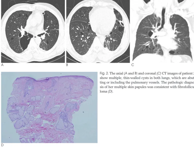
Fig. 2. The axial (A and B) and coronal (C) CT images of patient 2 show multiple, thin-walled cysts in both lungs, which are abut- ting or including the pulmonary vessels. The pathologic diagno- sis of her multiple skin papules was consistent with fibrofollicu- loma (D).
D
대한영상의학회지 2011;64:39-43
Birt-Hogg-Dube′증후군에서 나타난 자발성 기흉:
2 증례11고려대학교 의과대학 구로병원 영상의학과
2고려대학교 의과대학 구로병원 흉부외과
3고려대학교 의과대학 구로병원 병리과
배형주∙우옥희∙용환석∙강은영∙김현구2∙최영호2∙신봉경3∙김윤경
Birt-Hogg-Dube′(BHD) 증후군은 피부의 섬유모낭종, 신장종양, 다발성 폐낭종을 나타내는 드문 상염색체 우성 유전질환으로 자발성 기흉을 동반하는 경우가 있다. 이 질환은 기능이 잘 알려지지 않은 folliculin이라는 단백질을 합성하는 FLCN 유전자의 생식계열 변이 때문에 발생한다. BHD 증후군 환자들은 항상 피부, 신장, 폐의 세 가지 이상징후를 모두 나타내는 것은 아니다. 지금까지 대한민국에서 BHD 증후군의 폐 이상징후에 대한 증례보고는 없 었다. 이에 우리는 자발성 기흉으로 나타난 BHD 증후군의 두 증례를 보고하고자 한다. BHD 증후군의 폐 이상 소 견은 얇은 벽을 가진 다양한 크기와 모양의 양측성 다발성 폐낭종으로 구성되어 있었다.