820 Original Article
Korean Circulation J 2006;36:820-822
ISSN 1738-5520
ⓒ 2006, The Korean Society of Circulation CASE REPORT
A Case of Embolization Seen in Pulmonary Arteriovenous Malformation in a Patient with Osler-Rendu-Weber Syndrome
Ho-Jun Jang, MD, Min-Seok Kim, MD, Song-Yi Kim, MD, Jung-Kyu Han, MD, Yong-Jin Kim, MD, Byung-Hee Oh, MD and Hwan-Jun Jae, MD
Department of Internal Medicine, Seoul National University Hospital, Seoul, Korea ABSTRACT
Osler-Rendu-Weber Syndrome or hereditary hemorrhagic telangiectasia (HHT) is characterized by telangiecta- sia of the skin and of the mucous membranes and intermittent bleeding from vascular abnormalities; in about 20% of the patients pulmonary arteriovenous malformation is present. Pulmonary arteriovenous malformation is a congenital anomaly in the lung which causes shunting of venous blood in the pulmonary artery to systemic circulation, resulting in cyanosis, polycythemia and clubbing. Recently we experienced a case of multiple pul- monary arteriovenous malformation associated with the telangiectatic change of the cerebral artery in a 16-year- old male patient, which was confirmed by pulmonary angiography. (Korean Circulation J 2006;36:820-822) KEY WORDS:Osler-Rendu-Weber syndrome;Arteriovenous malformation;Telangiectasia, hereditary he-
morrhagic.
Introduction
Osler-Rendu-Weber syndrome or hereditary hemo- rrhagic telangiectasia is an autosomal dominant dis- ease, characterized by telangiectasia of the skin and of the mucous membranes with intermittent bleeding from vascular abnormalities. About 20% of patients dis- playing this syndrome are known to have a pulmonary arteriovenous malformation.1) Pulmonary arteriove- nous malformation is a rare congenital anomaly in the lung which causes shunting of venous blood in the pulmonary artery to systemic circulation.2) The syn- drome develops gradually without symptoms during infancy or childhood, and is usually identified inci- dentally through the 3rd and 4th decades of life. How- ever, cyanosis, polycythemia and clubbing can develop and is characterized by the size of the lesion or severity of the malformation. Massive hemoptysis and parado- xical embolism may also occur.3-5)
We present here a case of multiple pulmonary arte- riovenous malformation associated with the telangie- ctatic change of the cerebral artery in a 16-year-old
male patient with mild dyspnea on exertion, who was successfully treated with coil embolization. We also of- fer a brief review and discussion of the medical liter- ature that has described this rare condition.
Case
A 16-year-old man was admitted to our hospital complaining of mild dyspnea on exertion. He had an evaluation of the upper respiratory tract a year earlier in another hospital, including a chest X-ray, which revealed multiple nodules in both lungs. He was refe- rred to our hospital and computed tomography of the chest showed the presence of multiple arteriovenous malformations in the lung: three in right lower lobe and one in left lower lobe. He was admitted to the hospital for definite treatment after a long period of observation. There was no other significant medical or family history related to the condition.
On admission, his blood pressure was 100/70 mm Hg, he had a heart rate of 94/min, and a respiratory rate of 22/min. On a chest examination, his breathing sound was clear and his heartbeat wa regular, without murmur nor thrill. There was no tenderness across the entire abdomen, and the liver and spleen were not palpated. An extremity examination revealed clubbing on both hands. The skin was unremarkable without any telangiectatic change.
Results of laboratory tests were as follows: CBC re-
Received:November 2, 2006 Accepted:November 4, 2006
Correspondence:Yong-Jin Kim, MD,Department of Internal Medicine, Seoul National University College of Medicine, 28 Yongon-dong, Chongno- gu, Seoul 110-744, Korea
Tel: 82-2-2072-1963, Fax: 82-2-2072-2577 E-mail: [email protected]
Ho Jun Jang, et al:A Case of Embolization for Pulmonary Arteriovenous Malformation·821
vealed WBC 7400/mm3, Hb 19.0 g/dL, platelets 305,000/mm3. Urea and electrolytes, liver function test, urine and stool examination were normal. ABGA under room air showed a pH of 7.448, pCO2 31.0 mm Hg, pO2 67.6 mmHg, HCO3- 21.1 mmol/L, SaO2
95.7% and did not show improvement with the ad- ministration of 2 L/min of oxygen: pO2 66.3 mmHg, SaO2 96.3%. Gastric fiberoscopy and nasal examinat- ion were unremarkable.
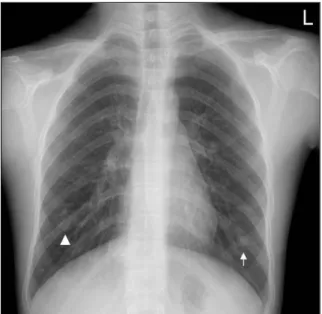
A chest X-ray demonstrated nodules in both lower lobes, with a connection to vascular marking(Fig. 1).
A computed tomography of the chest revealed three nodules in the right lower lobe, with maximal size of 3.1 cm, and one nodule in left lower lobe(Fig. 2). A brain magnetic resonance image showed a small sized enhancing lesion at the left cingulated gyrus, sugge- sting a small vascular malformation including telangie- ctasia(Fig. 3). A pulmonary artery angiography reve- aled contrast pooling lesions with early venous drain-
age in both lungs, confirming the diagnosis of pulmo- nary arteriovenous malformation(Fig. 4).
We performed a therapeutic embolization to feeding arteries with two steel coils in the left lower lobe and with a total of nine coils in theright lower lobe. There was no residual contrast pooling lesion nor draining vein seen after treatment(Fig. 5). Post-embolization ABGA was improved: pH 7.425, pCO2 42.4 mmHg, pO2 97.8 mmHg, HCO3- 27.3 mmol/L, SaO2 98.7%.
Discussion
Osler-Rendu-Weber syndrome was first described as a hereditary epistaxis by Babington in 1865. It was identified as one disease entity after descriptions by Rendu(1896), Osler(1901) and Weber(1907). In 1909, Hanes6) coined the term ‘hereditary hemorrhagic tela- ngiectasia’ in acknowledgement of the three features that defined the disorder. Goldman, et al.7) reported
Fig. 1. Chest X-ray shows 3 cm sized well demarcated lobular mass in right lower lung field (arrow head) and 1 cm sized nodule in left lower lung field (arrow).
Fig. 2. Computed tomography of chest reveals markedly enhanced lobulating masses in both lung fields.
Fig. 4. Pulmonary arteriograms reveal pulmonary arteriovenous fi- stulas in both lungs. Contrast pooling lesions and early venous drain- age patterns are noted.
Fig. 3. A 5 mm sized enhancing lesion at the left cingulate gyrus area, suggesting small vascular anomaly or malformation including telan- giectasia.
822·Korean Circulation J 2006;36:820-822
that hereditary hemorrhagic telangiectasia is associated with 30-60% of patients with pulmonary arterioven- ous malformation and appears to be a non sex-linked, autosomal dominant trait. The prevalence is about 1/8,000 to 1/100,000.2) Telangiectasia of the skin and mucous membranes causes intermittent bleeding from vascular abnormalities, such as nasal bleeding, gastro- intestinal bleeding and hematuria.3)
Pulmonary arteriovenous malformation may be con- genital or acquired. Congenital pulmonary arteriovenous malformation results from a persistent anomalous con- nection with the pulmonary artery and vein via a pri- mitive splanchnic capillary bed.5) It may be secondary to metastasis from thyroid cancer, pulmonary schistosomia- sis, advanced liver disease, mitral stenosis and trauma.
The most common symptoms include hemoptysis and dyspnea. Cyanosis, clubbing and secondary poly- cythemia may occur according to the size of the lesion and the severity of the malformation.5) Symptoms de- velop commonly after the 3rd decade of life. In here- ditary hemorrhagic telangiectasia, extrapulmonary sy- mptoms such as nasal bleeding, gastrointestinal bleed- ing and intracranial bleeding may occur.3)
A diagnosis of pulmonary arteriovenous malforma- tion is usually made by clinical manifestations, physical examination, chest X-ray, chest computed tomography and magnetic resonance imaging, contrast echocardio- graphy and perfusion lung scintigraphy. A radiolo- gical exam will reveal well demarcated round or lobu- lated masses, occasionally connected to the communi- cating vessels. Pulmonary arteriography confirms the diagnosis, showing the number, size and contour of the lesions and vascular connections.10) Magnetic re- sonance imaging is useful during follow-up.10)
Congenital pulmonary arteriovenous malformation is a progressive disorder with a high complication rate at the onset of the symptoms. Because complications such as brain abscess, hemothorax, infective endocar- ditis and paradoxical embolism can be fatal if present,
treatment is recommended whether the symptoms are present or not.5)
Treatment consists of surgical resection and non- surgical intervention by therapeutic embolization th- rough selective pulmonary artery catheterization. Sur- gical resection, the only treatment option available up to the 1970s, is limited to the patient with multiple lesions, pulmonary hypertension and severe obstru- ctive lung disease. Therapeutic embolization, first in- troduced by Taylor and colleagues9) in 1978, is effec- tive for those complicated patients and yields a better result and lower complication rate. Surgical treatment should be reserved considering potential risks of the operation and decrease of the lung function.8) While balloon and steel coils can be used for embolization, the use of a steel coil has advantages of easy insertion and permanent occlusion.5) The most common complica- tion of therapeutic embolization is the pulmonary in- farction syndrome.
Prognosis is related to the association of the syn- drome with cyanosis, hereditary hemorrhagic telangie- ctasia and complications. A good prognosis is seen fo- llowing either surgical or non-surgical treatment.1)
Our patient was asymptomatic except for mild dys- pnea on exertion and had no familial history of the syn- drome. However, the presence of multiple pulmonary arteriovenous malformation in association with cerebral arteriovenous malformation confirmed the diagnosis of Osler-Rendu-Weber syndrome. After therapeutic coil embolization, there was no residual contrast pooling le- sions nor draining vessels with a subsequent improve- ment of ABGA, suggesting a good prognosis.
REFERENCES
1) Baum GL, Wolinsky E. Textbook of Pulmonary Disease. 7th ed.
Boston: Lippincott Williams & Wilkins; 2004. p.1405.
2) Murray JF, Nadel JA. Textbook of Respiratory Medicine. 3th ed.
New York: W.B. Saunders; 2000. p.1713.
3) Brewis RA, Corrin B, Geddes DM, Gibson GJ. Respiratory Me- dicine, 2nd ed. Philadelphia: W. B. Saunder;1995. p.1526.
4) Hatfield DR, Fried AM. Therapehtic embolization of diffuse pul- monary arteriovenous malformations. AJR Am J Roentgenol 1981;137:861-3.
5) Anderson RH, Macartney FJ, Shinebourne EA, Tynan M. Pedi- atric Cardiology. 2nd ed. London: Churchill Livingstone; 2002.
p.1472.
6) Hanes FM. Multiple hereditary telangiectases causing hemo- rrhage (hereditary hemorrhagic telangiectasia). Bull Johns Ho- pkins Hosp 1909;20:63-73.
7) Goldman A. Arteriovenous fistula of the lung: its hereditary and clinical aspect. Am Rev Tuberc 1948;57:266.
8) Kim TH, Moon CI, Choi JW, et al. Congenital aortopulmonary fistula presenting as and exertional dyspnea. Korean Circ J 2000;
30:1291-4.
9) Terry PB, Barth KH, Kaufman SL, White RI Jr. Balloon embo- lization for treatment of pulmonary arteriovenous fistulas. N Engl J Med 1980;302:1189-90.
10) Dines DE, Seward JB, Bernatz PE, Gomes MR. Pulmonary ar- teriovenous fistula. Mayo Clin Proc 1974;49:460-5.
Fig. 5. Post-embolization with steel colis. Angiogram shows good em- bolization of pulmonary arteriovenous malformation without resi- dual contrast pooling lesions.