• 검색 결과가 없습니다.

Birt-Hogg-Dubé Syndrome Manifesting as Spontaneous Pneumothorax: A Novel Mutation of the Folliculin Gene

N/A
N/A
Protected

Academic year: 2021

Share "Birt-Hogg-Dubé Syndrome Manifesting as Spontaneous Pneumothorax: A Novel Mutation of the Folliculin Gene"

Copied!
5
0
0

로드 중.... (전체 텍스트 보기)

전체 글

(1)

Received: October 24, 2016, Revised: November 14, 2016, Accepted: November 14, 2016, Published online: October 5, 2017

Corresponding author: Seok Whan Moon, Department of Thoracic and Cardiovascular Surgery, Seoul St. Mary’s Hospital, College of Medicine, The Catholic University of Korea, 222 Banpo-daero, Seocho-gu, Seoul 06591, Korea

(Tel) 82-2-2258-6320 (Fax) 82-2-594-8644 (E-mail) [email protected]

© The Korean Society for Thoracic and Cardiovascular Surgery. 2017. All right reserved.

This is an open access article distributed under the terms of the Creative Commons Attribution Non-Commercial License (http://creativecommons.org/

licenses/by-nc/4.0) which permits unrestricted non-commercial use, distribution, and reproduction in any medium, provided the original work is properly cited.

Seoul St. Mary’s Hospital, College of Medicine, The Catholic University of Korea

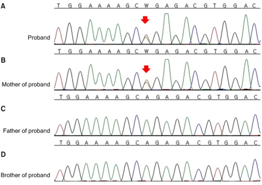
Birt-Hogg-Dubé syndrome (BHDS) is a rare disease with autosomal dominant inheritance that manifests through skin tumors, pulmonary cystic lesions, and renal tumors. A mutation of FLCN located on chromo- some 17p11.2, which encodes a tumor-suppressor protein (folliculin), is responsible for the development of BHDS. We report the case of a patient presenting with spontaneous pneumothorax, in whom a familial ge- netic study revealed a novel nonsense mutation: p.(Arg379*) in FLCN.

Key words: 1. Birt-Hogg-Dubé syndrome 2. Pneumothorax

3. FLCN

4. Thoracoscopy

5. Video-assisted thoracic surgery

Case report

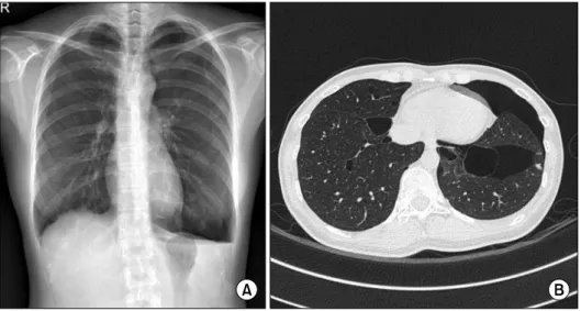
A 32-year-old female presented with an in- cidentally found pneumothorax during a workup for a vocal cord mass, after having experienced the symptom of voice change for several months. A chest X-ray and high-resolution chest tomography (HRCT) scan showed a left-side pneumothorax with a small amount of pleural effusion. In addition, several var- iably-sized thin-walled air cysts were found, mostly located on the basal or medial side of the bilateral lungs (Fig. 1). However, she had neither a history of smoking nor of pneumothorax. She had no history of underlying disease, such as an immunologic or colla- gen vascular disorder. Of note, her maternal relatives had a family history of pneumothorax. Because the multiple cysts in the bilateral lungs could increase

the risk of recurrence, surgical repair was planned.

Video-assisted thoracoscopic surgery (VATS) was

done for the single-stage resection of bilateral multi-

ple cystic lesions. A double-lumen endotracheal tube

was placed for bilateral sequential one-lung ven-

tilation under general anaesthesia. The patient was

placed in the lateral decubitus position, and 2-ports

VATS exploration was performed using a 30

o

thor-

acoscope (Karl Storz endoscope; Karl Storz, Tuttlingen,

Germany) for multiple wedge resections using

endostaplers. Both bulla plication and ligation were

also performed to remove multiple discrete small le-

sions, and sequential VATS for the contralateral side

was conducted. There were multiple large cysts, es-

pecially at the base of the lower lobes, and some

cysts were assessed as being at the stage of impend-

ing rupture (Fig. 2). After a air-leak test using saline

(2)

Fig. 1. (A) A chest X-ray showing a left-sided pneumothorax. (B) A chest computed tomography scan reveals left-sided pneumothorax with the lower lung predominant, and sev- eral variably-sized thin-walled air cysts in both lungs.

solution, 20-Fr chest tubes were placed in the bi- lateral pleural space through the scope ports.

Mechanical pleurodesis was not performed. The ex- amination of the gross specimen revealed multiple, variably-sized subpleural cysts on both lungs, and the largest one measured up to 6.5×4.6 cm.

The HRCT and operative findings were uncommon;

it is rare for young females to present with numer- ous thin-walled cysts in the bilateral lungs, especially in the lower lung fields. Furthermore, her grand- mother had a history of pneumothorax at a relatively young age. After obtaining an informed consent for a genetic analysis with the suspicion of Birt-Hogg-Dubé syndrome (BHDS), genomic DNA was isolated from peripheral blood leukocytes using the QIAmp DNA Mini Kit (Qiagen, Hamburg, Germany). Polymerase chain reaction (PCR) was carried out using gene-spe- cific primers for all coding exons and splice junctions of FLCN as previously described [1]. PCR amplicons were bidirectionally sequenced using a BigDye Terminator version 3.1 cycle sequencing kit (Applied Biosystems, Foster City, CA, USA) on an ABI PRISM 3100 Genetic Analyzer (Applied Biosystems). The chromatograms were analyzed using Sequencher ver.

5.0 (Gene Codes, Ann Arbor, MI, USA) and all muta- tions were described according to the Human Genome Variation Society nomenclature [2]. The RefSeq ID NM_144997.5 was used for alignment. Direct se- quencing of the PCR products revealed a hetero- zygous, novel nonsense mutation, c.1135A>T; p.Arg379*

in exon 10 of FLCN. This mutation has not been pre- viously reported and is absent from locus-specific da-

tabases [3,4] as well as public databases (HGMD;

ExAC; 1000 Genomes; dbSNP; and the ethnically spe- cific Korean Reference Genome database, KRGDB). A subsequent familial genetic study was performed in both parents and an older brother of the proband, revealing an identical heterozygous mutation in the proband’s mother (Fig. 3).

The bilateral chest tubes were removed on post- operative day 1, and the patient was discharged on postoperative day 2 with complete resolution of the pneumothorax. The vocal cord mass was excised a month later, and was confirmed as a glomus tumor, measuring 2.0×1.2 cm. The patient has been followed for 12 months after the operation, and neither re- currence of pneumothorax nor other symptoms have been noted.

Discussion

BHDS is a rare inherited autosomal dominant sys-

temic disorder, manifesting through skin lesions, pul-

monary cystic lesions, and renal tumors. The Euro-

pean BHD consortium reported BHD patients should

fulfill one major or two minor criteria (major cri-

teria: (1) at least five fibrofolliculomas or trichodisco-

mas, at least one histologically confirmed, of adult

onset; (2) pathogenic FLCN germline mutation; minor

criteria: (1) bilateral basally located multiple lung

cysts with no other apparent cause, with or without

spontaneous primary pneumothorax; (2) early-onset

[<50 years] or multifocal or bilateral renal cancer, or

renal cancer of mixed chromophobe and oncocytic

(3)

Fig. 2. (A−F) Thoracoscopic findings showing wedge resection and ligation of multiple cystic lesions. RUL, right upper lobe; LUL, left up- per lobe; LLL, left lower lobe; RLL, right lower lobe.

histology; (3) first-degree relative with BHD). The precise prevalence of BHDS remains unknown due to its considerable heterogeneity in terms of clinical symptoms, even within a family entity, and many pa- tients with BHDS are underdiagnosed [5]. The skin-related findings, a triad of fibrofolliculomas, tri- chodiscomas, and acrochordons, usually develop in the third to fourth decades of life. Generally, the scalp, face, forehead, or neck is involved, with the manifestations growing in number and size, but the skin lesions alone are not diagnostic [6]. Toro et al.

[7] first described the relationship between cystic le-

sions and pneumothorax, presenting an incidence

rate of 33%–38%. Pulmonary cystic lesions have

been reported in 77%–89% of BHDS patients, and

usually manifest as multiple mostly well-defined,

air-filled cysts varying in size and shape, from round

to oval, lentiform, and often with a multiseptated ap-

pearance in the largest cyst. The large cysts tend to

be located in the lower lobes of the bilateral lungs,

and are asymptomatic, but pose a risk of sponta-

neous pneumothorax. The differential diagnoses in-

clude an unusual manifestation of bullous emphyse-

ma, lymphangioleiomyomatosis (LAM), pulmonary

(4)

Fig. 3. Direct sequencing analysis of FLCN. (A) The proband was het- erozygous for a nonsense mutation, c.1135A >T, p.Arg379*. (B) The same base change was also identified in her mother, but not in the pro- band’s father (C) or brother (D).

The base substitution mutation is indicated by the red arrows.

Langerhans cell histiocytosis (LCH), and lymphocytic interstitial pneumonia. LAM shows small to medium cysts in comparison with BHDS, and LCH demon- strates irregularly shaped cysts, including nodules, predominant in the upper lung [8]. The risk of kid- ney tumors is high, and approximately 15%–25% of patients develop multiple, bilateral, slowly-growing tumors that are diagnosed at the median age of 48 years old. In some family pedigrees, either kidney tu- mors or spontaneous pneumothorax can be found without skin manifestations [9].

The mutation of the folliculin gene (FLCN), which encodes a tumor suppressor protein and is located on the short arm of chromosome 17 (17p11.2), is re- sponsible for BHDS with an autosomal dominant pat- tern, and was first identified in 2001. It is known that this gene may play a key role in pulmonary cystic lesions, and that it is expressed in stromal cells and pneumocytes. The mutation is detected in about 88% of patients using direct sequencing of the whole coding exon. However, cases have been re- ported with genetic deletions or duplications that were not detected by direct sequencing chromosome analysis in approximately 3%–5% of patients [10].

Numerous mutations have been described, and dal Sasso et al. [6] reported that up to 142 unique DNA mutations have been implicated. The 2 most common pathogenic variants are c.1285dupC and c.1285delC,

and all pathogenic variants predict protein truncation [11], as is the case in this report of a novel non- sense mutation. In accordance with the tumor sup- pressor role of folliculin, the fact that all germline pathogenetic variants lead to protein truncation sug- gests that the loss of the folliculin protein is the ba- sis of tumor formation in BHDS [12]. In Korea, few reports have been published about the mutations found in pneumothorax patients with BHDS [10,13,14].

In our case, we found a novel nonsense mutation, p. (Arg379*) in exon 10, as the cause of BHDS in a young female patient with multiple, bilateral pulmo- nary cystic lesions, and the mother of the proband showed the same mutation. In order to detect the re- currence of pneumothorax and renal malignancies, radiologic surveillance of the chest and abdomen should be performed in family members, as well as in the patient with BHDS. In conclusion, we report a surgically managed, rare case of BHDS that presented with incidentally found pneumothorax, and a familial genetic study that revealed a novel nonsense mutation.

Conflict of interest

No potential conflict of interest relevant to this ar-

ticle was reported.

(5)

database (LSDB) for mutations in the folliculin (FLCN) gene. Hum Mutat 2010;31:E1043-51.

4. Wei MH, Blake PW, Shevchenko J, Toro JR. The folliculin mutation database: an online database of mutations asso- ciated with Birt-Hogg-Dube syndrome. Hum Mutat 2009;

30:E880-90.

5. Menko FH, van Steensel MA, Giraud S, et al. Birt-Hogg- Dube syndrome: diagnosis and management. Lancet Oncol 2009;10:1199-206.

6. Dal Sasso AA, Belem LC, Zanetti G, et al. Birt-Hogg-Dube syndrome: state-of-the-art review with emphasis on pul- monary involvement. Respir Med 2015;109:289-96.

7. Toro JR, Pautler SE, Stewart L, et al. Lung cysts, sponta- neous pneumothorax, and genetic associations in 89 fami- lies with Birt-Hogg-Dube syndrome. Am J Respir Crit Care Med 2007;175:1044-53.

2012;499:339-42.

11. Schmidt LS, Nickerson ML, Warren MB, et al. Germline BHD-mutation spectrum and phenotype analysis of a large cohort of families with Birt-Hogg-Dube syndrome. Am J Hum Genet 2005;76:1023-33.

12. Vocke CD, Yang Y, Pavlovich CP, et al. High frequency of somatic frameshift BHD gene mutations in Birt-Hogg- Dube-associated renal tumors. J Natl Cancer Inst 2005;

97:931-5.

13. Kim EH, Jeong SY, Kim HJ, Kim YC. A case of Birt- Hogg-Dube syndrome. J Korean Med Sci 2008;23:332-5.

14. Park G, Seo HJ, Jang SJ, Shin BS, Hong R, Lee SK. A case of

primary spontaneous pneumothorax with a three nucleo-

tide deletion mutation of the FLCN gene. Korean J Thorac

Cardiovasc Surg 2010;43:824-8.

수치

Fig. 1. (A) A chest X-ray showing a  left-sided pneumothorax. (B) A chest computed tomography scan reveals  left-sided pneumothorax with the  lower lung predominant, and  sev-eral variably-sized thin-walled air  cysts in both lungs.
Fig. 2. (A−F) Thoracoscopic findings showing wedge resection and ligation of multiple cystic lesions
Fig. 3. Direct sequencing analysis  of FLCN. (A) The proband was  het-erozygous for a nonsense mutation,  c.1135A >T, p.Arg379*

참조

관련 문서