Basic data on the hematology, serum biochemistry, urology, and organ weights of beagle dogs
So-Young Choi
1, Jae-Sik Hwang
2, Ill-Hwa Kim
1, Dae-Yeon Hwang
3, Hyun-Gu Kang
1*
1
Veterinary Medical Center, Chungbuk National University, Cheongju, Korea
2
Biotoxtech Co., Ltd., Ochang, Korea
3
Department of Biomaterials Science, College of Natural Resources & Life Science, Pusan National University, Miryang, Korea
This study was conducted to provide basic data on physiological and hematological characteristics, and organ weights of beagle dogs. A total of 237 beagle dogs were used to determine differences in physiological and hematological parameters, and organ weights depending on sex and age. The respiratory rate of both sexes tended to increase as they grew older and the female heart rate was slightly higher than that of males. Male and female body weights increased rapidly to 33 weeks old followed by a gradual increase to 41-weeks-old. The relative weight of the brain was negatively correlated with body weight, whereas the weight of reproductive organs was positively correlated with body weight. The platelet count of female dogs was slightly higher than that of males. The red blood cell, hemoglobin, and hematocrit of both sexes increased non-significantly with age. In the leukocyte differential count, the neutrophils, and eosinophils of both sexes tended to increase as they grew older, whereas basophils, lymphocytes, and monocytes decreased. In the serum biochemical profiles, alkaline phosphatase was slightly higher in males than females, while the total cholesterol of female dogs at 9-months-old was higher than that of males at the same age. Other biochemical components, including alanine aminotransferase, blood urea nitrogen, creatinine, triglyceride, and total protein increased non- significantly with age in both sexes. To conclude, we observe no significant physiological or hematological differences with sex or age, although decreasing and increasing trends were detected with some parameters. These data provide valuable reference indices of the normal physiological and hematological characteristics of beagle dogs, which should prove useful in toxicological and pharmacological studies.
Key words: Physiology, hematology, serum biochemistry, age, organ weight, beagle dog
Received 19 August 2011; Revised version received 15 November 2011; Accepted 28 November 2011
The drug development process is typically divided into three major steps: discovery, preclinical development, and clinical trials. The preclinical development of a new drug generally includes studies of efficacy, pharmacology, and experimental toxicology to define the dose, route, and frequency to be investigated in subsequent studies. Efficacy studies demonstrate that treatment with drug candidates has the desired therapeutic effect. Efficacy studies also help to identify the best drug candidates for further development [1].
The regulatory requirements of international guidelines
generally demand toxicological and pharmacological studies in at least two laboratory animal species, a rodent and a nonrodent [1]. Beagle dogs are frequently used as model non-rodents for studying the efficacy and safety of newly developed drugs and chemicals, because of similarities in their anatomical morphology and physiology compared to humans [2]. In biomedical research, it is well recognized that individual animal variability can affect study results. To minimize animal variability, researchers should be provided with normal, healthy, well-adapted animals that represent a http://dx.doi.org/10.5625/lar.2011.27.4.283
*Corresponding author: Hyun-Gu Kang, Veterinary Medical Center, Chungbuk National University, 52 Naesudongro (Gaesin-dong), Heungdeok-gu, Cheongju, Chungbuk 361-763, Korea
Tel: +82-43-261-3359; Fax: +82-43-261-3224; E-mail: [email protected]
This is an Open Access article distributed under the terms of the Creative Commons Attribution Non-Commercial License (http://creativecommons.org/licenses/
by-nc/3.0) which permits unrestricted non-commercial use, distribution, and reproduction in any medium, provided the original work is properly cited.
physiological model characterized by a narrow range of physical, behavioral, and clinical parameters [3-5]. Basic data of the test animal's blood, urine, and blood chemical values have detected many differences depending on sex and age, and the conditions where animals are maintained [7-9]. Thus, animals may be genetically identical but environmental effects during development cannot be ignored and these differences mean that is essential to establish basic data [10]. The establishment of standard basic data is important in the biosciences, because it can reduce and refine the use of laboratory animals [11].
Baseline parameters for hematology, urine, and blood chemical values play an important role in estimating target organ effects when it is difficult to observe the changes in tissues or when they are likely to be ignored during histopathological examinations [3-5]. These basic data can provide a foundation for interpreting certain test results more accurately [10,12]. Thus, it is necessary to acquire different types of basic data for laboratory animals to facilitate the reliable interpretation of a broad range of research studies using model animals. There have been some studies to acquire basic data for laboratory animals [10,12,13] but the volume of data is not great, so many tests are conducted based on foreign basic data [13,14].
The current study aimed to provide basic data on physiological and hematological characteristics, and organ weights for beagle dogs. This study analyzed whether there were differences in physiological and hematological parameters, and the organ weights of male and female beagle dogs. Thus, basic data was generated that should prove valuable to research in fields relevant to basic medical sciences where basic data are required to evaluate tests on dogs.
Materials and Methods
Animal husbandry and maintenance
Male and female beagle dogs (5 months old) were obtained from Samtako (Beijing Marshall Biotechnology, Beijing, China)
and housed in stainless net cages (80W×90L×65H cm) or a fenced room (130W×372L×125H cm) (1 dog per cage).
Quarantine and acclimatization periods were 10 days. The animals were housed in a room maintained at a temperature (23±3
oC) and a relative humidity (55±10%). The light and dark cycle was 12:12 h (light from 08:00 to 20:00). The temperature and humidity of the housing rooms was measured every hour and no changes were detected that could be considered to have any effect on the tests. The study evaluated 237 beagles including 148 (male 74, female 74) aged 6 months, 52 (male 27, female 25) aged 7 months, and 37 (male 20, female 17) aged 9 months. The 7-month-old and 9-month- old beagles were used as a control group in the toxicity test.
The number of beagle dogs in each evaluation class is shown in Table 1.
Food and water intakes
Dry food for dogs (Oriental Yeast, Tokyo, Japan) was provided at a rate of 300 g per day, so no food-related factors could have affected the study. Water was sterilized and disinfected using a membrane filter with an UV ray flowing-water sterilizer, and provided ad libitum. Water examination was conducted by the Research Institute of Public Health and Environment (Daejeon, Korea) twice each year. No pollutants were detected in the water that could have influenced the evaluations conducted in this study.
Blood collection
Dogs were fasted overnight prior to blood collection. Blood samples were collected from the cephalic veins using a syringe with a 21- or 22-gauge needle. The blood samples were collected in CBC bottles containing EDTA-2K (Green Cross Medical Industry, Yongin, Korea) and used for hematological examination. For the serum biochemical evaluation, 3 mL blood samples were centrifuged at 3,000 rpm for 10 min within 1 h of collection. Sera were stored at −80
oC in a freezer prior to analysis. All processes used in the animal testing, including blood-gathering and collection, were reviewed by Table 1. Number of beagle dogs in each evaluation class
Evaluation parameter
Number of beagle dogs
6 months old 7 months old 9 months old
Male Female Male Female Male Female
Physical examination 55 56 - - 21 18
Body weight gain 1280 1280 56 55 42 20
Organ weights 13 13 10 10 15 15
Hematology 64 63 17 17 12 12
Serum biochemistry 74 74 27 25 20 17
Urinalysis 09 09 09 08 06 05
the Institutional Animal Care and Use Committees before they were conducted. Blood collection was conducted after the animals adapted to their feeding conditions.
Hematology
Blood samples were analyzed within 20 min using automatic blood analyzers (T-540 Coulter Counter, Coulter Electronics, Hialeah, FL, USA; ADVIA120 Hematology System, Bayer Healthcare LLC, Tarrytown, NY, USA). The following parameters were determined: white blood cell (WBC) count, differential leukocyte count, red blood cell (RBC) count, hemoglobin concentration, hematocrit, platelet count, mean corpuscular volume, mean corpuscular hemoglobin, and mean corpuscular hemoglobin concentration.
Serum biochemistry
The following serum biochemical parameters were evaluated using an automatic analyzer (Shimadzu CL-7200, Shimadzu, Kyoto, Japan): alkaline phosphatase (ALP), alanine aminotransferase (ALT), aspartate aminotransferase (AST), total bilirubin (TB), total cholesterol (TC), triglycerides (TG), phospholipids, total protein (TP), albumin, albumin/globulin (A/G) ratio, glucose, blood urea nitrogen (BUN), creatinine, creatine phosphokinase (CPK), calcium, and inorganic phosphorus. Sodium, potassium, and chloride were measured using an automatic electrolyte analyzer (Ciba-Corning 644 Na/K/Cl Analyzer, Ciba-Corning, Medfield, MA, USA).
Statistical analysis
Data processing and statistical analysis were performed using SAS version 9.2 (SAS Institute Inc., Cary, NC, USA). Values determined for different sexes and ages were compared using two-way ANOVA (SAS Version 9.2). A one-way ANOVA test (SAS Version 9.2) was applied at each time interval to compare the reproductive organ weight within each sex. Duncan’s multiple-comparison test was used to assess the statistical significance of differences among the study groups. A level of P<0.05 was accepted as statistically significant.
Results
Physiological values
A significant increase in the body temperature was observed in male dogs (P<0.001). The mean body temperature in 6-month-old male dogs was 38.3±0.4
oC, whereas that in 9-month-old male dogs was 39.0±0.3
oC. In both sexes, the respiratory rate increased significantly with age (P<0.001), while the heart rate of females was slightly higher than that of males (Table 2).
Body weight gain and organ weights
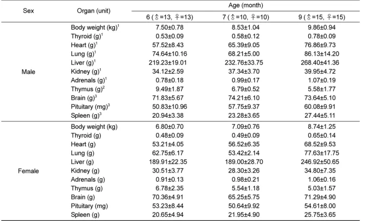
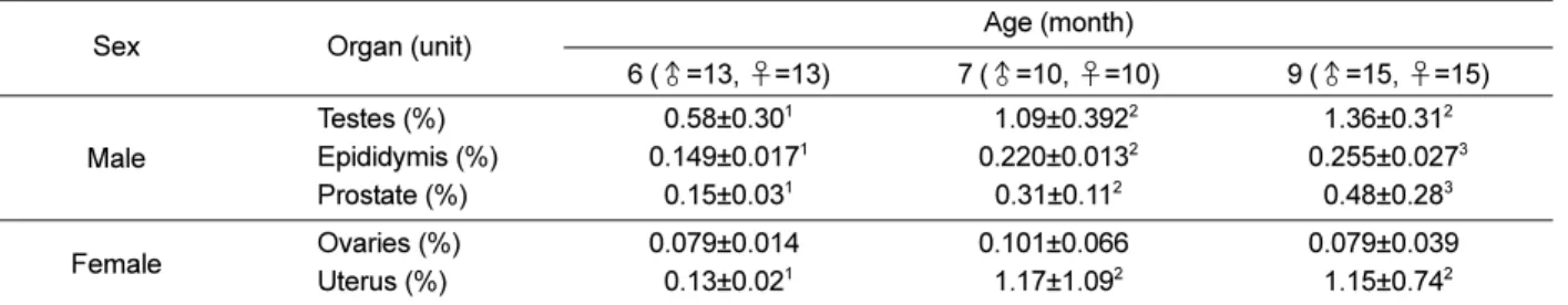
The body weight of male and females increased rapidly up to 33 weeks (0.18±0.07 kg/week for male dogs, 0.14±0.06 kg/week for female dogs), before increasing gradually up to 41 weeks (0.14±0.06 kg/week for males, 0.11±0.08 kg/week for females). The body weight of males was significantly higher than that of females (Figure 1, P<0.001). The absolute and relative weights of the thyroid, heart, lung, liver, kidney, and adrenals increased significantly as both sexes grew older, whereas male organs weighed significantly more than those of females (Table 3, P<0.001). The thymus gradually decreased in weight as both sexes grew older (P<0.001). Brain, pituitary, and spleen weights increased significantly with age (Tables 3 and 5, P<0.001). In absolute and relative terms, the reproductive organ weight of males and females, including the epididymis, prostate, and uterus, increased significantly as they grew older (P<0.05), whereas the weight of the ovaries did not increase significantly with age (Table 4). The absolute weight of testes increased significantly from 6 to 9 months, while their relative weight increased significantly from 6 to 7 months (P<0.05).
Table 2. Body temperature, pulse rate, and respiratory rate of male and female dogs
Sex Items
Age (month) 6
(♂=55, ♀=56) 9 (♂=21, ♀=18) Male
Body temperature (
oC)
138.3±0.40 39.0±0.30 Heart rate (/min) 83.4±15.2 97.5±15.6 Respiratory rate (/min)
120.7±9.20 26.2±5.80
Female
Body temperature (
oC) 38.4±0.50 38.6±0.50 Heart rate (/min) 130.2±21.80 121.8±16.70 Respiratory rate (/min)
121.4±8.30 25.2±4.60
1
Indicates that the body temperature and respiratory rate increased significantly with age (P<0.001).
Figure 1. Changes in body weights of male and female dogs.
Hematological parameters
The results of hematological examination are shown in Tables 7 and 8. The platelet count of female dogs was slightly higher than that of males. The RBC, hemoglobin, and hematocrit of both sexes increased non-significantly with age. In the leukocyte differential count, the neutrophils and eosinophils of both sexes tended to increase with age, whereas basophils, lymphocytes, and monocytes decreased.
Biochemical parameters
The results of the serum biochemical profiles are shown in Table 9. The ALP of male beagles was slightly higher than that of females, while the TC in female dogs aged 9 months
was higher than that of males at the same age. The remaining biochemical parameters, including, ALT, BUN, creatinine, TG, and TP, increased non-significantly with age in both sexes.
Urological parameters
The amount of urine and sodium ions tended to increase as dogs grew older. However, there were large individual differences (Table 10).
Discussion
Beagle dogs are the most frequently used nonrodent animals in biomedical research, because of similarities in their Table 3. Changes in the absolute organ weights of male and female dogs
Sex Organ (unit) Age (month)
6 (♂=13, ♀=13) 7 (♂=10, ♀=10) 9 (♂=15, ♀=15)
Male
Body weight (kg)
17.50±0.78 8.53±1.04 9.86±0.94
Thyroid (g)
10.53±0.09 0.58±0.12 0.78±0.09
Heart (g)
157.52±8.430 65.39±9.050 76.86±9.730
Lung (g)
174.64±10.16 68.21±5.000 86.13±14.20
Liver (g)
1219.23±19.010 232.76±33.750 268.40±41.360
Kidney (g)
134.12±2.590 37.34±3.700 39.95±4.720
Adrenals (g)
10.78±0.18 0.99±0.17 1.07±0.19
Thymus (g)
29.49±1.87 6.79±0.52 5.58±1.77
Brain (g)
371.83±5.670 74.21±6.100 73.64±5.100
Pituitary (mg)
350.83±10.96 57.75±9.370 60.08±9.910
Spleen (g)
320.94±3.380 23.28±3.650 27.44±5.110
Female
Body weight (kg) 6.80±0.70 7.09±0.76 8.74±1.25
Thyroid (g) 0.48±0.09 0.49±0.09 0.65±0.14
Heart (g) 53.21±4.050 56.52±6.350 68.52±9.530
Lung (g) 62.75±6.170 53.42±2.140 77.63±17.75
Liver (g) 189.91±22.350 189.00±28.700 246.92±50.650
Kidney (g) 30.51±3.770 28.30±3.260 34.80±7.350
Adrenals (g) 0.91±0.13 0.98±0.21 1.06±0.16
Thymus (g) 6.78±2.35 5.54±1.18 5.03±1.57
Brain (g) 70.36±4.910 65.25±5.750 71.29±4.900
Pituitary (mg) 53.23±8.440 50.64±9.920 54.61±8.000
Spleen (g) 20.65±4.940 21.95±4.900 25.75±3.650
1
Indicates that the body and organ weights increased significantly with age and there were significant differences between sexes (P<0.001).
2Indicates that organ weights decreased significantly with age and there were significant differences between sexes (P<0.001).
3Indicates that organ weights increased significantly with age (P<0.02).
Table 4. Changes in absolute reproductive organ weights of male and female dogs
Sex Organ (unit) Age (month)
6 (♂=13, ♀=13) 7 (♂=10, ♀=10) 9 (♂=15, ♀=15) Male
Testes (g) 4.39±2.60
19.48±3.85
213.21±2.94
30
Epididymis (g) 1.11±0.14
11.87±0.21
22.50±0.36
3Prostate (g) 1.08±0.28
12.74±1.02
24.79±2.94
3Female Ovaries (g) 0.52±0.08
00.70±0.44
00.88±0.72
0Uterus (g) 0.78±0.16
17.99±6.96
211.39±7.64
30
1,2,3
Values with different superscripts in the same row were significantly different (P<0.05).
anatomical morphology and physiology to humans [2]. In biomedical research, it is well recognized that individual animal variability can affect study results. Therefore, highly defined dog models should be used in biomedical studies [3]. In clinical or non-clinical tests, the normal range of blood constituent is a standard means of diagnosis when identifying diseases or abnormalities. Thus, they are very important values, although they can vary significantly depending on the individual environment (e.g., species, sex, age, and conditions) or genetic characteristics [13,15-17]. This means it is essential to reduce errors by determining the normal range as accurately as possible to facilitate the exact diagnosis of diseases or confirming abnormalities. The current study was conducted to determine
basic physiological data on the body weight, organ weight, body temperature, heart rate, and hematological, biochemical and urological parameters for male and female beagle dogs.
In the physical examination of male and female beagle dogs aged 6 and 9 months, the body temperature and respiratory rate did not differ significantly between the sexes, although the respiratory rate of both sexes tended to increase as they grew older, while the heart rate of females was slightly higher than that of males (Table 2). Ferasin et al. [18] reported that dogs aged less than 1 year appeared to have a significantly higher heart rate than older dogs, while the heart rate of healthy dogs undergoing routine clinical examination was related to their age.
Table 5. Changes in the relative organ weights of male and female dogs
Sex Organ (unit) Age (month)
6 (♂=13, ♀=13) 7 (♂=10, ♀=10) 9 (♂=15, ♀=15)
Male
Body weight (kg)
17.50±0.78 8.53±1.04 9.86±0.94
Thyroid (%)
10.071±0.008 0.068±0.014 0.080±0.015
Heart (%)
10.77±0.06 0.77±0.09 0.78±0.09
Lung (%)
11.02±0.12 0.81±0.11 0.85±0.15
Liver (%)
12.95±0.36 2.74±0.33 2.73±0.41
Kidney (%)
10.46±0.05 0.44±0.03 0.41±0.06
Adrenals (%)
10.104±0.020 0.116±0.019 0.110±0.020
Thymus (%)
20.127±0.017 0.080±0.013 0.056±0.001
Brain (%)
30.97±0.11 0.88±0.12 0.75±0.09
Pituitary (%)
30.007±0.001 0.007±0.001 0.006±0.001
Spleen (%)
30.28±0.04 0.27±0.04 0.28±0.04
Female
Body weight (kg) 6.80±0.70 7.09±0.76 8.74±1.25
Thyroid (%) 0.071±0.013 0.070±0.012 0.075±0.013
Heart (%) 0.79±0.07 0.80±0.08 0.79±0.13
Lung (%) 0.94±0.09 0.76±0.06 0.86±0.08
Liver (%) 2.81±0.39 2.68±0.45 2.85±0.66
Kidney (%) 0.43±0.03 0.40±0.04 0.40±0.06
Adrenals (%) 0.135±0.025 0.141±0.038 0.125±0.029
Thymus (%) 0.100±0.026 0.078±0.018 0.058±0.025
Brain (%) 1.04±0.13 0.93±0.08 0.83±0.10
Pituitary (%) 0.008±0.002 0.007±0.001 0.006±0.001
Spleen (%) 0.31±0.08 0.31±0.07 0.30±0.05
1
Indicates that body and organ weights increased significantly with age and there were significant differences between sexes (P<0.001).
2Indicates that organ weights decreased significantly with age and there were significant differences between sexes (P<0.001).
3Indicates that organ weights increased significantly with age (P<0.02).
Table 6. Changes in the relative reproductive organ weights of male and female dogs
Sex Organ (unit) Age (month)
6 (♂=13, ♀=13) 7 (♂=10, ♀=10) 9 (♂=15, ♀=15) Male
Testes (%) 0.58±0.30
101.09±0.392
21.36±0.31
2Epididymis (%) 0.149±0.017
10.220±0.013
20.255±0.027
3Prostate (%) 0.15±0.03
10.31±0.11
20.48±0.28
3Female Ovaries (%) 0.079±0.014
00.101±0.066
00.079±0.039
0Uterus (%) 0.13±0.02
11.17±1.09
21.15±0.74
21,2,3
Values with different superscripts in the same row were significantly different (P<0.05).
The body weight of male and female dogs increased rapidly up to 33 weeks, followed by a gradual increase to 41 weeks.
The body weight was significantly different between the sexes (Figure 1). Their body weight increased by 0.18±0.07 kg/
week in male dogs and by 0.14±0.06 kg/week in female dogs. Nunamaker et al. [19] reported that the body weight increased by 19.7 g/day in a control group of dogs. The relative organ weights were similar to those reported for a control group in a previous report [20]. The relative reproductive organ weights of both sexes increased rapidly up to 7 months (Table 6). These results indicate that puberty may be reached at 7 months of age.
Age-related differences were found in terms of RBC count, hemoglobin, hematocrit, neutrophils, eosinophils, basophils, lymphocytes, monocytes, ALT, BUN, creatinine, TG, and TP.
Sex-related differences were found for ALP and TC (Tables 7, 8, and 9). However, these parameters were not significantly related to age or sex. These results were slightly different to those reported by Jeong et al. [12]. The number of eosinophils was higher than that reported by Jeong et al [12], but similar to that reported by Wolford et al. [21]. The number of lymphocyte was also higher than that reported by Jeong et al. [12] and Wolford et al. [21]. This was considered to be a consequence of transportation stress, but it may also be Table 7. Hematological parameters of male and female dogs
Sex Parameter Age (month)
6 (♂=64, ♀=63) 7 (♂=17, ♀=17) 9 (♂=12, ♀=12)
Male
White blood cell (10
3/mm
3) 9.29±2.14 9.15±1.77 9.01±1.27
Red blood cell (10
6/mm
3)
16.63±0.53 6.91±0.63 7.29±0.94
Hemoglobin (g/dL)
214.9±1.20 15.5±1.50 16.5±2.10
Hematocrit (%)
242.8±3.4 0 44.7±4.20 46.7±6.40
Platelet (10
3/mm
3)
3327.3±59.30 314.8±61.70 294.3±66.70
Mean corpuscular volume (fL) 64.6±2.20 64.7±2.40 64.1±2.80
Mean corpuscular hemoglobin (pg) 22.4±0.60 22.5±0.80 22.6±0.70
Mean corpuscular hemoglobin concentration (g/dL) 34.6±1.40 34.7±1.30 35.4±1.80
Female
White blood cell (10
3/mm
3) 8.43±1.47 8.87±1.42 9.18±2.66
Red blood cell (10
6/mm
3) 6.79±0.52 6.91±0.63 7.21±0.58
Hemoglobin (g/dL) 15.4±1.10 16.3±1.20 16.8±1.20
Hematocrit (%) 44.4±3.50 44.7±4.20 47.2±4.40
Platelet (10
3/mm
3) 332.5± 58.3 319.8±45.10 304.4±51.50
Mean corpuscular volume (fL) 65.3±2.40 64.7±2.40 65.5±4.00
Mean corpuscular hemoglobin (pg) 22.5±0.70 22.9±0.80 23.3±0.80
Mean corpuscular hemoglobin concentration (g/dL) 34.5±1.3 0 34.7±1.30 35.6±1.50
1
Indicates that the parameter increased significantly with age in both sexes (P<0.0001).
2Indicates that the parameter increased significantly with age, while the parameter was significantly higher in females compared with males.
3Indicates that the parameter decreased significantly with age.
Table 8. Differential leukocyte counts of male and female dogs
Sex Parameter Age (month)
6 (♂=64, ♀=63) 7 (♂=17, ♀=17) 9 (♂=12, ♀=12)
Male
Neutrophils (%)
152.2±6.7 54.4±8.00 55.6±4.9 0
Lymphocytes (%)
235.1±5.9 34.8±7.40 33.4±3.10
Eosinophils (%)
305.0±2.1 5.1±2.1 5.3±2.7
Monocytes (%)
305.2±1.3 5.1±0.8 4.5±1.1
Basophils (%)
401.1±0.6 0.7±0.4 0.8±0.7
Female
Neutrophils (%) 53.8±5.3 54.2±4.40 58.3±4.80
Lymphocytes (%) 36.3±5.0 35.4±4.90 31.3±3.80
Eosinophils (%) 03.9±1.9 3.9±1.9 5.1±3.8
Monocytes (%) 04.2±0.9 4.3±1.1 4.0±1.1
Basophils (%) 01.2±0.7 0.8±0.6 0.9±0.7
1