246
책임저자 : 박정수, 서울시 서대문구 성산로 250
120-752, 연세대학교 의과대학 외과학교실 Tel: 02-2228-2100, Fax: 02-313-8289
E-mail: [email protected] 게재승인일:2007년 11월 30일
수질암과 유두암이 공존하는 갑상선암
연세대학교 의과대학 1외과학교실 및 2내분비 연구소, 3관동대학교 제일병원 외과
이학민1ㆍ이용상1,2ㆍ이해경3ㆍ윤지섭1,2ㆍ정종주1,2ㆍ남기현1,2ㆍ장항석1,2ㆍ정웅윤1,2ㆍ박정수1,2
Coexistence of Medullary and Papillary Thyroid Carcinoma
Hak-Min Lee, M.D.
1, Yong-Sang Lee, M.D.1,2, HaeKyoung Lee, M.D.
3, Ji-Sup Yun, M.D.1,2, Jong Ju Jeong, M.D.1,2, Kee-Hyun Nam, M.D.1,2, Hang-Seok Chang, M.D.1,2, Woong Youn Chung, M.D.1,2 and Cheong SooPark, M.D.
1,2Purpose: The simultaneous occurrence of two distinct neo-
plasms derived from different cells of origin is a recognized entity. It presents uncommonly in all organs,and very rarely in the thyroid gland.Methods: Six patients with concurrent medullary thyroid car-
cinoma (MTC) and papillary thyroid carcinoma (PTC) were seen between January 2000 and June 2007. Clinicopatho- logical features were evaluated, and follow-up for the pa- tients was obtained.Results: All patients were female, with ages ranging from
39 to 63 years (mean age, 55.4 years). Five of six patients were incidentally detected during a medical check-up. The medullary carcinomas measured from 0.5 to 4.0 cm in di- ameter (mean diameter, 1.72 cm), while the papillary carci- nomas ranged from 0.2 to 1.4 cm in diameter (mean diame- ter, 0.72 cm). Treatment consisted of a total thyroidectomy (n=5) and hemithyroidectomy (n=1) with central compart- ment node dissection. In twopatients, prophylactic bilateral neck dissection was also added. Supplemental radioactive iodine treatment was administered in five patients. During a mean follow-up period of 41.8 months, two patients showed recurrence in the lateral neck nodes, one recur- rence from a papillary carcinoma and the other recurrence from a medullary carcinoma. After re-operation for the re- current lesions, allof the patients were alive andfree of disease.Conclusion: Attention should be given to the possibility of
the simultaneous occurrence of MTC and PTC. Treatment for concurrent MTC and PTC should be based on the treat-ment for MTC, considering the more aggressive behavior of the tumor than PTC. (Korean J Endocrine Surg
2007;7:246-251)
Key Words: Simultaneous occurrence, Medullary thyroid
carcinoma, Papillary thyroid carcinoma중심 단어:동시 발현, 갑상선 수질암, 갑상선 유두암
1Department of Surgery and 2Endocrine Research Institute, Yonsei University College of Medicine, 3Department of Surgery, Cheil General Hospital & Women’s Healthcare Center, Kwandong University, Seoul, Korea
서 론
갑상선 수질암(medullary thyroid carcinoma)과 갑상선 유 두암(papillary thyroid carcinoma)은 발생학적으로 그 기원이 달라서, 갑상선 유두암은 혀의 중앙 내배엽 원기(median en- dodermal anlage)로부터 나오는 소포상 상피 세포(follicular epithelial cell)에서 유래되며, 수질암은 4번째 인두낭(pha- ryngeal pouch)에서 나오는 아가미끝소체(ultimobrachial body) 의 소포곁 C-세포(parafollicular C cell)에서 기원한다.(1) 갑상선 수질암은 전체 갑상선암의 5% 이하의 빈도를 보 이며, 반대로 유두암은 전체 갑상선암의 90% 정도를 차지 한다.(2) 특히 갑상선 수질암과 유두암이 동반되어 나타나 는 경우는 매우 드물어, 영어로 보고된 문헌에 20예 밖에 안되는 것으로 되어 있다.(3)
기원이 다른 두 종류의 갑상선암은 서로 전혀 다른 임상 양상을 보이나 예후는 갑상선 수질암의 경과에 따르는 수 가 많다.(2)
적은 유병률로 인해 갑상선 수질암과 유두암이 동반된 경우에 대한 임상적 특성 등에 대한 연구는 아직까지 충분 히 이루어지지 못했으며, 특히 그 치료에 대해서는 아직 논 란의 여지가 많다.
이에 저자들은 최근 7년간 경험한 갑상선 내에 수질암과 유두암이 동시에 존재했던 증례들을 분석하여 보고하고자 한다.
Table 1. Clinicopathologic characteristics of patients (n=6) 55.5 years
Age (range, 39∼63 years)
Symptoms and signs
Incidentaloma 5 (83.3%)
Clinical cancer 1 (16.7%)
Find needle aspiration cytology
Medullary carcinoma 2 (33.3%)
Papillary carcinoma 3 (50.0%)
Benign neoplasm 1 (16.7%)
Operation method (initial)
Less than total thyroidectomy 1 (16.7%)
Total thyroidectomy 3 (50.0%)
Total thyroidectomy with
lateral neck dissection 2 (33.3%) Location of masses
Same lobe 3 (50.0%)
Different lobe 3 (50.0%)
Size of mass
1.72 cm Medullary carcinoma
(range, 0.5∼4.0 cm)
0.72 cm
Papillary carcinoma
(range, 0.2∼1.4 cm) Extracapsular invasion (PTC) 2 (33.3%) Serum CEA concentration
15.4 pg/ml
Preoperative
(range, 1.7∼39.9 pg/ml)
6.5 pg/ml
Postoperative
(range, 0.9∼20.1 pg/ml) Serum calcitonin concentration
277.0 pg/ml Preoprative
(range, 29.0∼653.6 pg/ml) 25.9 pg/ml
Postoperative
(range, 5.1∼102.8 pg/ml) Recurrences in lateral neck node 2 cases (33.3 %) Papillary carcinoma 1 Medullary carcinoma 1
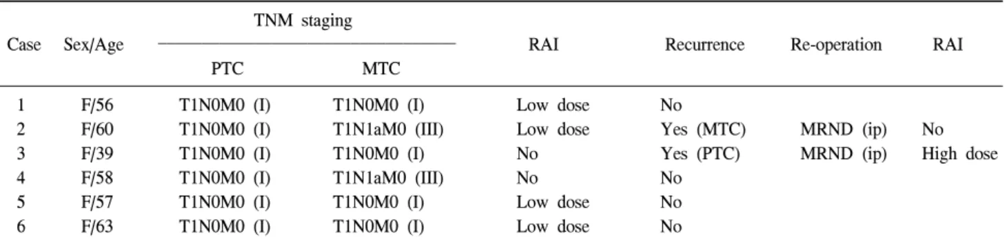
Table 2. Summary of clinical course of patients with concurrent MTC and PTC (n=6) TNM staging
Case Sex/Age RAI Recurrence Re-operation RAI
PTC MTC
1 F/56 T1N0M0 (I) T1N0M0 (I) Low dose No
2 F/60 T1N0M0 (I) T1N1aM0 (III) Low dose Yes (MTC) MRND (ip) No
3 F/39 T1N0M0 (I) T1N0M0 (I) No Yes (PTC) MRND (ip) High dose
4 F/58 T1N0M0 (I) T1N1aM0 (III) No No
5 F/57 T1N0M0 (I) T1N0M0 (I) Low dose No
6 F/63 T1N0M0 (I) T1N0M0 (I) Low dose No
PTC = papillary thyroid carcinoma; MTC = medullary thyroid carcinoma; RAI = radioactive iodine; MRND = modified radical neck dissection; ip = ipsilateral.
방 법
2000년 1월부터 2007년 6월까지 연세대학교 의과대학 외 과와 관동대학교 제일병원 외과에서 갑상선암으로 수술 받 은 환자 중 갑상선 수질암과 유두암이 동시에 발견된 6예 (연세대학교 5예, 관동대학교 1예)의 환자를 대상으로 후향 적 분석을 하였다. 평균 추적 기간은 41.8개월(범위, 9∼100 개월)이었다(Table 1).
임상 양상은 최초 내원 시 주증상, 나이, 성별, 수술 범위, 수술 후 방사성 요오드(radioactive iodine, RAI) 치료 여부, 원격전이 여부, 수술 전 및 후의 혈청 암종배아항원(carci- noembryonic antigen, CEA), 칼시토닌(calcitonin) 수치 등을 조사하였다. 병리조직학적 소견으로 각 종양의 크기와 경 부 림프절 전이 여부를 조사하였다.
절제된 조직은 10% 포르말린 용액에 24시간 동안 고정한 뒤 5μm의 두께로 얇게 잘라 헤마톡실린(hematoxylin)과 에 오신(eosin)으로 염색하여(H&E 염색) 관찰하였다. 면역조직 화학 염색은 cytokeratin-19, galectin-3, thyroglobulin, calcito- nin, CEA, chromogranin A 등을 이용하였다.
모든 환자들은 갑상선 자극 호르몬 억제를 위해 갑상선 호르몬 제제를 투여하였고, 수술 후 3개월 혹은 6개월 간격 으로 추적 관찰하였다.
결 과
진단 당시 평균 연령은 55.4세(범위, 39∼63세)였고, 6예 모두 여성이었다. 모든 경우에서 가족력 및 두경부 방사선 노출 또는 방사성 요오드 치료를 받은 과거력은 없었다. 임 상암이 1예(16.7%), 우연암이 5예(83.3%)였으며, 수술 전 세 침흡인검사에서 갑상선 수질암으로 진단된 경우가 3예 (50.0%), 유두암이 2예(33.3%), 그리고 여포상 종양으로 진 단된 경우가 1예(16.7%)였다(Table 1).
Fig. 1. Papillary thyroid carcinoma. (A, B) Cells are cuboidal and columnal, characterized by a basophilic cytoplasm and show nuclear grrove and nuclear inclusion. (C, D) Immunohistochemical staining, galectin-3 and cytokeratin-19.
수술은 1예는 갑상선 엽 및 협부절제술(hemithyroidecto- my), 5예는 갑상선 전절제술(total thyroidectomy)과 중앙 경 부림프절 절제술이 시행되었고, 2예는 갑상선 전절제술 외 에 추가로 예방적 양측 측경부 림프절 절제술이 시행되었 다(Table 2).
병리조직학적 소견으로 갑상선 수질암과 갑상선 유두암 이 같은 엽에 존재하는 경우가 3예(50.0%)였고, 각각 다른 엽에 존재하는 경우가 3예(50.0%)였다. 갑상선 수질암의 평 균 크기는 1.72 cm (범위, 0.5∼4.0 cm)였고, 유두암은 0.72 cm (범위, 0.2∼1.4 cm)였다(Table 1). 림프절 전이는 2예에 서 중앙 구획 림프절 전이가 확인되었는데 모두 갑상선 수 질암에 의한 전이였다. 예방적 측경부 림프절 절제술이 시 행된 2예에서는 림프절 전이 소견은 없었다.
갑상선 유두암은 6예 모두 1기(T1N0M0)였으나, 수질암 에서는 4예는 1기(T1N0M0), 2예는 3기(T1N1aM0)로 수질암 에서 좀 더 진행된 결과를 보였다(Table 2).
수술 전 혈청 암종배아항원(CEA)과 칼시토닌 수치는 각
각 평균 15.4 (1.7∼39.9) pg/ml, 277.0 (29.0∼653.6) pg/ml였 고, 수술 후에는 각각 평균 6.5 (0.9∼20.1) pg/ml, 25.9 (5.1∼
102.8) pg/ml으로 감소하였다(Table 1).
추적 관찰 중 2예에서 재발이 있었다. 한 예는 일차 수술 후 지속적인 혈청 칼시토닌 수치의 상승이 있어 영상 검사 를 시행한 결과 일측 측경부에 림프절 전이 소견이 있어, 측경부 림프절 청소술을 시행한 결과 수질암의 전이가 있 었던 것으로 확인되었으며, 재수술 이후에는 혈청 칼시토 닌 수치가 정상 범위로 감소하였다. 다른 예는 정기적인 추 적 검사상 측경부 림프절 전이 소견 있어 측경부 림프절 절제술을 시행한 결과 유두암의 재발로 확인되었다.
갑상선 전절제술을 시행한 5예 중 4예는 저용량 방사성 요오드 치료(30∼100 mCi)를, 측경부 림프절 전이가 있었던 유두암 1예에서는 고용량 방사성 요오드 치료(150 mCi)를 시행하였다. 갑상선 엽절제술이 시행된 1예는 수술 후 재발 및 전이의 증거가 없고, 환자가 원하지 않아 완결 갑상선 절제술 및 추가치료를 시행하지 않고 추적관찰 중이다.
Fig. 2. Medullary thyroid carcinoma. (A, B) The MTC component consisted of nests of predominantly round cells with ample, finely granular amphophilic cytoplasm and ovoid to round nuclei. (C, D) Immunohistochemical staining, calcitonin and CEA.
1예의 일시적 저칼슘혈증 외에 특별한 다른 합병증은 없 었다. 추적 기간 동안 사망한 환자는 없었으며, 현재 6명 모 두 건강하게 생존하고 있다.
고 찰
갑상선 수질암과 유두암이 동시에 존재하는 것은 매우 드물지만 두 가지 형태로 존재할 수 있다. 하나는 세계보건 기구(World Health Organization, WHO) 갑상선 종양 분류에 서 소개된 것과 같이 이중의 분화를 보이는 혼합된 종괴로 존재하는 것이고,(2) 다른 하나는 공간적으로 구별되며 각 각 개별적으로 동시에 존재하는 것인데,(1,4-7) 본 연구 대 상은 모두 후자에 해당된다.
갑상선 수질암은 외배엽인 신경 능선(neural crest)으로부 터 기원하며, 갑상선 유두암은 내배엽(endoderm)에서 기원 한다.(2) 이처럼 각각 신경외배엽(neuroectoderm)과 내배엽 기원(endoderm origin)인 서로 상반되는 암종이 동시에 존재
하는 현상에 대해 여러 가설들이 있으나, 어떠한 가설도 아 직 완벽하지는 못하다. 첫째, 아직 무엇으로 분화될지 결정 되지 않은 줄기세포가 각각 소포곁 C-세포와 소포상 상피 세포로 분화한다는 것이다.(8,9) 소포곁 C-세포들은 발생학 적으로 신경 능선으로부터 나와 아가미끝소체로 이동하게 되고 4∼5번째 인두낭을 거쳐 갑상선으로 구체화되는 데,(10,11) 이 세포들의 티로글로블린과 칼시토닌에 대한 면 역학적인 반응은 아가미끝소체가 소포곁 C-세포뿐만 아니 라 소포상 상피 세포들과도 연관이 있다는 것을 의미한다.
두 번째 가설은 서로 다른 두 가지 세포를 각각 갑상선 수 질암과 유두암으로 전환시키는 공통된 자극원이 존재한다 는 것이다. 세 번째는, 두 가지 갑상선암종이 우연히 발견되 었다는 것이다.
유전학적으로도 두 암은 서로 달라서, 갑상선 수질암은 유두암과 마찬가지로 대부분은 산발적으로 발생하지만, RET 전암유전자(proto-oncogene)의 이상으로 인해 발생하는 경우도 있다. 갑상선 수질암과 유두암이 종시에 존재하는
경우에 대한 RET 종양유전자의 유전적인 분석은 매우 혼란 스럽게 만드는 결과를 보여주고 있는데,(12-15) 유전적 분 석을 이용한 7개의 연구 중 2개에서만이 exon 14 (codon 804)와 exon 13 (codon 790)에서 각각 점 돌연변이(germline point mutation)를 보였고,(13,15) 어떤 연구에서도 종양세포 에서 RET 유전자의 체세포 돌연변이(somatic mutation)를 보 인 경우는 없었다.(14)
병리조직학적으로 갑상선 유두암은 유두(papillae) 구조 와 특징적인 핵의 변화를 보인다. 핵은 둥글고, 윤곽이 미묘 하게 불규칙하며, 고랑(groove)을 가지고 있다. 또한, 세포질 의 함입에 의한 가성봉입체(pseudo-inclusion)도 특징적이다 (Fig. 1).(2) 갑상선 수질암은 C-세포 기원이므로, 병리조직 검사에서 C-세포를 확인하면 갑상선 수질암을 진단할 수 있다. C-세포는 대체로 소포상 세포보다 크고, 큰 핵을 가지 고 있는데, 둥글고 긴 모양의 핵을 갖는 방추형 세포와 다각 형 세포들이 관찰되며 세포질 내에 과립들이 관찰된다 (Fig. 2). 그러나 일반적인 H&E 염색만으로는 두 암종을 진 단하기 어려운 경우가 있는데, 이러한 경우에는 면역조직 화학 염색이 도움이 된다. 갑상선 유두암은 thyroglobulin, galectin-3, cytokeratin-19 등이 도움이 되며, 수질암은 calci- tonin, CEA, chromogranin A 등이 도움이 된다(Fig. 1, 2).(16) 수술 전 진단은 세침흡입 세포검사를 통해서 이루어지는 데, 갑상선 유두암은 세침흡인 세포검사를 통해서 손쉽게 진단할 수 있으나,(17) 수질암은 수술 전 세포검사에서 명 확하게 진단하기 어려운 경우가 많다. 하지만 본 연구 대상 과 같이 두 종류의 암종이 동시에 발현된 경우에는 수술 전에 두 암이 동시에 진단되는 경우는 드물며, 갑상선 수질 암이 먼저 진단되고, 갑상선 유두암은 수술 후 잠재성 병변 으로 병리조직검사에서 우연히 발견되는 경우가 흔하 다.(18,19)
갑상선 수질암에서는 특징적으로 혈청 암종배아항원과 칼시토닌 수치가 증가된 경우가 많아, 수술 전 혈청 암종배 아항원 및 칼시토닌 수치가 증가되어 있으면 갑상선 수질 암을 의심해야 한다.(20-22) 또한, 수질암이 재발하거나 전 이되는 경우에도 혈청 칼시토닌 혹은 암종배아항원 수치가 증가되므로, 유두암에서 티로글로브린과 마찬가지로 수술 이후에도 지속적인 추적관찰이 필요하다. 본 연구에서도 갑상선 수질암이 측경부에 재발한 환자의 경우, 일차 갑상 선 전절제술 이후에도 지속적인 혈청 칼시토닌 수치의 상 승이 있었으며, 추가적인 영상의학적 검사에서 측경부 림 프절 전이가 진단되어 재수술이 시행된 바 있다.
이처럼 두 가지 갑상선암이 공존하는 경우는 매우 드물 어 치료 경험이 많지 않지만 수질암은 유두암에 비해 악성 도가 높고 방사성 동위원소 치료와 같은 효과적인 추가치 료가 없어 수술적 치료의 의존도가 절대적이라는 점을 감 안한다면 수술적 치료는 수질암에 준하여 시행해야 할 것 이다.(23) 본 연구에서도 TNM 병기상 유두암은 6예 모두에
서 1기였으나, 갑상선 수질암은 4예에서는 1기, 2예에서는 3기로, 좀 더 진행된 양상을 보였다. 갑상선 수질암의 수술 범위에 대해서는 아직도 많은 논란이 있으나, 갑상선 전절 제술 및 중앙경부림프절 청소술을 기본으로 하고 있으며, 일부 저자들은 재발 및 지속적인 고칼시토닌혈증의 원인이 측경부 림프절 전이임을 감안하여 예방적으로 양측 측경부 림프절 절제술을 동시에 시행하자는 주장도 하고 있다.(24) 최근 저자들도 측경부 림프절 전이의 증거가 없더라도, 수 질암의 수술시 양측 측경부 림프절 절제술을 동시에 시행 하여 그 결과를 알아보고자 하고 있다.
이처럼 수술은 악성도가 높은 수질암에 준해야 하지만 유두암의 악성도도 반드시 고려하여야 하며, 특히, 수술 후 티로글로블린 측정을 통한 경과관찰을 위하여 방사성 요오 드 치료를 시행하는 것이 효과적이라고 생각된다. 방사성 요오드 치료는 기존 갑상선 유두암의 병기에 준하여 시행 하는데, 본 연구자들도 전절제술만 시행된 경우는 저용량 치료를 하였고, 측경부 림프절 전이가 확인된 경우는 고용 량 치료를 하였다.
예후는 보통 갑상선 수질암의 경과에 따를 것이라고 생 각되지만, 악성도가 높은 진행된 유두암이 동반된 경우는 이에 대한 영향도 고려해야 할 것이다.(21)
결론적으로, 서로 다른 양상의 갑상선 수질암과 유두암 이 함께 동반되어 있는 경우는 매우 드물지만 가능성을 항 상 염두에 두고 수술 전 혈청 암종배아항원 혹은 칼시토닌 수치를 확인하는 것이 중요하다. 수술적 치료는 수질암에 준하여 시행하는 것이 재발률을 낮추는데 도움이 될 수 있 으며, 유두암에 준하여 방사성 요오드 치료도 시행하는 것 이 효과적일 것으로 생각된다. 수술 후에는 주기적으로 영 상학적 검사 및 혈청 암종배아항원, 칼시토닌, 티로글로블 린 수치 측정을 통해 재발 여부를 확인하여야 할 것이다.
REFERENCES
1) Lamberg BA, Reissel P, Stenman S, Koivuniemi A, Ekblom M, Mäkinen J, et al. Concurrent medullary and papillary thyroid carcinoma in the same thyroid lobe and in siblings.
Acta Med Scand 1981;209:421-4.
2) Hedinger C, Williams ED, Sobin LH. The WHO histological classification of thyroid tumors: a commentary on the second edition. Cancer 1989;63:908-11.
3) Dionigi G, Castano P, Bertolini V, Boni L, Rovera F, Tanda ML, et al. Simultaneous medullary and papillary thyroid cancer: two case reports. J Med Case Reports 2007;1:133.
4) Gero MJ, Lipper S, Chernys AE, Silver L. Medullary and papillary carcinomas occurring as a collision tumor: report of a case. Clin Nucl Med 1989;14:171-4.
5) Gonzalez-Campora R, Lopez-Garrido J, Martin-Lacave I, Miralles-Sanchez EJ, Villar JL. Concurrence of a symptomatic
encapsulated follicular carcinoma, an occult papillary carci- noma and a medullary carcinoma in the same patient. Histo- pathology 1992;21:380-2.
6) Kobayashi K, Teramoto S, Maeta H, Ishiguro S, Mori T, Horie Y. Simultaneous occurrence of medullary carcinoma and papillary carcinoma of the thyroid. J Surg Oncol 1995;59:
276-9.
7) Pastolero GC, Coire CI, Asa SL. Concurrent medullary and papillary carcinomas of thyroid with lymph node metastases.
A collision phenomenon. Am J Surg Pathol 1996;20:245-50.
8) Ljungberg O, Bondeson L, Bondeson AG. Differentiated thyroid carcinoma, intermediate type: a new tumor entity with features of follicular and parafollicular cell carcinoma. Hum Pathol 1984;15:218-28.
9) Ljungberg O, Ericsson UB, Bondeson L, Thorell J. A compound follicular-parafollicular cell carcinoma of the thyroid: a new tumor entity? Cancer 1983;52:1053-61.
10) Hoyes AD, Kershaw DR. Anatomy and development of the thyroid gland. Ear Nose Throat J 1985;64:318-33.
11) Williams ED, Toyn CE, Harach HR. The ultimobranchial gland and congenital thyroid abnormalities in man. J Pathol 1989;159:135-41.
12) Mazziotti G, Rotondi M, Manganella G, Franco R, Capone PFRS, Colantuoni V, et al. Medullary thyroid cancer, papillary thyroid microcarcinoma and Graves’ disease: an unusual clinical coexistence. J Endocrinol Invest 2001;24:892-6.
13) Brauckhoff M, Gimm O, Hinze R, Ukkat J, Brauckhoff K, Dralle H. Papillary thyroid carcinoma in patients with RET proto-oncogene germline mutation. Thyroid 2002;12:557-61.
14) Fugazzola L, Cerutti N, Mannavola D, Ghilardi G, Alberti L, Romoli R, et al. Multigenerational familial medullary thyroid cancer (FMTC): evidence for FMTC phenocopies and association with papillary thyroid cancer. Clin Endocrinol (Oxf.) 2002;56:53-63.
15) Papi G, Corrado S, Pomponi MG, Carapezzi C, Cesinaro A, LiVolsi VA. Concurrent lymph node metastases of medullary and papillary thyroid carcinoma in a case with RET oncogene germline mutation. Endocr Pathol 2003;14:269-76.
16) Prasad ML, Pellegata NS, Huang Y, Nagaraja HN, dela Chapelle A, Kloos RT. Galectin-3, fibronectin-1, CITED-1, HBME1 and cytokeratin-19 immunohistochemistry is useful for the differential diagnosis of thyroid tumor. Mod Pathol 2005;18:48-57.
17) Schlumberger M. Papillary and follicular thyroid carcinoma.
Ann Endocrinol (Paris) 2007;68:120-8.
18) Rios A, Rodriguez JM, Ferri B, Balsalobre MD, Parrilla P.
Association of medullary and differentiated thyroid carcino- mas. Otolaryngol Head Neck Surg 2006;135:473-5.
19) Biscolla RP, Ugolini C, Sculli M, Bottici V, Castagna MG, Romei C, et al. Medullary and papillary tumors are frequently associated in the same thyroid gland without evidence of reciprocal influence in their biologic behavior. Thyroid 2004;
14:946-52.
20) Kudo T, Miyauchi A, Ito Y, Takamura Y, Amino N, Hirokawa M. Diagnosis of medullary thyroid carcinoma by calcitonin measurement in fine-needle aspiration biopsy specimens.
Thyroid 2007;17:635-8.
21) Elisei R, Rottici V, Luchetti F, Di Coscio G, Romei C, Grasso L, et al. Impact of routine measurement of serum calcitonin on the diagnosis and outcome of medullary thyroid cancer;
experience in 10,864 patients with nodular thyroid disorders.
J Clin Endocinol Metab 2004;89:163-8.
22) Pacini F, Fontanelli M, Fugazzola L, Elisei R, Romei C, Di Coscio G, et al. Routine measurement of serum calcitonin in nodular thyroid disease allows the preoperative diagnosis of unsuspected sporadic medullary thyroid carcinoma. J Clin Endocinol Metab 1994;78:826-9.
23) Behrand M, von Wasielewski R, Brabant G. Simultaneous medullary and papillary microcarcinoma of thyroid in a patient with secondary hyperparathyroidism. Endocr Pathol 2002;13:
65-73.
24) Gimm O, Ukkat J, Dralle H. Determinative factors of biochemical cure after primary and reoperative surgery for sporadic medullary thyroid carcinoma. World J Surg 1998;
22:562-7.