211
서 론
치근낭종은 세계보건기구(WHO) 기준에 의한 치성낭 종의 분류에서 상피성 낭 중에 염증성 낭에 속하고, 전체 치성낭종의 약 65%를 차지하는 흔한 낭종이다. 치근낭 종을 진단하고, 다른 치성낭종과 감별하기 위해 병력청 취 및 이학적 검사, 비 내시경 검사, 방사선학적 검사 등
이 필요하나, 일반적으로 무증상인 경우가 많으며, 치성 감염이 확인되지 않은 경우 다른 질환으로 오인될 수 있다. 저자들은 치근낭종이 치성감염의 병력없이 발생 하여 비순낭종으로 오인하였고, 이는 임상에서 쉽게 오인 될 수 있다고 판단되기에 문헌 고찰과 함께 보고하는 바이다.
증 례
71세 남자 환자가 우측 비순부의 동통과 우측 비익구 (naso-alar sulcus)의 종창을 주소로 본원 외래를 방문 하였다. 지속적으로 종창부위를 통해 분비물이 나왔고 발적이 동반되었다. 병력 청취상 상기 증상은 6개월 전부 터 서서히 발생하였으며, 타병원에서 절개배농술을 시행
피부누공을 동반한 비순낭종으로 오인된 치근낭종
고신대학교 의과대학 이비인후-두경부외과학교실
송준웅
·
장성욱·
김주연·
권재환A Radicular Cyst with Cutaneous Fistula which was Misunderstood as a Nasolabial Cyst
Jun Woong Song, MD, Seong Uk Jang, MD, Joo Yeon Kim, MD, PhD and Jae Hwan Kwon, MD, PhD Department of Otolaryngology-Head and Neck Surgery, Kosin University College of Medicine, Busan, Korea
- ABSTRACT -
A radicular cyst is an inflammatory, epithelial cyst that occurs in the maxillar bone, especially anterior segment.
For optimal treatment, differentiation with other odontogenic cyst is very important. In this report, there was a 71-years-old man who visited our clinic with right naso-alar sulcus swelling and pus-like discharge. We diagnosed as nasolabial cyst based on history, physical examination and PNS CT scan, and designed excision via Caldwell- Luc approach. However, In operation, we found an adhered cyst that induced by 13th canine root infection, and its pathologic findings were chronic inflammation. So, we made definite diagnosis to radicular cyst, and per- formed additional dental treatment. Radicular cyst is most common in odontogenic cyst. Nevertheless it can be easily misunderstanding to other odontogenic cyst, we present this report with a review of literatures. (J Clinical Otolaryngol 2014;25:211-214)
KEY WORDS:Radicular cystㆍOdontogenic cystㆍDental focal infection.
臨床耳鼻:第 25 卷 第 2 號 2014
• • • • • • • • • • • • • • • • • • • • • • • • • • • • • • • • • • • • • • • • • • • • • • • • • • • • • • • • • • • • • • • • • • • • • • • • • • • • • • • • • • • • • • • • • • • • • • • • • • • • • • • • • • • • • • • • • • • • • • • • • • • • • • • • • • • • • • • • • • • • • • • • • • • • • • • • • • • • • • • • • • • • • • • • • • • • • • • • • • • • • • • • • • • • • • • • • • • • • • • • • • • • • • • • • • • • • • •
J Clinical Otolaryngol 2014;25:211-214 증 례
논문접수일 :2014년 19월 29일 논문수정일 :2014년 10월 13일 심사완료일 :2014년 11월 13일
교신저자 :권재환, 602-702 부산광역시 서구 감천로 262 고신대학교 의과대학 이비인후-두경부외과학교실 전화 :(051) 990-6248・전송:(051) 245-8539 E-mail:[email protected]
J Clinical Otolaryngol 2014;25:211-214
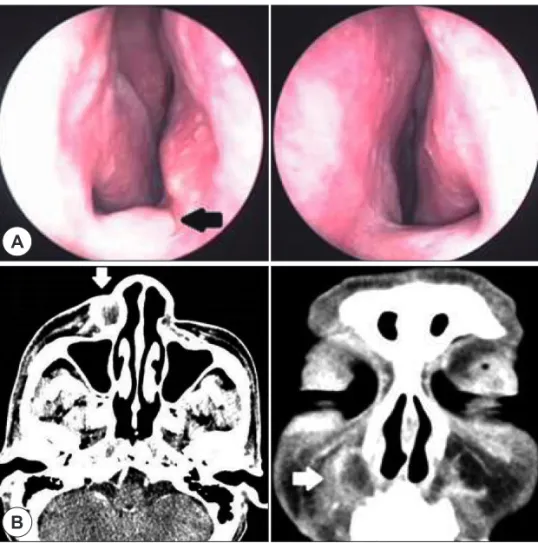
212 하였으나 호전 없이 재발하였다. 이전 치성감염 및 다른 치과적 병력은 없었다. 이학적 검사상 우측 비익구에 홍 반의 종창이 보였고, 종창에서 화농성 분비물이 분비되 었다(Fig. 1). 비 내시경 검사상 우측 비순부의 종창이 비저부를 경미하게 융기하고 있었고, 부비동 컴퓨터단 층촬영 상 우측 비순부에 18×17 mm 크기의 경계가 불 분명한 낭종성 병변이 관찰되었으며, 병변 안으로 이질 성의 조영 증강을 보였다. 주위 치근과의 연결성은 명 확하지 않았다(Fig. 2). 병력, 이학적 검사 및 영상학적 검사를 종합하여 우측 비순낭종 및 우측 비익구 피부누 공으로 진단하였으며, 수술적 절제를 계획하였다. 전신 마취하에 비익구 피부누공을 1 cm가량 쐐기절제하고 구강 내 Caldwell-Luc씨 술식을 이용하여 접근하였다.
술 중 우측 비순부에서 주변 조직과 유착된 낭종성 병 변이 관찰되었으며 화농성 분비물이 분비되었다. 낭종 은 13번 견치의 치근에서 우측 비익구 피부까지 관을 형 성하고 있었고, 주변 조직과 유착이 심해 박리가 어려웠 다. 13번 견치의 치근부에는 골미란, 염증 및 유착의 소 견이 보여, 13번 견치의 감염으로 인해 우측 치근낭종이
발생하고 우측 비익구의 피부누공이 동반되었음을 진 단하였다(Fig. 3). 낭종 및 동반된 피부누공을 절제하고 피부 봉합 후 수술을 마쳤다. 병리 조직 소견상 중층편 평상피와 침윤된 염증세포를 관찰할 수 있었으며, 이는 치근낭종의 조직소견과 일치하였다(Fig. 4). 술 후 1개 월째 치과 협진하에 13번 견치를 발치하였고, 현재 낭종 의 재발 소견없이 경과 관찰하고 있다.
고 찰
치근낭종은 세계보건기구(WHO) 기준에 의한 치성낭 종의 분류에서 상피성 낭 중에 염증성 낭에 속하고, 전체 치성낭종의 약 65%를 차지하는 흔한 낭종이며, 모든 치 아의 치근부위에서 발생가능하나 주로 상악부, 특히 전 치부에서 호발하는 것으로 알려져 있다.1-3) 진단 및 다 른 치성낭종과의 감별은 임상적 소견과 영상학적 소견, 그리고 조직학적 소견을 바탕으로 한다. 임상적으로 병 력청취를 통해 치성 감염의 병력 및 이전의 기왕력을 확 인해야 하며 이는 다른 발육성 낭과의 감별점이 된다. 일 반적으로 특이한 자각 증상이 없이 우연히 발견되나, 낭 종이 지속적으로 팽창할 경우 누공, 농양 등 안면 변형이 발생하고, 주위 골구조의 와해하여 병적 골절, 이차 감염 등이 발생한다.1,2,4) 영상학적으로 컴퓨터 단층촬영상 치 근주변의 중심부에 원형 혹은 타원형의 단방성, 방사선 투과성의 낭종으로 나타나며, 낭과 주위 조직과의 경계가 뚜렷하나 이차감염의 경우 경계가 불분명해질 수 있다.
조직학적으로 중층편평상피세포에 염증세포들이 침윤 된 만성 염증의 형태를 관찰할 수 있다.5)
치근낭종의 치료는 보존적 치료와 수술이 있다. 보존 적 치료인 치근관 치료(Root canal therapy)는 치근관의 감염인자를 제거하고, 치근관을 폐쇄하여 재감염을 예 방하는 치료로 치근낭종이 치근과 연결된 경우 시행할 수 있다.2,6) 수술은 조대술(Marsupialization)과 전적출술 (Enucleation)이 있다. 조대술은 낭의 일부를 제거하여 배액하는 수술로 낭의 크기가 매우 커서 전 적출이 어려 운 경우나 주위 구조물에 손상을 줄 경우 시행한다.7) 전 척출술은 낭이 수술적 접근이 가능할 정도로 크기가 작 거나 주위 해부학적 구조물의 손상을 주지 않을 경우 시 행하며 낭이 협부나 비강으로 확장된 경우 Caldwell-Luc Fig. 1. Pre-operation photograph shows cutaneous fistu-
la (arrow) that formed at the right naso-alar sulcus with pus-like discharge.
송준웅 외 : 피부누공을 동반한 비순낭종으로 오인된 치근낭종
213 씨 접근법, 낭이 경구개 확장없이 치은에 국한된 경우 구 순하 접근법, 낭이 경구개 확장을 한 경우 경구개 접근법 을 사용한다.8)
본 증례의 경우 치성감염 및 치과적 기왕력 등의 병력 이 없고, 컴퓨터단층촬영상 우측 비순부에 이질성의 조 Fig. 2. A : Preoperative endoscopy of nasal cavity shows mild eminence (arrow), result from nasolabial swelling, in right nasal cavity floor compared to left. B : Preoperative PNS CT scan shows 2 cm sized, diffuse heterogeneously en- hanced cystic lesion (arrow) in the right nasolabial area.
A
B
Fig. 3. Intraoperative photograph shows 13th canine bone
erosion (arrow) result from odontogenic infection. Fig. 4. Pathologic photograph shows that the cyst wall is lined by stratified squamous epithelium with infiltrating inflammatory cell (Hematoxylin & Eosin stain, ×100).
J Clinical Otolaryngol 2014;25:211-214
214 영 증강을 보이는 낭종성 병변이 확인되어 우측 비순낭 종 및 동반된 우측 비익구 피부누공으로 의진하였으나, 술 중 낭종이 13번 견치근의 중심부와 연결되어 있고 견 치근부에 염증이 있음을 관찰하여 치근낭종으로 진단하 게 되었다. 따라서 이전의 뚜렷한 치과병력이 없더라도 상악부에서 발생하는 낭종의 빈도가 치근낭종이 60%로 가장 흔하며 무증상으로 우연히 발견되는 경우가 많으 므로, 항상 치성감염의 가능성을 염두하여 진단 및 치료 하여야 할 것이다.
중심 단어:치근낭종・치성낭종・치성감염.
REFERENCES
1) Lee HM, Cho JH, Chu HS, Jeong JY, Lee JH, Hwang SJ, et al. A clinical study of nasolabial cyst. J Clinical Otolar- yngol 2001;12(2):240-3.
2) Caliskan MK. Prognosis of large cyst-like periapical lesions following nonsurgical root canal treatment: a clinical re- view. Int Endod J 2004;37(6):408-16.
3) Kim BH, Park SW. A case of bilateral nasolabial cysts. J Clinical Otolaryngol 2012;23(1):117-20.
4) Lee YC, Kim YK, Kwon SH, Yoon YJ. A case of infected dentigenous cyst with foreign body. Korean J Otorhinolar- yngol-Head Neck 1990;33(4):811-5.
5) Jin YH, Lee KI. Comparison of digital radiometric features between radicular cysts and periapical granulomas. Kore- an J Oral Maxillofac Radiol 1999;29(1):241-54.
6) Caliskan MK, Turkun M. Periapical repair and apical clo- sure of a pulpless tooth using calcium hydroxide. Oral Surg Oral Med Oral Pathol Oral Radiol Endod 1997;84(6):683-7.
7) Lapeer GL. The use of marsupialization in resolving a den- tigenous cystic lesion: a case presentation. J Can Dent As- soc 1985;51:569-70.
8) Tae K, Lee HC, Ryu RA, Song MN, Jeong JH, Cho SH, et al. Treatment of radicular cyst in maxilla. Korean J Oto- rhinolaryngol-Head Neck Surg 2007;50(9):789-94.