CASE REPORT
췌장종양으로 오인된 화농성 췌장농양: 4예 보고
김미진, 서의근1, 강은석, 김근모, 오영민, 조병하, 김형우, 지명진, 정지원, 박선미
충북대학교 의과대학 내과학교실, 하나병원 내과1
Pyogenic Pancreatic Abscess Mimicking Pancreatic Neoplasm: A Four-Case Series
Mi Jin Kim, Euikeun Seo1, Eun Seok Kang, Keun Mo Kim, Young Min Oh, Byung Ha Cho, Hyung-Woo Kim, Myoung Jin Ji, Ji Won Jeong and Seon Mee Park
Department of Internal Medicine, Chungbuk National University College of Medicine, Department of Internal Medicine, Hana General Hospital1, Cheongju, Korea
A pyogenic pancreatic abscess mimicking pancreatic neoplasm in the absence of acute pancreatitis is rare. We report four patients who each presented with a pancreatic mass at the pancreas head or body without acute pancreatitis. The presenting symptoms were abdominal pain, fever, or weight loss. Abdominal CT scans showed low-density round masses at the pancreas head or body with/without lymphadenopathy. In each case, a PET-CT scan showed a mass with a high SUV, indicating possible malignancy. Comorbid diseases were identified in all patients: chronic pancreatitis and thrombus at the portal vein, penetrating duodenal ulcer, distal common bile duct stenosis, and diabetes mellitus. Diagnoses were performed by laparoscopic biopsy in two patients and via EUS fine needle aspiration in one patient. One patient revealed a multifocal microabscess at the pancreatic head caused by a deep-penetrating duodenal ulcer. He was treated with antibiotics and a proton-pump inhibitor.
The clinical symptoms and pancreatic images of all the patients were improved using conservative management. Infective causes should be considered for a pancreatic mass mimicking malignancy. (Korean J Gastroenterol 2015;65:252-257) Key Words: Pancreas; Abscess; Neoplasms; Endosonography
Received July 27, 2014. Revised September 22, 2014. Accepted September 29, 2014.
CC This is an open access article distributed under the terms of the Creative Commons Attribution Non-Commercial License (http://creativecommons.org/licenses/
by-nc/3.0) which permits unrestricted non-commercial use, distribution, and reproduction in any medium, provided the original work is properly cited.
Copyright © 2015. Korean Society of Gastroenterology.
교신저자: 박선미, 362-711, 청주시 서원구 1순환로 776, 충북대학교병원 소화기내과
Correspondence to: Seon Mee Park, Department of Internal Medicine, Chungbuk National University Hospital, 776, 1sunhwan-ro, Seowon-gu, Cheongju 362-711, Korea. Tel: +82-43-269-6019, Fax: +82-43-273-3252, E-mail: [email protected]
Financial support: None. Conflict of interest: None.
INTRODUCTION
Pancreatic abscess usually occurs by way of an infection of a pseudocyst or pancreatic necrosis in acute pancreatitis.
It rarely presents via a perforation of the bowel into the pan- creas,1,2 infected bile reaching the pancreas via the pancre- atic duct, hematogenous spread from a distant site, or lym- phatic spread from the intestinal tract. A pancreatic abscess usually presents as abdominal pain, fever, and leukocytosis.3 However, some cases manifest with abdominal pain or weight loss. A pyogenic pancreatic abscess mimicking pan-
creatic neoplasm in the absence of acute pancreatitis is rare and difficult to diagnose.4 We report on four patients who each presented with a pancreatic mass at the pancreas head or body that mimicked malignancy but was diagnosed as a pancreatic abscess (Table 1).
CASE REPORT
1. Case 1
A 51-year-old woman was admitted following two days of abdominal pain. There was no history of fever or weight loss.
Table 1. Baseline and Clinical Characteristics of the Four Patients Case Sex/
age (yr)
Pancreas location
Size
(cm) Comorbidities Clinical symptoms
WBC (mm3)
CRP (mg/dL)
CA 19-9 (IU/ML)
Diagnostic
methods Patholgy/cytology Treatment Case 1 F/51 Body 2.7 DM Abdominal pain 9,100 0.94 Normal Laparoscopy Stromal fibrosis,
reactive lymph node hyperplasia
Antibiotics
Case 2 F/65 Head 5.0 DM, CP Abdominal pain, fever
14,200 24.44 Normal Laparoscopy Acute and chronic inflammation
Antibiotics Case 3 M/77 Head 1.2 DM, HTN
CBD stenosis
Epigastric/
back pain
10,610 6.98 121.51 EUS-FNA Pus Antibiotics
Case 4 M/61 Head 2.8 DU Abdominal pain, weight loss
8,900 2.22 Normal EUS Not performed Antibiotics, PPIs WBC, white blood cells; DM, diabetes mellitus; CP, chronic pancreatitis; HTN, hypertension; CBD, common bile duct; DU, duodenal ulcer;
FNA, fine needle aspiration; PPI, proton pump inhibitors.
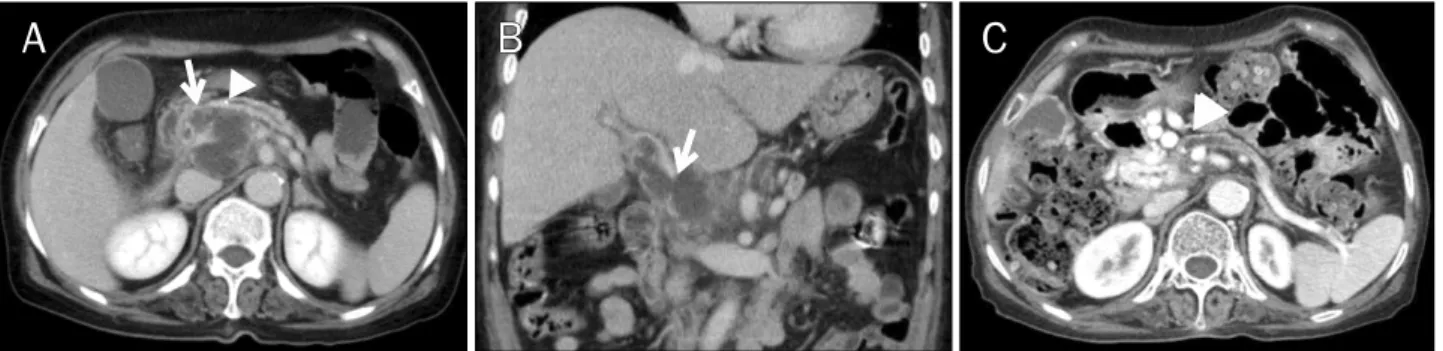
Fig. 1. Image studies of Case 1. (A) Axial view of the abdominal CT scan. A 2.7-cm ill-defined low-density mass (arrow) with peripancreatic fat infiltration was located at the pancreatic body. (B) Coronal view of the abdominal CT scan. An ill-defined, soft tissue density lesion around the celiac axis, common hepatic artery, splenic artery, and portal vein was noted (arrow). (C) PET-CT scan. Hot uptake (SUV 3.8) at the pancreatic body, indicating possible malignancy, was observed (arrow).
Her past medical history included a five-year history of type II dia- betes mellitus. She denied any smoking or alcohol consump- tion. The abdomen was not tender. Her vital signs were normal.
Blood tests showed white blood cells (WBC) 9,100/mm3, hemo- globin (Hb) 14.9 g/dL, platelets 228,000/mm3, AST 220 IU/L, ALT 64 IU/L, ALP 212 IU/L, albumin 3.4 g/dL, bilirubin 0.7 mg/dL, amylase 17 U/L, lipase 78 U/L, and CRP 0.94 mg/dL.
CA 19-9 was normal. An abdominal CT scan showed a 2.7-cm ill-defined low-density mass with peripancreatic fat infiltra- tion located at the pancreatic body (Fig. 1A). An ill-defined soft tissue density lesion around the celiac axis, common hepatic artery, splenic artery, and portal vein was noted (Fig.
1B), which was suggestive of lymphadenopathy with vascular invasion. A PET-CT scan showed hot uptake (SUV 3.8) at the pancreatic body and peripancreatic lymph nodes, which in- dicated possible malignancy (Fig. 1C). Based on the image findings, borderline respectable pancreatic cancer was diagnosed. Diagnostic laparoscopy was performed for evalu-
ation of the tumor’s extent and a pathologic diagnosis. The laparoscopy revealed an encapsulating lesion in the pancre- atic body. An excisional biopsy of the mass and lymph node was performed. Pathology showed glandular atrophy, stro- mal fibrosis, and reactive lymph node hyperplasia. Seven days after diagnosis, the patient was discharged following treatment with intravenous antibiotics. A CT scan four months later showed the resolution of the abscess after a six-week course of oral antibiotics. The patient was well at the two-year follow-up.
2. Case 2
A 65-year-old woman was admitted following five days of abdominal pain and fever. Her past medical history revealed a 10-year history of type II diabetes mellitus. She denied any smoking or alcohol consumption. Her vital signs were blood pressure 140/75 mmHg, pulse rate 63/min, respiratory rate 18/min, and body temperature 37.2oC. Abdominal tender-
Fig. 3. Image studies of Case 3. (A, B) Axial and coronal views of the abdominal CT scan. A 1.2-cm well-defined low-density mass at the pancreatic head was observed (arrows). (C) Endoscopic ultrasonography. A 1.2-cm-sized anechoic round mass was seen at the pancreas head (arrows).
Fig. 2. Image studies of Case 2. (A) Axial view of the abdominal CT scan. A 5.0-cm well-defined low-density mass with heterogenous enhancement at the pancreas head (arrow), parenchymal atrophy, and a calcifying stone in the dilated pancreatic duct (arrowhead). (B) Coronal view of the abdominal CT scan. The portal vein and common hepatic artery were encased by a low-density mass (arrow). (C) Axial view of the abdominal CT scan after treatment. Resolution of the abscess was noted (arrowhead).
ness was detected in the upper abdomen. The laboratory findings were WBC 14,200/mm3, Hb 12.6 g/dL, platelets 181,000/mm3, AST 30 IU/L, ALT 20 IU/L, ALP 324 IU/L, albu- min 3.2 g/dL, bilirubin 0.6 mg/dL, amylase 16 U/L, lipase 9 U/L, and CRP 24.44 mg/dL. CA 19-9 was normal. An abdomi- nal CT scan showed a 5.0-cm well-defined low-density mass with heterogenous enhancement at the pancreas head and mild pancreatic duct dilatation with a calcifying stone (Fig.
2A). The portal vein and common hepatic artery were en- cased by the mass (Fig. 2B). Based on the CT findings, the di- agnosis was suspected pancreatic neoplasm against the background of chronic pancreatitis. A laparoscopy was per- formed for evaluation of the tumor extent and pathologic diagnosis. A pancreatic biopsy revealed acute and chronic in- flammation with abscess formation. Three weeks later, the patient was discharged following two weeks’ treatment with intravenous antibiotics. A repeat CT scan two months later showed resolution of the abscess (Fig. 2C).
3. Case 3
A 77-year-old man was admitted with a one-week history of both epigastric and back pain. His past medical history in- cluded an eight-year history of hypertension, type II diabetes mellitus, and acute cholangitis caused by bile duct stones. He had been treated with ERCP five years previously. He was a 40-packs-a-year smoker and a social drinker. The abdomen was not tender. His vital signs were blood pressure 140/80 mmHg, pulse rate 76/min, respiratory rate 20/min, and body temperature 36.7oC. Blood tests showed WBC 10,610/mm3, Hb 15.6 g/dL, platelets 156,000/mm3, AST 20 IU/L, ALT 25 IU/L, ALP 94 IU/L, albumin 3.7 g/dL, bilirubin 0.58 mg/dL, and CRP 6.98 mg/dL. CA 19-9 was elevated (121.51 IU/mL;
reference range, 0-37 IU/mL). An abdominal CT scan showed a 1.2-cm well-defined low-density mass at the pancreatic head without lymphadenopathy (Fig. 3A, B). Based on the clin- ical findings, pancreatic adenocarcinoma was suggested.
EUS showed an approximately 1.2 cm-sized anechoic round mass (Fig. 3C). A small quantity of yellow fluid, which was con-
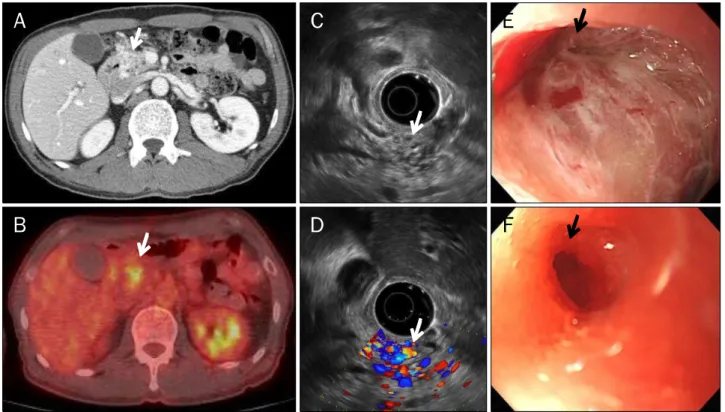
Fig. 4. Image studies of Case 4. (A) Axial view of the abdominal CT scan. A 1.7-cm multifocal ill-defined low-density lesion with multiple enhancing tubular structures at the head of the pancreas was observed (arrow). (B) PET-CT scan. Hot uptake (SUVmax 3.8) at the pancreatic head indicated possible malignancy or inflammation (arrow). (C, D) Endoscopic ultrasonography. A 2.8×1.6 cm-sized round isoechoic lesion with internal anechoic tubular structures was observed using vascular doppler (arrows). (E) Endoscopic finding. A 3.0-cm round deep-penetrating duodenal ulcer with a central pit was observed (arrow). (F) Endoscopic finding one year later. The duodenal ulcer had improved; however, the duodenal fistula remained (arrow).
firmed as pus and thus suggestive of an abscess, was drained by EUS-fine needle aspiration (FNA). The patient showed improvement after six weeks on antibiotics, and a re- peat CT scan showed resolution of the abscess.
4. Case 4
A 61-year-old man was admitted for abdominal pain and weight loss. Upper abdominal pain and anorexia had devel- oped two months prior, and the symptoms had become se- vere one month later, with a more recent weight loss of 15 kg, dizziness, and general weakness. He was a 40-packs-a-year smoker and a teetotaler. His vital signs were normal. Blood tests showed a WBC 8,900/mm3, Hb 9.9 g/dL, platelets 372,000/mm3, AST 16 IU/L, ALT 19 IU/L, ALO 195 IU/L, albu- min 4.2 g/dL, bilirubin 0.25 mg/dL, amylase 17 U/L, lipase 25 U/L, and CRP 2.22 mg/dL. CA 19-9 was normal. An ab- dominal CT scan showed a 1.7 cm-sized ill-defined low den- sity lesion with multiple enhancing tubular structures in the head of the pancreas, which was suggestive of a hyper-
vascular pancreatic tumor or engorged collateral vessels (Fig. 4A). A PET-CT scan showed hot uptake (SUVmax 3.8) at the pancreatic head, indicating possible malignancy or in- flammation (Fig. 4B). EUS using a vascular doppler showed an approximately 2.8×1.6 cm-sized round isoechoic lesion with internal anechoic tubular structures (Fig. 4C, D).
Endoscopy showed a 3.0-cm round deep-penetrating duode- nal ulcer with a central pit (Fig. 4E). We diagnosed a multifocal pancreatic abscess due to a duodenal fistula caused by a deep-penetrating duodenal ulcer, and treated the patient with antibiotics and proton-pump inhibitors. The clinical symptoms and duodenal ulcer were improved after a four-week course of antibiotics. One year later, the duodenal fistula remained (Fig. 4F), as did an air-containing low-attenu- ating lesion in the pancreatic head.
DISCUSSION
Pancreatic abscesses are commonly associated with
pancreatitis. A pyogenic pancreatic abscess in the absence of pancreatitis is very rare.4 Other causes include an ex- tension abscess from nearby structures, hematogenous spread from a distant site, and lymphatic spread from the in- testinal tract. There are a few reports of seeding from ad- jacent organs, fish bone perforations of the gastric wall into the pancreas,1 secondary to gastric or duodenal diverticula perforations,5-7 and pancreatic tail abscesses directly due to bacterial peritonitis.6 Pancreatic abscess formation can oc- cur by means of hematogenous spread during a transient epi- sode of bacteremia that has resolved before presentation. A few cases of Salmonella typhi,7-9 Corynebacterium coyleae,10 and Klebsiella pneumoniae infection have been reported.4 Our cases were not associated with acute pancreatitis.
However, three cases revealed possible underlying etiol- ogies: penetrating duodenal ulcer, pancreatic stone, and dis- tal common bile duct stenosis. Only longstanding diabetes mellitus was found in one patient. Poor diabetes control is known to cause compromised immunity resulting in in- creased risk for infections.11 Infections without obvious sour- ces are particularly common among patients with diabetes mellitus.
Our four cases showed various clinical spectrums of pan- creatic abscesses. Not all patients developed typical symp- toms of pancreatic abscesses, such as fever, abdominal pain, and leukocytosis; All patients developed abdominal pain and only one patient developed fever. One patient pre- sented with weight loss without fever, and his symptoms were difficult to differentiate from pancreatic ductal adeno- carcinoma. Similar to other reports of pancreatic abscess mimicking malignancy, our initial diagnosis was a tumor of the pancreatic head, especially given the elevated CA 19-9.
Leukocytosis is the only laboratory test that can indicate the presence of an abscess. However, only two out of the four pa- tients showed mild leukocytosis.
Image studies including abdominal CT and PET-CT scans are not helpful in differentiating between a pancreatic ab- scess and pancreatic neoplasm. In our cases, abdominal CT scans showed low-density round masses with lymphadenop- athy and vascular infiltration, mimicking invasive ductal adenocarcinomas. A high-SUV mass was not helpful in differ- entiating an inflammatory mass from a malignancy. However, contrast-enhanced EUS has been reported to be very advantageous. A homogenous, low-density, round mass
without enhancement is beneficial in the diagnosis of a pan- creatic abscess.12
Nevertheless, pathological examination is still required for diagnosis. We used laparoscopic exploration in two cases as we did not have the opportunity to perform EUS at that time.
Before the widespread use of EUS, it was reported in the liter- ature that percutaneous cystic drainage with the assistance of a laparoscopy could be used to drain a pancreatic abscess.13 However, in the EUS era, a pancreatic abscess can be easily diagnosed and treated with EUS images and drainage.12 EUS has proved to be very sensitive and specific for the diagnosis of pancreatic lesions, especially with the use of fine needle aspiration cytology. It may even obviate the need for biopsy and unnecessary interventions in the future.
In this study, we aspirated pus by EUS-FNA in one case and detected a microabscess with duodenal fistula in another case using a EUS image. EUS-FNA could not be performed on one patient with a microabscess due to a penetrating duode- nal ulcer because of high vascularity and the abscess was too small. This patient improved on antibiotics and proton-pump inhibitors.
In conclusion, our cases highlight the rare condition of a pyogenic pancreatic abscess mimicking an underlying pan- creatic malignancy. In similar cases, infective causes should be considered in order to avoid any unnecessary major surgeries.
REFERENCES
1. Goh BK, Jeyaraj PR, Chan HS, et al. A case of fish bone perfo- ration of the stomach mimicking a locally advanced pancreatic carcinoma. Dig Dis Sci 2004;49:1935-1937.
2. An CH, Kim KH, Kim JS, Kim JI. Pancreatic abscess caused by gastric perforation. ANZ J Surg 2007;77:709.
3. Saxon A, Reynolds JT, Doolas A. Management of pancreatic abscesses. Ann Surg 1981;194:545-552.
4. Chong VH. Isolated pyogenic pancreatic abscess mimicking a neoplasm. JOP 2008;9:309-312.
5. Safioleas M, Stamatakos MK, Mouzopoulos GJ, et al. Pancreatic abscess due to perforation of duodenal diverticulum. Chirurgia (Bucur) 2006;101:523-524.
6. Hu J, Sheu MH, Yang WC, Li JC, Ng YY. Peritonitis and pancreatic abscess in a CAPD patient. Perit Dial Int 2002;22:430-431.
7. Seo KS, Oh HM, Hong JH, et al. A case of pancreatic abscess due to Salmonella typhi. Korean J Med 1998;54:101-104.
8. Arya M, Arya PK. Pancreatic abscess caused by s. typhi. Indian J Med Microbiol 2001;19:18-19.
9. Garg P, Parashar S. Pancreatic abscess due to Salmonella typhi.
Postgrad Med J 1992;68:294-295.
10. Taguchi M, Nishikawa S, Matsuoka H, et al. Pancreatic abscess caused by Corynebacterium coyleae mimicking malignant neoplasm. Pancreas 2006;33:425-429.
11. Eliashiv A, Olumide F, Norton L, Eiseman B. Depression of cell-mediated immunity in diabetes. Arch Surg 1978;113:1180- 1183.
12. Chase MP, Yarze JC, Gumustop B, Leach RP. Endoscopic ultra-
sound-guided aspiration and oral antibiotic therapy as definitive treatment of an asymptomatic pancreatic abscess. Pancreas 2009;38:475-476.
13. Horvath KD, Kao LS, Wherry KL, Pellegrini CA, Sinanan MN. A technique for laparoscopic-assisted percutaneous drainage of infected pancreatic necrosis and pancreatic abscess. Surg Endosc 2001;15:1221-1225.