논문접수일 :2011년 08월 31일 논문수정일 :2011년 09월 19일 심사완료일 :2011년 10월 21일
교신저자 :정진혁, 471-701 경기도 구리시 교문동 249-1 한양대학교 의과대학 이비인후-두경부외과학교실 전화 :(031) 560-2368·전송:(031) 566-4884 E-mail:[email protected]
서 론
악성 림프종은 면역계의 림프구에서 발생하는 악성 종 양으로 신체 어느 부위에서나 발생할 수 있지만 주로 림프 절 침범이 많고, 림프절 외에서 발생하는 경우 위장관, 비강 및 부비동, 비인두, 구개편도, 구강, 골, 뇌, 후두 등에 호발 한다.1,2) 두경부의 경우에도 흔히 경부 림프절을 침범하지 만 약 10%가 비림프절을 침범하는 것으로 알려져 있다.3)
비부비동에 발생하는 악성 림프종은 대부분 비호지킨
림프종(Non-Hodgkin’s lymphoma)으로 주로 NK/T 세 포 림프종이다. B 세포 림프종은 극히 드물고 주로 비부 비동 이외의 두경부에 호발한다.4-6)
저자들은 시력소실을 주소로 내원한 환자에서 부비동 의 거대한 악성 B 세포 림프종을 발견하고, 시력을 회복 하고자 빠르게 진단하고 치료하여 좋은 결과를 보인 환 자를 보고하고자 한다.
증 례
72세 여자로 내원 1달 전부터 안면 부종과 약간의 비폐 색 증상이 있었으나 특별한 치료 없이 지내다가 내원 1일 전 갑작스러운 우측 시력 소실을 보여 안과 진료 후 이비 인후과로 의뢰되어 응급 입원하였다.
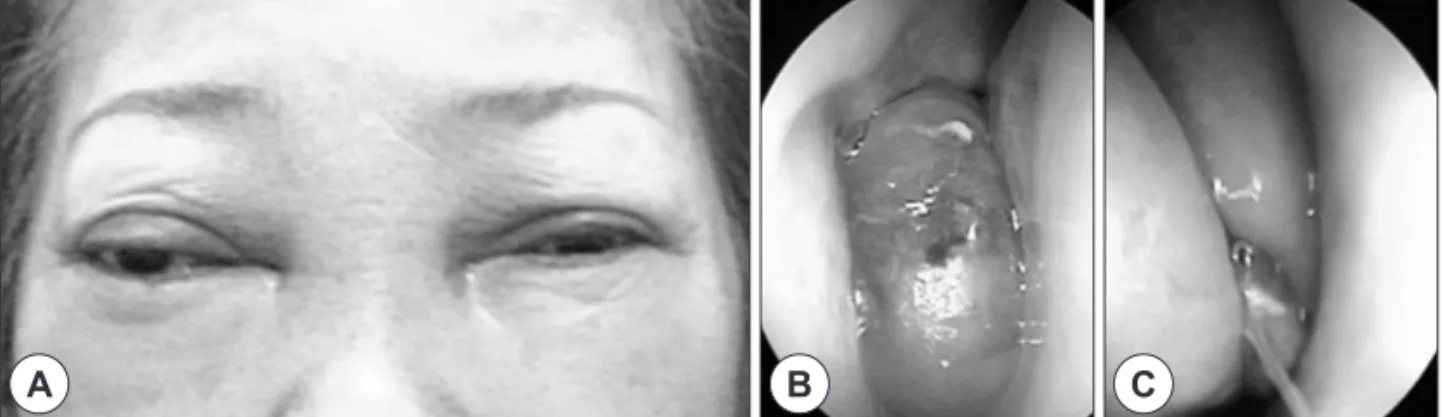
기저 질환으로 당뇨가 있었고, 이학적 검사상 안각간 부 위의 부종과 안각격리증(Telecanthus)이 관찰되었고(Fig.
시력소실을 동반한 거대한 부비동 미만성 대세포 림프종 1예
한양대학교 의과대학 이비인후-두경부외과학교실
김종민 · 조석현 · 김경래 · 정진혁
A Case of Huge Diffuse Large B-Cell Lymphoma of Paranasal Sinus with Visual Loss
Jong Min Kim, MD, Seok Hyun Cho, MD, Kyung Rae Kim, MD and Jin Hyeok Jeong, MD Department of Otolaryngology-Head and Neck Surgery, College of Medicine, Hanyang University, Seoul, Korea
-
ABSTRACT -
Non-Hodgkin’s lymphoma of nasal cavity or sinuses is rare. Usually, it is a NK/T cell lymphoma. The B cell lymphoma is extremely rare in Koreans. B cell lymphoma usually occurs in western people in the paranasal si- nuses. Because it presents as a mass in the paranasal sinus, unlike NK/T cell lymphoma, B cell lymphoma can cause sinusitis like symptoms or eye symptoms such as optic neuropathy, telecanthus, exophthalmos. We report a rare case of a huge diffuse large B cell lymphoma in the paranasal sinus with acute visual loss in a 72 year- old female. The patient was successfully treated due to a fast diagnosis and immediate treatment via chemo- therapy.
(J Clinical Otolaryngol 2011;22:250-254)
KEY WORDS
:Malignant lymphomaㆍParanasal sinusㆍOptic neuropathy.1A), 비강 내시경 소견 상 양측 중비도 및 상비도는 쉽게 출혈되는 연조직 종물로 차있었다(Fig. 1B, C). 당시 안과 검사상 우측은 빛감지(light perception)가 불가능했으며, 좌안 시력은 0.6로 측정되었다. 우안에서 구심성 동공장 애(relative afferent pupillary defect ; RAPD)가 관찰되 었으나, 양안 모두 안저검사는 정상 소견을 보였다. 안구 돌출은 우측이 좌측에 비해 4 mm 돌출되어 있었다.
안와 CT상 양쪽 비강 및 사골동, 안와, 두개내를 침범 하고 있는 거대한 균질 음영의 연조직 종물이 보였고, 부 비동 격벽을 포함한 주위의 심한 골파괴 소견이 보였으며 (Fig. 2A, B), 두부 MRI상 T1 강조 영상과 T2 강조 영상 모두 저 신호강도를 보이고 조영증강이 잘되는 종물이 관 찰되었다(Fig. 2C, D).
내원 당일에 우측 비강 내에서 조직생검을 시행하였 고, 시력소실을 고려하여 병리과의 도움을 받아 응급 면 역 염색을 시행한 결과, 내원 2일째에 조직검사 결과로 dif- fuse large B cell lymphoma{CD20(+), CD79a(+), Bcl- 2(+), Bcl-6(+), Mum-1(+), Ki-67(+)}를 확인할 수 있었다
(Fig. 3).
조직검사 결과를 확인한 내원 2일째 바로 혈액종양내 과로 전과되어, 내원 3일째 흉부 및 복부 CT를 시행하였 고 결과 특이 소견 보이지 않았으며, 골수 천자 생검 및 척 수액 천자 세포 검사에서는 림프종의 침범이나 악성세포 가 관찰되지 않았다.
혈액 검사 소견상 LDH : 511(211~423 U/L), CK : 795 (30~180 U/L), ß2-myoglobin : 2.1(0~2.4 mg/L), ALP : 109(42~98 U/L), CEA : 3.38(0~2.5 ng/mL)로 측정되었 고, 그 외의 혈액검사는 정상소견을 보였다.
내원 4일째 바로 항암치료{R-CHOP for 5days : Ritu- ximab(375 mg/m2), Cyclophosphamide(750 mg/m2), Do- xorubicin(50 mg/m2), Vincristine(1.4 mg/m2), Prednisone (100 mg)}를 시작하였다. 내원 9일째 우측은 10 cm 거리 에서 손가락 수를 헤아릴 수 있을 만큼 시력 회복되었고, 좌측 시력은 0.5로 측정되었다. 내원 12일째 비강 내시경 상 보이지 않았던 해부학적 정상적인 구조물들을 확인할 수 있었다(Fig. 4A, B). 2차 항암치료가 끝난 후인 내원 후
A B C
Fig. 1. Physical examination and endoscopic finding. A : Intercanthal swelling and telecanthus are observed. B : A large, soft, pinkish mass is noted in the right nasal cavity and easily bleeds. C : A small mass is noted in the left middle meatus of the left nasal cavity.
Fig. 2. Orbit CT & PNS MRI. A huge mass with homogenous enhancement and bony destruction is observed in the eth- moid sinus, nasal cavity, orbit, and intracranium (A : Coronal CT. B : Axial CT). Intracranial extension with dural enhance- ment is observed in MRI, but brain parenchyme is not involved (C : T2WI. D : T1WI with enhancement).
A B C D
47일째 이학적 검사에서 안각간의 부종 및 안각간 격리증 은 사라졌으며(Fig. 4C), 안와 CT에서도 이전에 보이던 중 앙 돌출의 안면 종물의 확연한 호전 소견이 확인되었고, 이전 종양에 의한 골파괴로 보이지 않았던 부비동 주위 의 골이 다시 보였다.
약 4개월에 걸쳐 R-CHOP regimen 6회 시행하였고, 항 암치료 종료 후로부터 3주 및 5개월 후에 시행한 PET-CT 에서 재발이나 전이 소견 관찰되지 않았다. 항암치료 종료
후 3개월째 비강 내시경상 이전에 관찰되던 연조직은 완 전히 사라졌으며, 4개월 후 시행한 안과적 검사에서 우측 은 20 cm 거리에서 손가락 수 헤아릴 수 있을 만큼 시력 회 복되었고, 좌측은 1.0으로 측정되었다. 함암치료 종료 후 로부터 1년 6개월째 시행한 안와 CT에서는 이전 종물은 완전히 치유되었고, 부비동과 안와 주위의 얇은 골들도 완 전히 다시 보였다(Fig. 5A, B). 2011년 8월 현재 항암치료 종료 후 2년째 완전 관해 상태로 추적 관찰 중이다.
Fig. 3. Histological features. Diffuse proliferation of atypical lymphoid cells are noted and there are large-sized cells with prominent nucleoli (A : H-E stain, ×100), These diffusely proliferative cells express CD79a (B), Bcl-6 (C) positivity in immunohistochemical stains.
A B C
C
A B
Fig. 4. Physical examination and endoscopic finding after treatment. Previously observed masses of nasal cavity have disappeared. Normal anatomic structure is observed (A : Right nasal cavity. B : Left nasal cavity). C : Intercanthal swelling and telecanthus have improved.
Fig. 5. PNS CT after treatment. Masses which were observed in paranasal sinuses, nasal cavity, intracranium and orbit have disappeared and destructed bone has become visible again like normal bony structure. A : Enhanced coronal CT. B : Non-enhanced axial CT.
A B
고 찰
비부비동 원발 악성 림프종은 전체 두경부 악성 림프 종 중 약 10%에서 발생하며 예후가 좋지 않은 질환이다.3) NK/T 세포 림프종은 주로 아시아, 아프리카인에서 생기 고, 대부분 부비동보다는 비강에서 기원하며, 종괴를 형 성하기 보다는 점막 병변으로 나타나며, 비폐색, 비점막의 광범위한 궤양 및 가피, 비출혈, 비중격 천공을 흔히 동반 하며 급속도로 진행하여 예후가 불량하다.7,8) 반면 B 세포 림프종은 서양인에서 주로 발생하며, 대부분 부비동에서 기원하고 NK/T 세포 림프종에 비해 덜 파괴적이고, 주 로 종괴를 형성하며, 종괴로 인해 안구를 침범하여 안구 증상을 흔히 일으키며, 점막 비후나 자연공 폐쇄로 인한 부비동염을 유발시킨다.
비부비동에서 악성 림프종이 발생한 경우에 임상증상 은 비특이적이나 주로 비폐색, 비출혈, 비루, 안면부종, 치 성통증, 시력장애 등의 증상이 나타날 수 있다.9) 본 증례 와 같이 사골동에 발생하는 경우 인접해 있는 안와 및 두 개내에 침범하여 두통, 복시, 시력장애 등을 야기 할 수 있 고, 더욱 진행하면 중추신경계 증상도 나타날 수 있다.
진단 과정에서 보는 CT 소견상 특이한 점이 있는데, B 세포 림프종의 경우 종괴를 형성하고 이로 인한 CT상 골 파괴를 보이는데 비부비동 상피세포악성종양에서 보이 는 파괴적인 소견과는 확연히 차이가 나는 녹아 없어진 느낌의 소견을 보인다는 것이다.10) 또한 본 증례와 같이 종물이 거대해서 골 흡수 소견이 아주 심한 경우에도 항 암치료 후 사골동 격벽 같은 아주 얇은 골조직을 포함 한 모든 골 조직이 다시 정상으로 돌아온다는 것도 상피 세포악성종양과는 확연한 차이를 보이는 것으로 림프종 세포의 골파괴 기전도 흥미를 유발시킨다.10)
치료로는 항암약물치료를 시행하며, 주로 CHOP(Cy- clophosphamide, Doxorubicin, Vincristine, Prednisone) 을 사용하며, 최근 B 세포 특이 표지자의 단클론 항체인 Rituximab을 병용 투여하는 R-CHOP을 사용하여 치료 성적을 높이고 있다.1) 방사선치료도 같이 시행하기도 하 나 정립된 치료는 아니며 치료에 반응이 없거나 재발하 는 경우 골수이식 하기도 한다.
비부비동염 B 세포 림프종의 경우 비특이적인 비 증상
보다 특이적인 안구 혹은 시신경 증상으로 병원을 내원 하는 경우가 많은데, 이런 경우 진단과 치료가 늦어질 경 우 시력 회복은 물론 악성 림프종의 치료에도 영향을 미 쳐 나쁜 결과를 가져올 수 있다. Takahiko 등11)은 본 증 례와 같이 급성 시력소실을 주소로 내원한 환자에서 초기 에 특발성 시신경염으로 생각하여 스테로이드 치료를 하 였으나 호전 없어 이후 늦게 조직검사를 시행하고 항암 화학요법을 시행하였지만 영구적인 시력소실을 가져왔 다고 보고하면서 비부비동 기원의 악성 림프종에 의한 시 신경 병변의 경우 빠른 진단과 치료를 해야 한다고 강조 하였다. 본 증례의 경우 부비동에서 기원하여 비 증상보 다 안구 증상을 일으켜 내원한 환자로, 시력 소실을 동반 하고 있어 시간을 지체하지 않고 응급으로 CT와 MRI를 촬영하고, 병리과의 협조를 얻어 2일 만에 면역조직염색 결과를 얻어 바로 혈액종양 내과에서 빠르고 적절한 항 암요법으로 시력도 호전되고 종양도 완전 관해를 이룬 환자로 Takahiko 등이 강조하는 빠른 진단과 치료를 보여 주는 좋은 예라고 생각된다.11)
중심 단어:악성 림프종・부비동・시신경병증.
REFERENCES
1) Tae K, Lee HS, Seo IS, Lee YS, Cho SH, Choi JH, et al.
Hodgkin’s and non-Hodgkin’s lymphoma of head and neck.
Korean J Otolaryngol-Head Neck Surg 2003;46(4):324-30.
2) Choi CY, Jo YK, Lee BH, Lee YW, Lee KD, Yu TH. Ana-
lysis of treatment in the patient with non-Hodgkin’s lym- phoma of the head and Neck. Korean J Otolaryngol-Head Neck Surg 1997;40(12):1820-5.
3) Advani R, Jacobs CD. Lymphoma of the head and neck. In:
Bailey BJ, editor. Head and neck surgery-Otolaryngology, 3rd edition. Philadelphia: Lippincott-Raven Publishiers;
2006. p.1621-8.
4) Logsdon MD, Ha CS, Kavadi VS, Cabanillas F, Hess MA, Cox JD. Lymphoma of the nasal cavity and paranasal si-
nuses. Cancer 1997;80(3):477-88.
5) Woo JS, Kim JM, Lee SH, Chae SW, Hwang SJ, Lee HM.
Clinical analysis of extranodal non-Hodgkin’s lymphoma in sinonasal tract. Eur Arch Otorhinolaryngol 2004;261(4):
197-201.
6) Quraishi MS, Bessell EM, Clark D, Jones NS, Bradley PJ.
Non-Hodgkin’s lymphoma of the sinonasal tract. Laryngo- scope 2000;110(9):1489-92.
7) Fellbaum C, Hansmann ML, Lennert K. Malignant lym-
phomas of the nasal cavity and paranasal sinuses. Vir-
chows Arch A Pathol Anat Histopathol 1989;414(5):399-
405.
8) Shohat I, Berkowicz M, Dori S, Horowitz Z, Wolf M, Ta- icher S, et al. Primary non-Hodgkin’s lymphoma of the si-
nonasal tract. Oral Surg Oral Med Oral Pathol Oral Ra- diol Endod 2004;97(3):328-31.
9) Juman S, Robinson P, Balkissoon A, Kelly K. B-cell non-
Hodgkin’s lymphoma of the paranasal sinuses. J Laryngol Otol 1994;108(3):263-5.
10) Matsumoto S, Shibuya H, Tatera S, Yamazaki E, Suzuki S.
Comparison of CT findings in non-Hodgkin lymphoma and squamous cell carcinoma of the maxillary sinus. Acta Ra- diol 1992;33(6):523-7.
11) Hayashi T, Watanabe K, Tsuura Y, Tsuji G, Koyama S, Yo- shigi J, et al. Sight-threatening optic neuropathy is associ-