2003; 8(2): 63-72
INTRODUCTION
There is a growing body of evidence from pre- clinical and clinical studies that synthetic or natural DNA damaging agents are able to induce apoptosis of human colon cancer cells.1,2) Among these, minor groove binder has attracted attentions for DNA
targeting therapeutics for colon cancer chemotherapy.3,4) Of minor groove binders, echinomycin, one of pro- totype DNA bisintercalator earned comparable effi- cacy to that of 5-FU in the clinical trials to target colon cancer patients.5,6) However, mechanism in- volved in cytotoxicity of echinomycin on colon cancer cell was unclear. Previously we have shown that echinomyicn exerted potent cyotoxicity on human
The Role of MAP Kinase Pathways in Echinomycin and Novel Analogue-induced Apoptosis of HT-29 Cells
Su Jung Park1, Ju Youn Park1*, Kwang Yong Shim3, Yun-Bong Kim4 Seong Ho Kim4, Yong Hae Kim4 and Soo Kie Kim1,2
1Dept. of Microbiology, 2Institute of Basic Medical Science, Wonju College of Medicine,
3IFBB, Yonsei Univ., Wonju 220-701, Korea, 4Center for Molecular Design and Synthesis, Dept of Chemistry, KAIST, Daejon 305-701, Korea
Echinomycin, typical DNA minor groove binder had comparable efficacy compared to 5-FU in phase II trial for colon cancer treatment. To improve echinomycin's drawback, we synthesized YK-2000 series (echinomycin analogues). Echinomycin and YK-2000 enabled to induce the apoptosis on HT-29 colorectal cancer cell line. Apoptosis in HT-29 cell triggered by echinomycin and YK-2000 were proven through DNA laddering, PARP (poly-(ADP-ribose) polymerase) cleavage and flow cytometric analysis. Such an apoptotic pattern displayed by YK-2000 was quite different from that of echinomycin. Next, to explore signaling pathway of echinomycin and YK-2000, we examined phosphorylation of extracellular signal-regulated kinase1/2 (ERK1/2), stress-activated protein kinase/c-Jun N-terminal kinase (SAPK/JNK) and p38 MAP kinase. However, it is not clearly that the mechanism of cancer cell death induced by echinomycin and YK-2000. Here, we report that one of major apoptotic signaling pathway induced by echinomycin and YK-2000 may be MAP kinase pathway in HT-29 human colon cancer cells.
ꠏꠏꠏꠏꠏꠏꠏꠏꠏꠏꠏꠏꠏꠏꠏꠏꠏꠏꠏꠏꠏꠏꠏꠏꠏꠏꠏꠏꠏꠏꠏꠏꠏꠏꠏꠏꠏꠏꠏꠏꠏꠏꠏꠏꠏꠏꠏꠏꠏꠏꠏꠏꠏꠏꠏꠏꠏꠏꠏꠏꠏꠏꠏꠏꠏꠏꠏꠏꠏꠏꠏꠏꠏ Key Words: Echinomycin, YK-2000, Apoptosis, MAP kinase, PARP
Corresponding author:Soo Kie Kim, Dept. of Microbiology, Wonju College of Medicine, Yonsei Univ., Wonju 220-701, Korea Tel: 82-33-741-0323, Fax: 82-33-748-2709, E-mail: [email protected]
Yong Hae Kim, Dept of Chemistry, KAIST, Daejon 305-701, Korea Tel: 82-42-869-2818, Fax: 82-42-869828589, E-mail: [email protected]
*Equally contributed as 1stauthor
Received:January 15, 2003, Accepted:March 1, 2003
colon cancer cell lines.7) As a possible mechanism of potent cytotoxicity, we first reported that echinomycin enabled to induce apoptosis in murine leukemic cells in vitro.8) Moreover, we demonstrated that methyl sulfonium derivative of echinomycin had an equiv- alent or better activity in mouse leukemic and mela- noma model.9) However, the mechanism of echino- mycin beside classical bisintercalation into tumor DNA base was unknown.3) Currently, we presented the evidence that echinomycin and it's derivative may trigger MAP kinase pathway in mouse leukemic cells, leading to synergistic apoptosis.9)
Likewise, antitumor agent-induced apoptosis is occurred by different mechanism such as the nucle- otide biosynthesis inhibition,10) signal transduction inhibition and direct DNA damage.11) It has been well-documented that different signaling pathway such as MAP kinase, Ras and caspase pathway were in- volved with antitumor agent-induced apoptosis.12) Stress-activated protein kinase/c-Jun N-terminal kinse (SAPK/JNK) is controlled by Ras/Rac 1/MEKK- SEK1 cascade.13,14) SAPK/JNK cascades were acti- vated by DNA damaging agents such as mitomycin C, 9-nitro-camptothecin and cis-platinum.15,16) Of several signal pathways, we hypothesized that MAP kinase pathway would be strongly involved with apo- ptotic phenomena in solid tumor cells when treated with echinomycin. Since Ech-7, echinomycin deriva- tive was developed in 19989) another improved analogue YK-2000 has been availabsle.
Recent reports demonstrated that one of the early reactions occurring in the cell after the damage of its DNA is the poly-(ADP-ribose)polymerase (PARP) and that PARP is involved in different cellular functions including DNA repair, DNA replication, and programmed cell death. Cleavage of PARP leads to an activation of Ca2+/Mg2+ dependent endonuclease17∼19) involved in DNA fragmentation in apoptosis. Inter- nucleosomal DNA fragmentation is a characteristic event in apoptotic death.18) Caspases are known to cleave several structural and regulatory proteins, such as poly (ADP-ribose) polymerase (PARP), lamine, and
DFF45. Specifically, an involvement of caspase-3 has been proposed in DNA fragmentation shown during apoptosis.18)
In the present study, we investigated the impact of echnomycin and YK-2000 treatment on the activity of the MAP kinase pathway in the regulation of res- ponses and the characterization of the mechanism by which drug induces apoptosis. Here we report that triggering of MAP kinase pathway activation through echnomycin and YK-2000 treatment may induce apoptosis of HT 29 cells.
MATERIALS AND METHODS 1) Materials
YK-2000 was provided by Dr. Y. H. Kim (KAIST, Korea). RPMI-1640 and Fetal Bovine Serum (FBS) were purchased from Gibco BRL Life Technologies, Inc. (Gaithersburg, MD). Reagents for SDS-PAGE and protein determination were purchased from Bio-Rad (Richmond, CA). Antibody to phospho- SAPK/JNK, phospho-ERK1/2 and phospho-p38 were purchased from Cell Signaling Technology, Inc.
(Beverly, MA). Poly-ADP-ribose polymerase (PARP) antibody was purchased from Santa Cruz Biotech- nology, Inc. (Santa Cruz CA). Peroxidase-labelled goat anti-rabbit IgG and anti-mouse IgG were pur- chased from Sigma Chemical Co. (St. Louis, MO).
Annexin V-FITC kit was purchased from Clontech Laboratories, Inc. (East Meadow circle Palo Alto, CA). All the other chemical and reagents were the highest grade and commercially available.
2) Cell lines and cell culture
Human colon cancer cell line HT-29 were pur- chased from American Type Culture Collection (ATCC) (Rockville, MD). HT-29 cells were routinely maintained in RPMI-1640 supplemented with 10%
FBS, streptomycin and penicillin. Cells were cultured at 37oC in a humidified atmosphere of CO2 and the medium was changed every two days.
3) Cell viability assay
Drug effect on cellular viability was evaluated using an assay based on the cleavage of the yellow dye MTT to purple formazan crystals by dehydro- genase activity in mitochondria, a conversion that occurs only in living cells. One hundred μl of cell suspensions (2×10⁴ cells/well) were put into each well of 96-well flat-bottomed microtiter plates, and each plate was incubated for 24 h at 37oC and 5%
CO2 atmosphere. After incubation, 100μl reagent solutions or media at the desired concentrations were distributed into each well. The microtiter plates were incubated for 72 h. There after, 20μl of the MTT dye (5 mg/ml, sigma) were added and the plates were incubated at 37oC for 4 h. To dissolve formazan, 150μl DMSO was added. The plates were subjected to measurement at 540 nm by spectrophotometer. IC50
values were determined by plotting the logarithm of the drug concentration versus the growth rate of the treated cells.
4) DNA fragmentation
Various concentrations of echinomycin or YK-2000 were added to 5×10⁶ HT-29 cells and incubated for 24 h. Cells were washed twice in a solution of ice cold PBS and harvested by centrifugation at 14,000 rpm for 10 min. The pellets were lysed with 500μl lysis buffer (1% Triton X-100, 50 mM Tris-HCl, pH 7.5, 20 mM EDTA) in 1 h on ice. Lysates were harvest by 14,000 rpm for 10 min and supernatants were collected and incubated for 3 h at 55oC with 50μl proteinase K (1 mg/ml), 10μl RNase A (10 mg/ml), and 10μl 10% SDS. Then, DNA was extracted by phenol:chloroform:isoamylalcohol (25:24:1). After ethanol precipitation, pellets were resuspended in 30μl TE buffer (10 mM Tris-HCl, pH 8.0, 1 mM EDTA).
Each DNA sample was electrophoresed thorough a 1.8% agarose gel contained with Ethidium bromide (0.5μg/ml) and gel was visualized by UV fluores- cence.
5) Apoptosis assessment by annexin-V/propi- dium iodide staining
Apoptosis in HT-29 cells were measured by annexin-V/propidium iodide kit. Briefly, after drug treatment the cells were washing in PBS and resuspended in 400μl binding buffer (containing annexin-V fluorescein and propidium iodide). After incubation at room temperature for 30 min, cells were analyzed by flow cytometry (Becton Dickinson, NJ).
6) Immunoblot analysis
Test cells were washed with ice-cold PBS and lysed in ice-cold RIPA buffer containing 50 mM Tris-HCl (pH 7.4), 150 mM NaCl, 1 mM EDTA, 1% Triton X-100, 1 mM Na3VO4, 1 mM PMSF, 1 mM NaF.
And then, the cell suspension was kept on ice for 30 min. The lysate was cleared by centrifuge at 15,000 rpm for 10 min at 4oC. The lysate was resolved by SDS-polyacrylamide gel electrophoresis, and then transferred to a nitrocellulose membrane. After block- ing, membranes were incubated at 37oC for 1 h with a primary antibody (1:1000) and for 1 h with the corresponding secondary horseradish peroxidase- conjugated secondary antibody (1:2000). The mem- brane was then developed in ECL reagent and exposed to x-ray film.
RESULTS
1) In vitro cytotoxic effect of YK-2000 Cytotoxicities were evaluated by MTT method
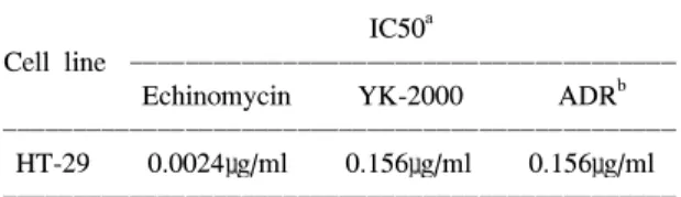
Table 1. Cytotoxic effect measurement of echinomycin and YK-2000 on various cell lines by MTT assay
ꠧꠧꠧꠧꠧꠧꠧꠧꠧꠧꠧꠧꠧꠧꠧꠧꠧꠧꠧꠧꠧꠧꠧꠧꠧꠧꠧꠧꠧꠧꠧꠧꠧꠧꠧꠧꠧꠧꠧꠧꠧꠧꠧꠧꠧꠧꠧꠧ
IC50a
Cell line ꠏꠏꠏꠏꠏꠏꠏꠏꠏꠏꠏꠏꠏꠏꠏꠏꠏꠏꠏꠏꠏꠏꠏꠏꠏꠏꠏꠏꠏꠏꠏꠏꠏꠏꠏꠏꠏꠏꠏ
Echinomycin YK-2000 ADRb
ꠏꠏꠏꠏꠏꠏꠏꠏꠏꠏꠏꠏꠏꠏꠏꠏꠏꠏꠏꠏꠏꠏꠏꠏꠏꠏꠏꠏꠏꠏꠏꠏꠏꠏꠏꠏꠏꠏꠏꠏꠏꠏꠏꠏꠏꠏꠏꠏ HT-29 0.0024μg/ml 0.156μg/ml 0.156μg/ml ꠏꠏꠏꠏꠏꠏꠏꠏꠏꠏꠏꠏꠏꠏꠏꠏꠏꠏꠏꠏꠏꠏꠏꠏꠏꠏꠏꠏꠏꠏꠏꠏꠏꠏꠏꠏꠏꠏꠏꠏꠏꠏꠏꠏꠏꠏꠏꠏ
aIC50: Inhibition concentration
bADR: Adriamycin
against HT-29 cancer celll line, IC50 values of test compounds were calculated. IC50 value of YK-2000 on HT-29 cell is 0.156μg/ml. IC50 value of echinomycin was 80 fold lower than that of YK-2000 (Table 1).
2) DNA fragmentation induced by echino- mycin and YK-2000
To define dose-response and time kinetics in HT-29 colon cancer cell line treated with echinomycin and YK-2000. HT-29 cells were exposed to the escalating concentrations of echinomycin or YK-2000 for 24 h and subjected to DNA fragmentation. DNA laddering was typically appeared on HT-29 cells when treated with echinomycin (0.2μg/ml) for 24 h (Fig. 2A). In parallel PARP cleavage was clearly shown in HT-29 cells, which were treated with various dose (0.2∼10 μg/ml) of echinomycin during 24 h (Fig. 2B). Next, we examined the time kinetics of DNA fragmentation with echinomycin (2μg/ml). DNA fragmentation was detected after 6 h of treatment with echinomycin (Fig.
2C). Immunoblot for PARP cleavage revealed the cleavage of 85 kDa fragment from whole PARP after echinomycin treatment (Fig. 2D). Meanwhile, increase
concentration of YK-2000 induced apoptotic DNA fragmentation (Fig. 3A). Apoptotic DNA fragment- ation was detected after 24 h of treatment with YK-2000 (10μg/ml)(Fig. 3C). To confirm the cleav- age of endogenous nuclear protein PARP in apoptotic cells, immunoblot was performed with anti-PARP antibody (Fig. 3B and 3D), corresponding to intact PARP cleavage in YK-2000 treated cells. PARP cleavage were increased by treatment of YK-2000, indicating apoptosis induced by YK-2000.
3) Effect of echinomycin and YK-2000 on cell death
The annexin-V-FITC binding assay was performed to confirm the degree of apoptosis in echinomycin or YK-2000 mediated cell death. Annexin-V binds to phosphatidylserine-expressed cells on the outer mem- brane of cell, while propidium iodide stains DNA of cells with the damaged cell membrane. Echinomycin and YK-2000 definitely produced a number of death cells, (25% and 16%) following 19h-exposure to YK-2000 (10μg/ml) and echinomycin (2μg/ml) respectively (Fig. 4). Apoptosis inducibility of YK- 2000 was likely more potent in terms of IC50 and drug Fig. 1. Chemical structures of echinomycin and YK-2000.
concentration inducing apoptosis compared to echino- mycin.
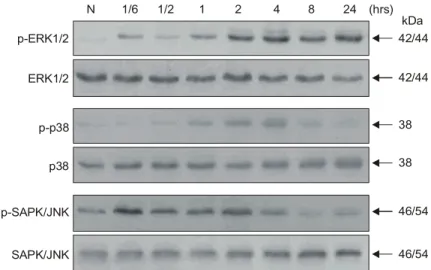
4) Echinomycin activates MAP kinase
HT-29 cells were treated with echinomycin (2μg/
ml) at regular time interval (10 min, 30 min, 1 h, 2 h, 4 h, 8 h and 24 h). Using phospho-specific anti- bodies against MAP kinase, ERK1/2 phosphorylation began quickly following echinomycin treatment and reached plateau at last 24 h. Meanwhile, SAPK/JNK was activated 10 min after echinomycin treatment and then the kinase activity was decreased. Contrary to p38 activation by YK-2000, phosphorylation of p38 began at 10∼30 min after echinomycin treatment and reached maximum peak at 4 h (Fig. 5).
5) Effect of YK-2000 on MAP kinase activ- ation
SAPK/JNK is part of a kinase cascade, which is strongly stimulated by various DNA-damaging agents.
To confirm MAP kinase activation by YK-2000, HT-29 cells were treated with YK-2000 (10μg/ml) and the phosphorylation status of three MAP kinse subfamilies (ERK1/2, p38 and JNK) in apoptotic cells were analyzed by immunoblotting with phospho-spe- cific antibodies. Surprisingly, ERK1/2 activity was peaked at 2 h after treatment of YK-2000. The SAPK/JNK and p38 activity after exposure of HT-29 cells to YK-2000 markedly decreased in activity and began at 4 to 8 h post treatment of YK-2000. Two MAP kinase (SAPK/JNK and p38) activation step Fig. 2. Induction of internucleosomal DNA fragmentation by echinomycin in HT-29. (A) HT-29 cells were cultured in media containing echinomycin at the indicated concentrations. HT-29 were treatment with echinomycin for the indicated time (24 hrs), after which total DNA was extracted and analyzed by agarose gel electrophoresis (M: DNA marker, N: naive HT-29). (B) Cleavage of 116 kDa PARP into an 85 kDa apoptotic fragment was monitored by immunoblot analysis. HT-29 cells were cultured with the indicated concentrations of echinomycin. After 24 h, cells were collected and analyzed for PARP cleavage. (C) Cells were cultured in media containing echinomycin (2μg/ml). At the indicated time points, cells were harvested and lysed. DNA fragmentation was examined by agarose gel electrophoresis (M: DNA marker, N: naive HT-29). (D) Cleavage of PARP after treatment with echinomycin (2μg/ml) for indicated time.
A
M N 0.2 1 2 10 ( g/ml)µ C
M N 6 12 24 48 (hrs)
B
N 0.2 1 2 10 ( g/ml)µ D
N 6 12 24 48 (hrs)
116 85
B-actin
kDa 116
85
B-actin kDa
forward with roughly similar time courses (Fig. 6).
DISCUSSION
Echinomycin is a novel antibiotic with diverse activity such as, DNA bisintercalation,20,21) CG base specific binding,5) potent cancericidal activity,22) tissue factor suppression23) and enhancement of peptide nucleic acid binding.24) To date, we proved that echinomycin is a strong apoptosis-inducer on mouse leukemia cells.9) Despite definite apoptosis inducibility on cancer cells by echinomycin, there is no clear evidence whether echinomycin, it's derivative or analogue triggers the ERK1/2, JNK or p38 kinases pathway or whether these compounds contribute to its apoptosis via MAP kinase pathway. In addition, the impact of echinomycin on human solid cancer cells Fig. 3. YK-2000 induced DNA fragmentation in HT-29 cells. (A) Cells were cultured in media containing YK- 2000 at the indicated concentrations (for 24 hrs) (M: DNA marker, N: naive HT-29). (B) Cleavage of 116 kDa PARP into an 85 kDa apoptotic fragment indicated concentration of YK-2000 (for 24 hrs).
(C) Cells were cultured in media containing YK-2000 (10μg/ml). At the indicated time points, cells were harvested. Purified DNA was separated by electrophoresis in 1.8% agarose gel at 100 V for 60 min. Ethidium bromide staining and UV illumination were used to visualize DNA fragmentation. (D) Cleavage of 116 kDa PARP into an 85 kDa apoptotic fragment after treatment with YK-2000 (10μg/ml) for indicated time.
A
M N 1 5 10 50 ( g/ml)µ C
M N 6 12 24 48 (hrs)
B
N 1 5 10 50 ( g/ml)µ D
N 6 12 24 48 (hrs)
116 85
B-actin
kDa 116
85
B-actin kDa
Fig. 4. Percentage of apoptosis in HT-29 cells treated with YK-2000 or echinomycin. HT-29 cells were treated with echinomycin (2μg/ml) or YK-2000 (10μg/ml) and incu- bated for 24 h. Results of flow cytometric analysis are presented as the mean±SE of two experiments.
was unknown. In the present study, we clearly demonstrated apoptosis on HT-29 colon cancer cell line with echinomycin and YK-2000, the echinomycin analogue as well as the role of MAP kinase pathway leading to apoptosis. YK-2000 was designed to circumvent echinomycin's hydrophobicity as well as to attenuate immune cell toxicity. Thus, we should not only expected the decreased cytotoxicity on cancer
cells but also more effective eradication of human colon cancer cells via induction of apoptosis.
Augmenting apoptosis through signaling pathway is essential to apoptosis- inducing antitumor agents.25) Apoptosis inducibility by YK-2000 was likely more potent than parent molecule, echinomycin.
Therefore, despite absence of in vivo study using YK-2000, our results warrant additional studies on the Fig. 5. Time-dependent changes in activities of ERK1/2, SAPK/JNK and p38 in HT-29 cells after exposure to echinomycin (2μg/ml). Immunoblots were probed with antibodies specific for total protein or activated phosphorylation form of ERK1/2, SAPK/JNK, p38.
N 1/6 1/2 1 2 4 8 24 (hrs)
p-ERK1/2
ERK1/2
p-p38
p38
p-SAPK/JNK
SAPK/JNK
42/44
42/44
38
38
46/54
46/54 kDa
Fig. 6. Time-dependent activation of ERK1/2 and reduction of SAPK/JNK and p38 by treatment of YK-2000 (10μg/ml) in HT-29 cells. Immunoblotting of phosphorylated ERK1/2, SAPK/JNK and p38.
N 1/6 1/2 1 2 4 8 24 (hrs)
p-ERK1/2
ERK1/2
p-p38
p38
p-SAPK/JNK
SAPK/JNK
42/44
42/44
38
38
46/54
46/54 kDa
therapeutic activity of YK-2000 in vivo. It should be pointed out that YK-2000 inhibited cell growth or decreased cell survival as shown in Table 1.
Most cancer chemotherapeutic agents induce apo- ptosis. However, signal transduction mechanisms reg- ulating apoptosis was not clearly defined, although proline-directed serine/threonine kinases of MAP kinase family were strongly implicated. Recent studies have shown that bisintercalator such as actinomycin, induce apoptosis in several types of solid cancers including human pancreas cancer cell line.26) Various chemically and biologically different agents have been found to mediate the induction of apoptosis through activation of p38.27∼30) Likewise, SAPK/JNK has been predominantly associated with stress responses such as apoptosis and cytokine release.31,32) During apoptosis process triggered by YK-2000, phosphorylation of ERK1/2 was increased, whereas phosphorylation of SAPK/JNK and p38 were downregulated in compari- son to control cell. These results suggest that acti- vation of ERK1/2 as well as downregulation of SAPK/JNK and p38 might mediate YK-2000 induced apoptotic process. Interestingly, MAP kinase pattern displayed by echinomycin and YK-2000 was similar but p38 phosphorylation is specifically different. In the naive colon cancer tissues, it was reported highly activation of SAPK/JNK and p38.33) In contrast to this pattern, SAPK/JNK and p38 were downregulated in apoptotic cells induced by YK-2000. This result is not consistent with other apoptosis models in which SAPK/JNK and p38 activation was crucial for apoptosis signaling. Such results suggest that higher SAPK/JNK and p38 activity is not required for induction of apoptosis in HT-29 cells by treatment of echinomycin and YK-2000. These and previous data support that cross-talk between ERK1/2 and p38 kinase signaling determines apoptosis.34,35) However, despite divergent role of two different signaling, it remained to be seen if which kinase would be survival or death signal in apoptosis induced by DNA damaging-bisintercalator such as echinomycin.
Another important implication in this study is that
cell death induced by bisintercalator may be mediated through MAP kinase pathway besides DNA binding.
Based on these findings, we suggest that YK-2000 may be a good candidate for additional evaluation as a cancer therapeutic agent for human colon cancer as well as other types of solid cancer implicated with aberrant expressions of MAP kinase.
These results indicate that echinomycin and novel synthetic echinomycin analogue may modulate MAP kinases activation in human HT-29 cell line.
ACKNOWLEDGEMENT
This work was supported by the academic research grant of Yonsei University Collage of Medicine for 2000.
REFERENCES
1) Rita D, Kambe M, Ishioka C, Kanamaru R. Induc- tion of p53-independent apoptosis associated with G2M arrest following DNA damage in human colon cancer cell lines. Jpn J Cancer Res 1997; 88: 39-43.
2) Peters GJ, van der Wilt CL, van Moorsel CJ, Kroep JR, Bergman AM, Ackland SP. Basis for effective combination cancer chemotherapy with antimetab- olites. Pharmacol Ther 2000; 87: 227-253.
3) Kaufmann SH, Earnshaw W. Induction of apoptosis by cancer chemotherapy. Exp Cell Res 2000; 256:
42-49.
4) Muss HB, Blessing JA, Baker VV, Barnhill DR, Adelson MD. Echinomycin (NSC 526417) in advan- ced ovarian cancer. A phase II trial of the Gyne- cologic Oncology Group. Am J Clin Oncol 1990; 13:
299-301.
5) Fletcher MC, Fox KR. Dissociation kinetics of echi- nomycin from CpG binding sites in different sequence environments. Biochemistry 1996; 35: 1064-1075.
6) Park YS, Kim YH, Kim SK, Choi SJ. A new antitumor agent: methyl sulfonium perchlorate of echinomycin. Bioorg Med Chem Lett 1998; 8: 731- 734.
7) Kim SK, Ahn CM, Kim TU, Choi SJ, Park YS, Shin WS, Koh CM. In vitro chemosensitivity test of SK- 302 on human colon carcinoma cell lines. Arch Pharm Res 1996; 19: 261-263.
8) Kim TU, Yang S, Kim SK. Cytotoxic and apoptotic effects of echinomycin on murine leukemia cells. J Biochem Mol Bio 1996; 29; 489-492.
9) Jeon H, Kim SS, Kim YS, Park YS, Kim YH, Choi SJ, Kim SK, Kim TU. Cytotoxic and apoptotic activities of echinomycin derivative (Echinomycin-7) on P388 murine leukemia cells. J Biochem Mol Biol 1998; 31: 560-564.
10) Kaufmann SH. Induction of endonucleolytic DNA cleavage in human acute myelogenous leukemia cells by etoposide, camptothecin, and other cytotoxic anti- cancer drugs: a cautionary note. Cancer Res 1989; 49:
5870-5878.
11) Kaufmann SH, Desnoyers S, Ottaviano Y, Davidson NE, Poirier GG. Specific proteolytic cleavage of poly (ADP-ribose) polymerase: an early marker of chemo- therapy-induced apoptosis. Cancer Res 1993; 53:
3976-3985.
12) Gardner AM, Johnson GL. Fibroblast growth factor- 2 suppression of tumor necrosis factor alpha-mediated apoptosis requires Ras and the activation of mitogen- activated protein kinase. J Biol Chem 1996; 273:
4928-4936.
13) Collett ED, Davidson LA, Fan YY, Lupton JR, Chapkin RS. n-6 and n-3 polyunsaturated fatty acids differentially modulate oncogenic Ras activation in colonocytes. Am J Physiol Cell Physiol 2001; 280:
1066-1075.
14) Coso OA, Chiariello M, Yu JC, Teramoto H, Crespo P, Xu N, Miki T, Gutkind JS. The small GTP-binding proteins Rac1 and Cdc42 regulate the activity of the JNK/SAPK signaling pathway. Cell 1995; 81: 1137- 1146.
15) Saleem A, Datta R, Yuan ZM, Kharbanda S, Kufe D.
Involvement of stress-activated protein kinase in the cellular response to 1-beta-D-arabinofuranosylcytosine and other DNA-damaging agents. Cell Growth Differ 1995; 6: 1651-1658.
16) Zanke BW, Boudreau K, Rubie E, Winnett E, Tibbles LA, Zon L, Kyriakis J, Liu FF, Woodgett JR. The stress-activated protein kinase pathway mediates cell death following injury induced by cis-platinum, UV irradiation or heat. Curr Biol 1996; 6: 606-613.
17) Bernges F, Burkle A, Kupper JH, Zeller WJ. Func- tional overexpression of human poly (ADP-ribose) polymerase in transfected rat tumor cells. Carcino- genesis 1997; 18: 663-668.
18) Janicke R, Ng P, Sprengart M, Porter A. Caspase-3 is required for-fodrin cleavage but dispensable for
cleavage of other death substrates in apoptosis. J Biol Chem 1998; 273: 15540-15545.
19) Le Rhun Y, Kirkland J, Shah G. Cellular responses to DNA damage in the absence of poly (ADP-ribose) polymerase. Biochem Biophys Res Commun 1998;
245: 1-10.
20) Chen H, Patel DJ. Solution structure of a quinomycin bisintercalator-DNA complex. J Mol Biol 1995; 246:
164-179.
21) Gao XL, Patel DJ. NMR studies of echinomycin bisintercalation complexes with d (A1-C2-G3-T4) and d (T1-C2-G3-A4) duplexes in aqueous solution: se- quence-dependent formation of Hoogsteen A1.T4 and Watson-Crick T1.A4 base pairs flanking the bisinter- calation site. Biochemistry 1998; 27: 1744-1751.
22) Cobb WR, Bogden AE, Reich SD, Griffin TW, Kelton DE, LePage DJ. Activity of two phase I drugs N-methylformamide (NSC-3051), Echinomycin (NSC- 526417) against fresh surgical explants of human tumors in the 6-day subrenal capsule (SRC) assay.
Invest New Drugs 1983; 1: 5-9.
23) Herbert JM, Lale A, Pereillo JM, Derocq JM, Casellas P. Echinomycin suppresses the pyrogenic effects of endotoxin and interleukin-1β in human endothelial cells and peripheral blood mononuclear cells. Thromb Haemost 1996; 75: 359-362.
24) Mollegaard NE, Bailly C, Waring MJ, Nielsen PE.
Quinoxaline antibiotics enhance peptide nucleic acid binding to double-stranded DNA. Biochemistry 2000;
39: 9502-9507.
25) Wu CH, Jeng JH, Wang YJ, Tseng CJ, Liang YC, Chen CH, Lee HM, Lin JK, Lin CH, Lin SY, Li CP, Ho YS. Antitumor effects of miconazole on human colon carcinoma xenografts in nude mice through induction of apoptosis and G0/G1 cell cycle arrest.
Toxicol Appl Pharmacol 2002: 180: 22-35.
26) Kleeff J, Kornmann M, Sawhney H, Korc M.
Actinomycin D induces apoptosis and inhibits growth of pancreatic cancer cells. Int J Cancer 2000; 86:
399-407.
27) Thrane EV, Refsnes M, Thoresen GH, Lag M, Sch- warze PE. Fluoride-induced apoptosis in epithelial lung cells involves activation of MAP kinases p38 and possibly JNK. Toxicol Sci 2001; 61: 83-91.
28) She QB, Bode AM, Ma WY, Chen NY, Dong Z.
Resveratrol-induced activation of p53 and apoptosis is mediated by extracellular-signal-regulated protein ki- nases and p38 kinase. Cancer Res 2001; 61: 1604- 1610.
29) Qiao D, Stratagouleas ED, Martinez JD. Activation and role of mitogen-activated protein kinases in deoxycholic acid-induced apoptosis. Carcinogenesis 2001; 22: 35-41.
30) Stadheim TA, Kucera GL. c-Jun N-terminal kinase/
stress-activated protein kinase (JNK/SAPK) is required for mitoxantrone- and anisomycin-induced apoptosis in HL-60 cells. Leuk Res 2002; 26: 55-65.
31) Basu S, Kolesnick R. Stress signals for apoptosis:
ceramide and c-Jun kinase. Oncogene 1998; 17: 3277- 3285.
32) Chuang SM, Wang IC, Yang JL. Roles of JNK, p38 and ERK mitogen-activated protein kinases in the growth inhibition and apoptosis induced by cadmium.
Carcinogenesis 2000; 21: 1423-1432.
33) Hardwick JC, van den Brink GR, Offerhaus GJ, van Deventer SJ, Peppelenbosch MP. NF-kappaB, p38 MAP kinase and JNK are highly expressed and active in the stroma of human colonic adenomatous polyps.
Oncogene 2001; 20: 819-827.
34) Montaner B, Perez-Tomas R. The cytotoxic prodi- giosin induces phosphorylation of p38-MAPK but not of SAPK/JNK. Toxicol Lett 2002; 24: 93-98.
35) Ding Q, Wang Q, Evers BM. Alterations of MAP kinase activities associated with intestinal cell dif- ferentiation. Biochem Biophys Res Commun 2001;
284: 282-288.