Apigenin Ameliorates Oxidative Stress-induced Neuronal Apoptosis in SH-SY5Y Cells
Yeo Jin Kim1, Eun Ju Cho1, Ah Young Lee2*, and Weon Taek Seo2*
1Department of Food Science and Nutrition and Kimchi Research Institute, Pusan National University, Busan 46241, Republic of Korea
2Department of Food Science, Gyeongsang National University, Jinju 52725, Republic of Korea
Received: September 9, 2020 / Revised: January 6, 2021 / Accepted: January 14, 2021
Introduction
Oxidative stress is defined as out of balance between the elimination and production of reactive oxygen spe- cies (ROS) and reactive nitrogen species (RNS) [1]. ROS and RNS, such as hydroxyl radical (·OH), nitric oxide radical (NO), hydrogen peroxide (H2O2), and superoxide
anion radical (O2−) are produced by external environ- mental factors and biochemical reactions [2]. Moderate levels of ROS and RNS play an essential role, either directly or indirectly, in health through mechanisms including cellular signaling, biosynthetic processes, and host defenses [3]. However, overproduction of ROS and RNS is thought to be responsible for damages to lipids, proteins, and DNA, leading to oxidative stress [4].
Oxidative stress can induce neuronal apoptosis and neuroinflammation in brain cells, resulting in neurode- generative diseases such as Alzheimer's disease (AD) and Parkinson's disease (PD) [5, 6]. The brain is espe- cially more vulnerable to oxidative stress than other organs, on account of its high oxygen consumption and The overproduction of reactive nitrogen species (RNS) and reactive oxygen species (ROS) causes oxidative damage to neuronal cells, leading to the progression of neurodegenerative diseases. In this study, we deter- mined the nitric oxide radical (NO), hydroxyl radical (·OH), and superoxide anion radical (O2−) scavenging activities of apigenin. Our results showed that apigenin exhibited remarkable, concentration-dependent
·OH, O2−, and NO radical scavenging activities. Particularly, apigenin indicated the strongest ·OH radical scavenging activity with 93.38% in the concentration of 100 µM. Furthermore, we also investigated the pro- tective effects of apigenin against hydrogen peroxide (H2O2)-induced oxidative stress in SH-SY5Y cells. The H2O2 treatment resulted in a significant decrease in cell viability, as well as an increase in lactate dehydro- genase (LDH) release and ROS production compared with the H2O2-nontreated SH-SY5Y cells. However, the cell viability significantly increased in the apigenin-treated group, as well as inhibited ROS generation and LDH release compared with the H2O2-induced control group. To elucidate the protective mechanisms of apigenin against oxidative stress in SH-SY5Y, we analyzed the apoptosis-related protein expression. The apigenin treatment resulted in the downregulated expression of apoptosis-related protein markers, such as cytochrome C, cleaved caspase-3, poly (ADP)-ribose polymerase (PARP), and B-cell lymphoma 2-associated X (Bax), as well as the upregulated expression of anti-apoptosis markers such as B-cell lymphoma 2 (Bcl-2).
In this study, we report that apigenin exhibits a neuroprotective effect against oxidative stress in SH-SY5Y cells. These results suggest that apigenin may be considered as a potential agent for neurodegenerative dis- ease prevention.
Keywords: Hydrogen peroxide, neuronal, neurodegenerative disease, neuroprotection, oxidative stress
*Corresponding authors A. Y. Lee
Tel: +82-55-772-3278, Fax: +82-55-772-3279 E-mail: [email protected]
W. T. Seo
Tel: +82-55-772-3276, Fax: +82-55-772-3279 E-mail: [email protected]
amount of polyunsaturated fatty acids. The neurons are especially sensitive to free radicals, which can lead to cell death [7].
SH-SY5Y neuronal cells have been widely used to characterize neuron-like behavior when investigating potential neuroprotective activity [8]. Recently, studies have confirmed that oxidative stress contributes to apop- tosis and inflammation in SH-SY5Y neuronal cells [9, 10]. H2O2 is related to pathological processes of chronic and acute neuronal toxicity [11]. ROS production and apoptosis-related protein expression of Bax, caspase-3 and cytochrome C are induced by H2O2 in SH-SY5Y cells [12]. Therefore, H2O2-induced SH-SY5Y cells are well used to research the promising agents with protective effects from oxidative stress.
Apigenin (4′,5,7-trihydroxyflavone), non-mutagenic and non-toxic natural plant flavonoid, is found widely in plants, vegetables, tea leaves, and fruits [13, 14]. Previous studies demonstrated that apigenin has beneficial effects, such as anti-oxidant [15], anti-inflammation [16], anti-apoptosis activity [17], and anti-cancer prop- erties [18]. In addition, administration of apigenin improved the memory deficit of amyloid beta (Aβ) trans- genic mice [19]. Apigenin attenuated lipopolysaccharide- induced myocardial damage by inhibiting inflammatory cytokines and their regulatory factor, nuclear factor kappa B [20]. Apigenin has also been reported to inhibit apoptosis through the regulation of mitogen-activated protein kinase (MAPK) induced by Aβ in neuronal cells [21]. However, the protective effect of apigenin for H2O2- induced neurotoxicity and neuronal apoptosis in SH- SY5Y cells have not been examined yet. Thus, the cur- rent study investigated the O2−, ·OH, and NO radical scavenging effects of apigenin in vitro. In addition, the protective effect and underlying mechanisms of apigenin against oxidative damage induced by H2O2 were con- firmed using SH-SY5Y neuronal cells.
Materials and Methods
Reagents
Dimethyl sulfoxide (DMSO), 3-(4,5-dimethylthiazol-2- yl)-2,5-diphenyltetrazolium bromide (MTT), and 2′,7′- dichloro-fluorescein diacetate (DCF-DA) were obtained from Sigma-Aldrich Inc. (USA). Sodium pentacyanoni- trosylferrate (III) dihydrate (SNP) and H2O2 were
supplied by Junsei Chemical Co. (Japan). The LDH cytotoxicity detection kit was obtained from Takara Bio (Japan). Trichloroacetic acid (TCA) was purchased from Biosesang Inc. (Korea). Radioimmunoprecipitation assay (RIPA) buffer was purchased from Elpis Biotech.
(Korea). Phenazine methosulfate (PMS) and NADH were obtained from Bio Basic Inc. (USA). Polyvinylidene fluoride (PVDF) membrane was obtained from Millipore Co. (USA). Thiobarbituric acid (TBA) was purchased from Acros Organics Inc. (USA).
Sample preparation
Apigenin (4′,5,7-trihydroxyflavone) was obtained from Sigma-Aldrich Inc. and had a purity > 95% based on HPLC analysis. Apigenin was dissolved in DMSO as a stock solution and diluted with dulbecco's modified eagle's medium (DMEM) prior to use.
·OH scavenging assay
·OH radical scavenging activity was determined follow to the method reported by Chung et al. [22]. Various con- centrations of apigenin (25, 50, 100, and 250 µM) were added to the reaction mixture containing 10 mM H2O2 and 10 mM FeSO4·H2O-EDTA with 10 mM 2-deoxyribose solution and incubated at 37℃ for 4 h. After incubation, 1.0% TBA and 2.8% TCA were added to the mixture and boiled for 20 min at 100℃. ·OH radical scavenging ability was measured at 490 nm by an RT-6100 microplate reader (Rayto Life and Analytical Sciences Co. Ltd., China).
O2− scavenging assay
O2− radical scavenging activity was estimated in accor- dance of the method reported by Nishikimi et al. [23].
Various concentrations of apigenin (25, 50, 100, and 250 µM) were added to the reaction mixture containing 100 µl of 0.1 M Tris-HCl (pH 7.4), 200 µl of 100 mM PMS, 400 µl of 500 mM NADH and 200 µl of 0.5 mM NBT. After 10 min, the absorbance was read at 560 nm utilizing an RT-6100 microplate reader (Rayto Life and Analytical Sciences Co. Ltd.).
NO scavenging assay
The NO radical scavenging activity of apigenin was measured following to the method reported by Marcocci et al. [24]. Various concentrations of apigenin (25, 50,
100, and 250 µM) diluted in MeOH were mixed with 10 mM SNP, and incubated at room temperature for 150 min. Griess reagent was added at a ratio of 1:1 in a 96-well plate for 30 min at room temperature. The absorbance was measured at 540 nm using an RT-6100 microplate reader (Rayto Life and Analytical Sciences Co. Ltd.).
Cell culture
SH-SY5Y cells were purchased from KCLB (Korea Cell Line Bank, Korea). The cells were cultured with 100 units/ml penicillin-streptomycin and 10% fatal bovine serum based DMEM at 37℃ with 5% CO2 in an incuba- tor. The culture medium was replaced three times per week. Cells were sub-cultured with 0.05% trypsin-EDTA in phosphate buffered saline (PBS). Cells were used for experimentation when confluence was ~ 80%.
MTT assay
Cells were seeded in a 96-well plate that at a density of 5 × 10⁴ cells/ml and cultured for 24 h. The cells were treated with different concentrations of apigenin (0.1, 0.5, and 1 µM) for 3 h, followed by 300 µM of H2O2 treat- ment for 24 h to induce oxidative stress. After incuba- tion, 5 mg/ml MTT solution was added and allowed to react for 4 h. The incorporated formazan crystals in the viable cells were solubilized using DMSO. The absor- bance was read at 540 nm using an RT-6100 microplate reader (Rayto Life and Analytical Sciences Co. Ltd.).
DCF-DA assay
To measure the ROS scavenging activity, the DCF-DA assay was used [25]. SH-SY5Y cells were seeded in 96- well plate at a density of 5 × 10⁴ cells/ml and cultured for 24 h. Apigenin was added to each well in diverse concen- trations (0.1, 0.5, and 1 µM) and incubated for 3 h. To induce oxidative stress, 300 µM of H2O2 was added for 24 h. After incubation, 80 µM DCF-DA was added and left to react for 30 min. Fluorescence was read using a fluorescence spectrophotometer (FLUOstar OPTIMA, BMG Labtech, Germany) at an excitation of 480 nm and an emission of 535 nm.
LDH release assay
SH-SY5Y cells were seeded in a 96-well plate at a
density of 5 × 10⁴ cells/ml. Apigenin (0.1, 0.5, and 1 µM) was added for 3 h, followed by treatment with 300 µM H2O2. The supernatant and reaction mix were added to a 96-well plate and incubated at room temperature for 30 min. Absorbance was read at 540 nm by an RT-6100 microplate reader (Rayto Life and Analytical Sciences Co. Ltd.).
Western blotting analysis
SH-SY5Y cells were seeded in a cell culture dish and incubated for 24 h. After incubation, the cells were pre- treated with apigenin (0.1, 0.5, and 1 μM) for 3 h and treated with 300 μM of H2O2 for another 24 h. Protein extracts from the SH-SY5Y cells were prepared using lysis buffer. Proteins were separated by electrophoresis using 10% or 13% sodium dodecyl sulfate polyacryl- amide gels (SDS-PAGE), then transferred to the PVDF membranes. Proteins were blocked using 5% skimmed milk for 60 min, and washed with PBS. Membranes were incubated with the primary antibody overnight at 4℃ [each antibody at a dilution of 1:1000; poly (ADP)- ribose polymerase (PARP), cleaved caspase-3, cyto- chrome C, B-cell lymphoma 2-associated X (Bax), B-cell lymphoma 2 (Bcl-2), and β-actin]. Membranes were then incubated with the appropriate HRP-conjugated second- ary antibodies for 1 h. Expression of protein was visual- ized utilizing a chemiluminescent imaging system (Davinci Chemi, Korea).
Statistical analysis
Results were stated as means ± standard deviation (SD). SPSS (version 25; SPSS Inc., USA) was used to perform analysis of variance (ANOVA), followed by
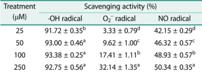
Table 1. ·OH, O2−, and NO radical scavenging activity of api- genin.
Treatment (µM)
Scavenging activity (%)
·OH radical O2− radical NO radical 25 91.72 ± 0.35b 03.33 ± 0.79d 42.15 ± 0.29d 50 93.00 ± 0.46a 09.62 ± 1.00c 46.32 ± 0.57c 100 93.38 ± 0.25a 17.41 ± 1.11b 48.93 ± 0.57b 250 92.75 ± 0.56a 32.14 ± 1.35a 50.34 ± 0.35a Values are mean ± SD.
a-dMeans are significantly different (p < 0.05) by Duncan’s multiple range test.
Duncan’s post-hoc tests. Significance was set at p < 0.05.
Results
Effect of apigenin on ·OH, O2−, and NO radical scavenging activity
The ·OH, O2−, NO radical scavenging activity of api- genin is shown in Table 1. All concentrations of apigenin (25, 50, 100, and 250 μM) showed more than 90% ·OH radical scavenging activities. Of note, the strongest ·OH radical scavenging activity was 93.38% at the concentra- tion of 100 μM. Apigenin also showed O2− radical scav- enging ability concentration-dependently. Apigenin revealed dose-dependent radical scavenging activity against NO. Furthermore, apigenin showed a NO radi- cal scavenging effect of 50.34% at 250 μM. Therefore, apigenin has antioxidant capabilities by the scavenging of ·OH, O2−, and NO radicals.
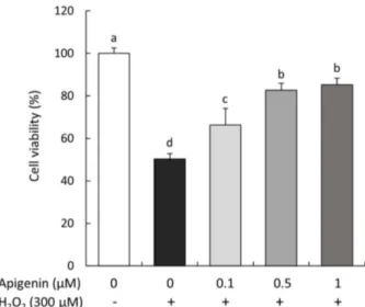
Protective effect of apigenin on cell viability in SH-SY5Y cells treated with H2O2
The effect of apigenin on cell viability of SH-SY5Y cells treated with H2O2 was investigated by the MTT assay (Fig. 1). The results indicated that H2O2-treated cells were significantly decreased cell viability to 50.26% com- pared to H2O2-non treated cells (100%). However, treat- ment with apigenin increased cell viability to 66.28%, 82.69%, and 85.16% at 0.1, 0.5, and 1 μM, respectively.
These results indicated that apigenin had protective effect from H2O2-induced cell death in SH-SY5Y cells.
Protective effect of apigenin on ROS release in SH-SY5Y cells treated with H2O2
To establish the protective effect of apigenin on ROS generation in H2O2-induced SH-SY5Y cells, the DCF-DA assay was performed. After 60 min, ROS production was increased in the H2O2-treated cells compared to the H2O2-non treated cells, indicating that H2O2 induced ROS overproduction (Fig. 2A). Compared to 100% of the
Fig. 2. Effect of apigenin on ROS production in SH-SY5Y cells treated with H2O2. Time course of change in intensity of DCF fluorescence during 60 min (A). Intensity of ROS produc- tion at 60 min (B). Values are mean ± SD. a-dMeans are signifi- cantly different (p < 0.05) by Duncan’s multiple range test.
Fig. 1. Effect of apigenin on cell viability in SH-SY5Y cells treated with H2O2. Values are mean ± SD. a-dMeans are signifi- cantly different (p < 0.05) by Duncan’s multiple range test.
300 μM H2O2-only treated group, ROS production in the non-treated group was 87.47% (Fig. 2B). Whereas treat- ment of the cells with apigenin showed inhibitory effects on ROS formation, showing 93.33%, 91.89%, and 90.82%
at concentrations of 0.1, 0.5, and 1 µM, respectively.
These results confirmed that ROS generation in SH- SY5Y neuronal cells treated with H2O2 was attenuated by apigenin.
Protective effect of apigenin on LDH elimination in SH- SY5Y cells treated with H2O2
The inhibitory effect of apigenin on LDH release in H2O2-treated SH-SY5Y cells was shown in Fig. 3. The levels of LDH were significantly increased in the group treated with only H2O2 (40.99%) compared to non- treated group (0.84%). However, treatment with api- genin (0.1, 0.5, and 1 μM) significantly inhibited LDH release by 27.49%, 25.18%, and 22.31%, respectively.
These finding suggest that apigenin has protective effect against neuronal oxidative damage caused by H2O2.
Protective effect of apigenin on apoptosis-related protein expressions in SH-SY5Y cells treated with H2O2
To confirm the protective mechanisms of apigenin against H2O2-induced apoptosis, we examined cyto- chrome C, cleaved caspase-3, cleaved PARP/PARP, and Bax/Bcl-2 protein expression by Western blotting in SH-
SY5Y cells treated with H2O2 (Fig. 4). In our results, the increase of Bax expression and decrease of Bcl-2 expres- sion were observed, in H2O2-treated control groups, resulting in significant increase in the Bax/Bcl-2 protein ratio. However, apigenin-treated groups showed a down- regulation of the Bax/Bcl-2 levels, which was similar to the H2O2-non treated group. In addition, the protein levels of cleaved PARP/PARP, cytochrome C, and cleaved caspase-3 were significantly increased in the H2O2-induced control cells compared with the H2O2-non induced cells. Nevertheless, the levels of cytochrome C, cleaved PARP/PARP, and cleaved caspase-3 protein expression were significantly decreased in apigenin- treated groups. In this study, our results indicated that apigenin protected from oxidative stress-induced neuro- nal cell death via regulation of the apoptosis signaling pathway.
Discussion
ROS and RNS play an important part in physiological functions, acting as molecular signals in the body. Over- production of ROS and RNS are causative of harmful reactions, which are associated with a high risk of oxida- tive damage to organs, resulting in the progression of degenerative diseases [26]. In particular, oxidative stress in the brain is strongly connected in neurodegen- erative diseases which are AD, PD, and Huntington’s disease [27]. The brain is insufficient for oxidative defense system and is more vulnerable to oxidative stress than other organs. Therefore, lipids, proteins, and DNA can be damaged by excessive production of ROS and RNS in the brain [7, 28]. Many previous studies have indicated that supplements of anti-oxidant may prevent the progression of neurodegenerative diseases, and may mitigate ROS-induced neuronal damage [27, 29]. Notably, natural anti-oxidants isolated from various plants are receiving more attention for the prevention and treatment of neurodegenerative disorders [30, 31].
Apigenin, one of the flavonoids with low toxicity, is found widely in plants, such as parsley, onions, oranges, and herb teas [13, 14]. Apigenin has been reported to have various beneficial effects including anti-apoptotic, anti- inflammatory, and anti-bacterial effects [14, 16, 32].
According to Balez et al. [33], apigenin possessed neuro- protective effects against inflammation, apoptosis in Fig. 3. Effect of apigenin on LDH release in SH-SY5Y cells
treated with H2O2. Values are mean ± SD. a-dMeans are signifi- cantly different (p < 0.05) by Duncan’s multiple range test.
induced pluripotent stem cell-derived neurons by atten- uating Ca2+ signaling and caspase-3/7 activity. In addi- tion, apigenin protected neuronal apoptosis by inhibiting the caspase-3 activity in H2O2-treated SH-SY5Y neuro- nal cells [34]. As mentioned above, oxidative stress is an imbalance of oxidant and antioxidant defense systems caused by stressors of external and internal in the body.
In addition, neurons in the brain are confirmed fastest degenerated, due to the lack of an antioxidant defense system than other institutions and susceptible to oxida- tive stress [35, 36]. Consistent with these reports, that oxidative stress caused by ROS interferes with cell func- tion and ultimately leads to cell death [37]. Previous
study has confirmed that AD patients had increased levels of oxidative stress in the cells which the most vul- nerable areas of the brain tissue [38]. Furthermore, oxidative stress affects synaptic transmission and ulti- mately could leads to a memory dysfunction due to oxi- dative stress [39]. Therefore, recent researches have been promoted on the prevention of diseases and protec- tive effect of neuronal cells based on the preventing or delaying of cell death. However, the protective effect of apigenin that inhibiting mitochondria-mediated apop- totic signaling pathway in SH-SY5Y neuronal cells has not been studied. The present study is focused on the radical scavenging of apigenin and its neuroprotective Fig. 4. Effects of apigenin on the levels of Bax/Bcl-2 (A) cytochrome C (B) cleaved caspase-3 (C) cleaved PARP/PARP (D) protein expression in SH-SY5Y cells treated with H2O2. Bax, B-cell lymphoma 2-associated X; Bcl-2, B-cell lymphoma 2; cyto- chrome C; cleaved caspase-3; PARP, poly (ADP)-ribose polymerase. Values are mean ± SD. a-eMeans are significantly different (p < 0.05) by Duncan’s multiple range test. β-actin was used as a loading control.
effects on oxidative stress-induced neuronal cell death by analyzing protein expressions participate in these effect.
The ·OH radical, which isthe most reactive ROS, can be formed by a Fenton-type reaction, leading to severe damage in adjacent biomolecules [40]. The O2− radical is a precursor of other major ROS such as ·OH radical and H2O2, which can damage biomolecules and alter physio- logical functions [41, 42]. Furthermore, overproduction of NO radical has been reported to induce DNA fragmen- tation, cell membrane damage, and neuronal cell death [43]. In this study, apigenin exerted strong ·OH, O2−, and NO radical scavenging activities in a concentration- dependently. Although lacking a hydroxyl group at posi- tion 3 and a catechol structure in the B-ring, apigenin has a double bond at the 2, 3 carbon that leads to a more reactive structure possessing anti-oxidant capacities [32, 44, 45]. Therefore, the strong radical scavenging ability imply attributed to the double bond at the 2, 3 carbon position.
SH-SY5Y cells are phenotypically and genetically homogeneous cells that can be used to study the rela- tionship between molecules and cell physiology [8].
Therefore, SH-SY5Y cells induced by oxidative stress are extensively used as a model system for investigating neuroprotective properties [9, 10]. Previous studies have indicated that H2O2 treatment significantly decreased cell viability via an oxidative-dependent apoptotic pro- cess [46, 47]. In this study, SH-SY5Y neuronal cells treated with H2O2 were used as an experimental model to examine the role of apigenin to protect against apopto- sis induced by oxidative stress. Apigenin-treated SH- SY5Y cells had an antioxidant effect by increasing cell viability and reducing ROS production. In addition, treatment with apigenin significantly decreased levels of LDH release, indicating that apigenin had protective effects against oxidative damage caused by H2O2.
The relative ratios of anti-apoptosis and pro-apoptosis Bcl-2 family proteins have been linked to various patho- logical conditions characterized by cell death [48].
Related proteins of the Bcl-2 family include anti-apopto- sis members (Bcl-2, Bcl-w, and Bcl-XL) and pro-apoptosis members (Bax, Bak, Bad, Bik, and Bid) [49]. The BH3 protein is a Bcl-2 homology protein and functions as a trans-dominant inhibitor of Bcl-2. However, the BH3 domain of Bax is heterodimerized with Bcl-2 for migra-
tion to the mitochondria via oligomerization to release cytochrome C, thereby causing apoptosis [50, 51]. There- fore, the ratio of the pro-apoptosis protein Bax to the anti-apoptosis protein Bcl-2 is important for regulating the release of caspase activity-inducing factors from mitochondria. Apigenin attenuated doxorubicin-induced cardiotoxicity via the reduction of oxidative stress and apoptosis in male rats, showing a marked decrease in the apoptotic protein Bax and caspase-3 as well as an increase in the anti-apoptosis marker Bcl-2 [52]. More- over, apigenin inhibited patulin-induced apoptosis in HEK 293 cells via up-regulation of Bcl-2 and down-regu- lation of Bax, leading to inhibition of caspase activation [53]. In present study, apigenin significantly reduced Bax level and increased Bcl-2 level. Therefore, these results showed that apigenin exerted neuroprotective effects against oxidative stress via the regulation of apoptosis-related protein in SH-SY5Y cells.
Cytochrome C is released into the cytosol from the mitochondrial intermembrane space, promoting caspase- 3 activation and eventually causing neuronal apoptosis [54]. In addition, caspase-3 is the most important end- cleaving enzyme in the process of apoptosis, cleaving the DNA repair enzyme, PARP, and inducing cell apoptosis [55]. When caspases are activated, that bind with PARP and cleave it, thereby preventing DNA repair and induc- ing apoptosis. Thus, PARP protein is considered as a marker of apoptosis. The results indicated that apigenin protected against H2O2 by down-regulation of cyto- chrome C, cleaved caspase-3, and PARP protein expres- sion in neuronal cells. A previous study indicated that apigenin protects against proteasome inhibitor-induced apoptosis in neuronal cells through regulation of cyto- chrome C, Bax/Bcl-2, caspases (-8, -9 and -3), and PARP [56]. Apigenin has been also reported to show neuropro- tective effects on subarachnoid hemorrhage-induced neuronal apoptosis via down-regulation of Bax and caspase-3 expression levels [57]. In present study, cyto- chrome C, PARP, and cleaved caspase-3 were up-regu- lated by treatment with H2O2 in SH-SY5Y cells.
However, apigenin treatment inhibited H2O2-induced cytochrome C, cleaved PARP/PARP, and cleaved caspase-3 protein expressions, suggesting that apigenin exerted its neuroprotective effect through down-regula- tion of the apoptotic signaling pathway. Apoptosis refers to a complex process involving condensation of the
nucleus, cytoplasm, and chromatin [58, 59]. Early and late apoptosis can be distinguished by the time of occur- rence of apoptosis in the central nervous system. In addition, early apoptosis is related to the proliferation site, whereas late apoptosis is related to the post-mitosis site [60]. Early cell death is characterized by exposure of the translocated phosphatidylserine (PS) to the outer plasma membrane surface [61]. Therefore, flow cytomet- ric analysis using annexin V and propidium iodide (PI), which is an externalized binding of PS, can confirm an indicator of early cell death. This staining can differenti- ate between early, late apoptotic and viable cells. Previ- ous study has confirmed that apigenin prevented rate of early apoptosis through double staining with annexin V- FITC and PI in oxygen and glucose deprivation/reperfu- sion-induced neuronal cell injury [62]. Although neuro- protective of apigenin on early-apoptosis and underlying mechanism should be further studied in H2O2-induced SH-SY5Y neuronal cells, apigenin may contribute to protecting neuronal cells against oxidative stress by reg- ulation of apoptosis signaling pathway involving Bax/
Bcl-2, cytochrome C release, and caspase activation.
In conclusion, apigenin showed effective ·OH, O2−, and NO radical scavenging activity. In particular, apigenin (25, 50, 100, and 250 μM) showed strong ·OH radical scavenging ability, more than 90%. Furthermore, api- genin increased the cell viability and decreased the ROS generation in H2O2-treated SH-SY5Y cells. The release of LDH induced by H2O2 was also reduced by treatment with apigenin. Moreover, apigenin exhibited anti-apop- totic effects against H2O2, as shown by down-regulation of cytochrome C, cleaved capase-3, cleaved PARP, and Bax expression levels, and up-regulation of Bcl-2 expres- sion level. In present study, we suggest that apigenin exerts a neuro-protective effect against oxidative stress in SH-SY5Y neuronal cells, and could potentially be used as a preventative agent in neurodegenerative disease.
Acknowledgments
This work was supported by Gyeongnam National University of Sci- ence and Technology Grant in 2020~2022.
Conflict of Interest
The authors have no financial conflicts of interest to declare.
References
1. Davalli P, Mitic T, Caporali A, Lauriola A, D'Arca D. 2016. ROS, cell senescence, and novel molecular mechanisms in aging and age- related diseases. Oxid. Med. Cell. Longev. 2016: 3565127.
2. Del Rio LA. 2015. ROS and RNS in plant physiology: an overview.
J. Exp. Bot. 66: 2827-2837.
3. Phaniendra A, Jestadi DB, Periyasamy L. 2015. Free radicals: prop- erties, sources, targets, and their implication in various diseases.
Indian J. Clin. Biochem. 30: 11-26.
4. Islam MT. 2017. Oxidative stress and mitochondrial dysfunction- linked neurodegenerative disorders. Neurol. Res. 39: 73-82.
5. Bhat AH, Dar KB, Anees S, Zargar MA, Masood A, Sofi MA, et al.
2015. Oxidative stress, mitochondrial dysfunction and neurode- generative diseases; a mechanistic insight. Biomed. Pharmacother.
74: 101-110.
6. Niu X, Zheng S, Liu H, Li S. 2018. Protective effects of taurine against inflammation, apoptosis, and oxidative stress in brain injury. Mol. Med. Rep. 18: 4516-4522.
7. Cobley JN, Fiorello ML, Bailey DM. 2018. 13 Reasons why the brain is susceptible to oxidative stress. Redox Biol. 15: 490-503.
8. Forster JI, Köglsberger S, Trefois C, Boyd O, Baumuratov AS, Buck L, et al. 2016. Characterization of differentiated SH-SY5Y as neu- ronal screening model reveals increased oxidative vulnerability.
J. Biomol. Screen 21: 496-509.
9. Park HR, Lee H, Park H, Jeon JW, Cho WK, Ma JY. 2015. Neuropro- tective effects of Liripope platyphylla extract against hydrogen peroxide-induced cytotoxicity in human neuroblastoma SH- SY5Y cells. BMC Complement. Med. Ther. 15: 171.
10. Zhang B, Cui Y, Wang L, Zhao L, Hou C, Zeng Q, et al. 2018. Auto- phagy regulates high concentrations of iodide-induced apoptosis in SH-SY5Y cells. Toxicol. Lett. 284: 129-135.
11. Nirmaladevi D, Venkataramana M, Chandranayaka S, Ramesha A, Jameel NM, Srinivas C. 2014. Neuroprotective effects of bikaverin on H2O2-induced oxidative stress mediated neuronal damage in SH-SY5Y cell line. Cell. Mol. Neurobiol. 34: 973-985.
12. Yang R, Wei L, Fu QQ, Wang H, You H, Yu HR. 2016. SOD3 amelio- rates H2O2 induced oxidative damage in SH-SY5Y cells by inhibit- ing the mitochondrial pathway. Neurochem. Res. 41: 1818-1830.
13. Shi MD, Shiao CK, Lee YC, Shih YW. 2015. Apigenin, a dietary flavonoid, inhibits proliferation of human bladder cancer T-24 cells via blocking cell cycle progression and inducing apoptosis.
Cancer Cell Int. 15: 33.
14. Chen XJ, Wu MY, Li DH, You J. 2016. Apigenin inhibits glioma cell growth through promoting microRNA-16 and suppression of Bcl-2 and nuclear factor-kappaB/MMP9. Mol. Med. Rep. 14: 2352- 2358.
15. Kowalska I, Adach W, Stochmal A, Olas B. 2020. A comparison of the effects of apigenin and seven of its derivatives on selected biomarkers of oxidative stress and coagulation in vitro. Food.
Chem. Toxicol. 136: 111016.
16. Amini R, Yazdanparast R, Ghaffari SH. 2015. Apigenin modulates the expression levels of pro-inflammatory mediators to reduce
the human insulin amyloid-induced oxidant damages in SK-N- MC cells. Hum. Exp. Toxicol. 34: 642-653.
17. Sun X, Min D, Wang Y, Hao L. 2015. Potassium aspartate inhibits SH-SY5Y cell damage and apoptosis induced by ouabain and H2O2. Mol. Med. Rep. 12: 2842-2848.
18. Sharma A, Ghani A, Sak K, Tuli HS, Sharma AK, Setzer WN, et al.
2019. Probing into therapeutic anti-cancer potential of apigenin:
recent trends and future directions. Recent. Pat. Inflamm. Allergy Drug. Discov. 13: 124-133.
19. Zhao L, Wang JL, Liu R, Li XX, Li JF, Zhang L. 2013. Neuroprotec- tive, anti-amyloidogenic and neurotrophic effects of apigenin in an Alzheimer's disease mouse model. Molecules 18: 9949-9965.
20. Li F, Lang F, Zhang H, Xu L, Wang Y, Zhai C, et al. 2017. Apigenin alleviates endotoxin-induced myocardial toxicity by modulating inflammation, oxidative stress, and autophagy. Oxid. Med. Cell.
Longev. 2017: 2302896.
21. Zhao L, Wang JL, Wang YR, Fa XZ. 2013. Apigenin attenuates cop- per-mediated beta-amyloid neurotoxicity through antioxidation, mitochondrion protection and MAPK signal inactivation in an AD cell model. Brain Res. 1492: 33-45.
22. Chung SK, Osawa T, Kawakishi S. 1997. Hydroxyl radical-scavenging effects of spices and scavengers from brown mustard (Brassica nigra). J. Biosci. Biotechnol. Biochem. 61: 118-123.
23. Nishikimi M, Appaji N, Yagi K. 1972. The occurrence of superox- ide anion in the reaction of reduced phenazine methosulfate and molecular oxygen. Biochem. Biophys. Res. Commun. 46: 849- 854.
24. Marcocci L, Packer L, Droy-Lefaix MT, Sekaki A, Gardes-Albert M.
1994. Antioxidant action of Ginkgo biloba extract EGb 761.
Methods Enzymol. 234: 462-475.
25. Cathcart R, Schwiers E, Ames BN. 1984. Detection of picomole levels of lipid hydroperoxides using a dichlorofluorescein fluo- rescent assay. Methods Enzymol. 105: 352-358.
26. Niedzielska E, Smaga I, Gawlik M, Moniczewski A, Stankowicz P, Pera J, et al. 2016. Oxidative stress in neurodegenerative diseases.
Mol. Neurobiol. 53: 4094-4125.
27. Cai L, Wang LF, Pan JP, Mi XN, Zhang Z, Geng HJ, et al. 2016. Neu- roprotective effects of methyl 3,4-dihydroxybenzoate against TBHP-induced oxidative damage in SH-SY5Y cells. Molecules 21:
1071.
28. Valko M, Jomova K, Rhodes CJ, Kuca K, Musilek K. 2016. Redox- and non-redox-metal-induced formation of free radicals and their role in human disease. Arch. Toxicol. 90: 1-37.
29. Enogieru AB, Haylett W, Hiss DC, Bardien S, Ekpo OE. 2018. Rutin as a potent antioxidant: implications for neurodegenerative disorders. Oxid. Med. Cell. Longev. 2018: 6241017.
30. Achete de Souza G, de Marqui SV, Matias JN, Guiguer EL, Bar- balho SM. 2020. Effects of Ginkgo biloba on diseases related to oxidative stress. Planta. Med. 86: 376-386.
31. Pezeshki-Nia S, Asle-Rousta M, Mahmazi S. 2020. Spinacia olera- cea L. extract attenuates hippocampal expression of TNF-alpha and IL-1beta in rats exposed to chronic restraint stress. Med. J.
Islam. Repub. Iran 34: 10.
32. Salehi B, Venditti A, Sharifi-Rad M, Kregiel D, Sharifi-Rad J, Durazzo A, et al. 2019. The therapeutic potential of apigenin. Int.
J. Mol. Sci. 20: 1305.
33. Balez R, Steiner N, Engel M, Muñoz SS, Lum JS, Wu Y, et al. 2016.
Neuroprotective effects of apigenin against inflammation, neu- ronal excitability and apoptosis in an induced pluripotent stem cell model of Alzheimer’s disease. Sci. Rep. 12: 31450.
34. Kang SS, Lee JY, Choi YK, Kim GS, Han BH. 2004. Neuroprotective effects of flavones on hydrogen peroxide-induced apoptosis in SH-SY5Y neuroblostoma cells. Bioorg. Med. Chem. Lett. 3: 2261- 2264.
35. Castelli V, Benedetti E, Antonosante A, Catanesi M, Pitari G, Ippoliti R, et al. 2019. Neuronal cells rearrangement during aging and neurodegenerative disease: metabolism, oxidative stress and organelles dynamic. Front. Mol. Neurosci. 28: 132.
36. Wang X, Michaelis EK. 2010. Selective neuronal vulnerability to oxidative stress in the brain. Front. Aging. Neurosci. 2: 12.
37. Zhang L, Yu H, Sun Y, Lin X, Chen B, Tan C, et al. 2007. Protective effects of salidroside on hydrogen peroxide-induced apoptosis in SH-SY5Y human neuroblastoma cells. Eur. J. Pharmacol. 564:
18-25.
38. Mattson MP, Pedersen WA, Duan W, Culmsee C, Camandola S.
1999. Cellular and molecular mechanisms underlying perturbed energy metabolism and neuronal degeneration in Alzheimer's and Parkinson's diseases. Ann. NY Acad. Sci. 893: 154-175.
39. Lee DZ, Chung JM, Chung K, Kang MG. 2012. Reactive oxygen species (ROS) modulate AMPA receptor phosphorylation and cell-surface localization in concert with pain-related behavior.
Pain. 153: 1905-1915.
40. AbdulSalam SF, Gurjar PN, Zhu H, Liu J, Johnson ES, Kadekaro AL, et al. 2017. Self-cyclizing antioxidants to prevent DNA damage caused by hydroxyl radical. Chembiochem. 18: 2007-2011.
41. Chiste RC, Freitas M, Mercadante AZ, Fernandes E. 2015. Superoxide anion radical: generation and detection in cellular and non-cellular systems. Curr. Med. Chem. 22: 4234-4256.
42. Sies H, Jones DP. 2020. Reactive oxygen species (ROS) as pleiotropic physiological signalling agents. Nat. Rev. Mol. Cell. Biol. 21: 363- 383.
43. Bagheri M, Nair RR, Singh KK, Saini DK. 2017. ATM-ROS-iNOS axis regulates nitric oxide mediated cellular senescence. Biochim.
Biophys. Acta. Mol. Cell. Res. 1864: 177-190.
44. Singh D, Khan MA, Siddique HR. 2019. Apigenin, a plant flavone playing noble roles in cancer prevention via modulation of key cell signaling networks. Recent. Pat. Anticancer. Drug Discov. 14:
298-311.
45. Wang M, Firrman J, Liu L, Yam K. 2019. A review on flavonoid apigenin: dietary intake, ADME, antimicrobial effects, and inter- actions with human gut microbiota. Biomed. Res. Int. 2019:
7010467.
46. Zhang L, Yu H, Sun Y, Lin X, Chen B, Tan C, et al. 2007. Protective effect of salidroside on hydrogen peroxide-induced apoptosis in SH-SY5Y human neuroblastoma cells. Eur. J. Pharmacol. 564: 18- 25.
47. Tian X, Gao L, An L, Jiang X, Bai J, Huang J, et al. 2016. Pretreat- ment of MQA, a caffeoylquinic acid derivative compound, pro- tects against H2O2-induced oxidative stress in SH-SY5Y cells.
Neurol. Res. 38: 1079-1087.
48. Kale J, Osterlund EJ, Andrews DW. 2018. BCL-2 family proteins:
changing partners in the dance towards death. Cell. Death. Differ.
25: 65-80.
49. Opferman JT, Kothari A. 2018. Anti-apoptotic BCL-2 family mem- bers in development. Cell. Death. Differ. 25: 37-45.
50. Siddiqui WA, Ahad A, Ahsan H. 2015. The mystery of BCL2 family:
Bcl-2 proteins and apoptosis: an update. Arch. Toxicol. 89: 289- 317.
51. Garner TP, Reyna DE, Priyadarshi A, Chen HC, Li S, Wu Y, et al.
2016. An autoinhibited dimeric form of BAX regulates the BAX activation pathway. Mol. Cell. 63: 485-497.
52. Zare MFR, Rakhshan K, Aboutaleb N, Nikbakht F, Naderi N, Bakhshesh M, et al. 2019. Apigenin attenuates doxorubicin induced cardiotoxicity via reducing oxidative stress and apopto- sis in male rats. Life. Sci. 232: 116623.
53. Zhong Y, Jin C, Gan J, Wang X, Shi Z, Xia X, et al. 2017. Apigenin attenuates patulin-induced apoptosis in HEK293 cells by modu- lating ROS-mediated mitochondrial dysfunction and caspase signal pathway Toxicon. 137: 106-113.
54. Qi H, Shuai J. 2016. Alzheimer's disease via enhanced calcium signaling caused by the decrease of endoplasmic reticulum- mitochondrial distance. Med. Hypotheses 89: 28-31.
55. Xu P, Cai X, Zhang W, Li Y, Qiu P, Lu D, et al. 2016. Flavonoids of Rosa roxburghii Tratt exhibit radioprotection and anti-apoptosis
properties via the Bcl-2(Ca2+)/Caspase-3/PARP-1 pathway.
Apoptosis 21: 1125-1143.
56. Kim A, Nam YJ, Lee MS, Shin YK, Sohn DS, Lee CS. 2016. Apigenin reduces proteasome inhibition-induced neuronal apoptosis by suppressing the cell death process. Neurochem. Res. 41: 2969- 2980.
57. Han Y, Zhang T, Su J, Zhao Y, Chenchen Wang, Li X. 2017. Api- genin attenuates oxidative stress and neuronal apoptosis in early brain injury following subarachnoid hemorrhage. J. Clin.
Neurosci. 40: 157-162.
58. Huang J, May JM. 2006. Ascorbic acid protects SH-SY5Y neuro- blastoma cells from apoptosis and death induced by beta- amyloid. Brain Res. 1097: 52-58.
59. Guillemain I, Fontès G, Privat A, Chaudieu I. 2003. Early pro- grammed cell death in human NT2 cell cultures during differenti- ation induced by all-trans-retinoic acid. J. Neurosci. Res. 71: 38-45.
60. Ross ME. 1996. Cell division and the nervous system: regulating the cycle from neural differentiation to death. Trends Neurosci.
19: 62-68.
61. Li P, Zhao QL, Wu LH, Jawaid P, Jiao YF, Kadowaki M, et al. 2014.
Isofraxidin, a potent reactive oxygen species (ROS) scavenger, protects human leukemia cells from radiation-induced apoptosis via ROS/mitochondria pathway in p53-independent manner.
Apoptosis 19: 1043-1053.
62. Guo H, Kong S, Chen W, Dai Z, Lin T, Su J, et al. 2014. Apigenin mediated protection of OGD-evoked neuron-like injury in differ- entiated PC12 cells. Neurochem. Res. 39: 2197-2210.