Ethanol Extract of Glycyrrhiza uralensis Protects Against Oxidative Stress-induced DNA Damage and Apoptosis in Retinal Pigment Epithelial Cells
So Young Kim
1,2, Jeong-Hwan Kim
3, Sung Ok Kim
4, Seh-Kwang Park
3, Ji-Won Jeong
5, Mi-Young Kim
3, Hyesook Lee
1,2, JaeHun Cheong
6and Yung Hyun Choi
1,2*
1Department of Biochemistry, Dong-eui University College of Korean Medicine, Busan 47227, Korea
2Anti-Aging Research Center, Dong-eui University, Busan 47227, Korea
3Research and Development Department, BGN CARE Co., Ltd., Busan 47195, Korea
4Department of Food Science and Biotechnology, College of Engineering, Kyungsung University, Busan 48434, Korea
5BGN Eye Clinic, Busan 47195, Korea
6Department of Molecular Biology, College of Natural Sciences, Pusan National University, Busan 46241, Korea Received August 22, 2019 /Revised September 16, 2019 /Accepted September 16, 2019
Age-related macular degeneration (AMD) is one of the leading causes of blindness in the elderly pop- ulation, and damage to retinal pigment epithelial (RPE) cells due to oxidative stress contributes to the development of AMD. Glycyrrhiza uralensis Fischer is one of the most widely used herbal medicines for the treatment of various diseases in Asian countries. Although recent studies indicated that treat- ment with G. uralensis can protect cells from oxidative stress, its mechanisms in RPE cells remain unknown. We evaluated the effect of a G. uralensis ethanol extract (GU) on H
2O
2-induced oxidative injury in ARPE-19 RPE cells. The GU pretreatment attenuated reactive oxygen species (ROS) gen- eration induced by H
2O
2, which was associated with induced expression of nuclear factor eryth- roid-derived-2-like 2 (Nrf2) and heme oxygenase-1 (HO-1). GU also suppressed H
2O
2-induced DNA damage and mitochondrial dysfunction. The inhibitory effect of GU on H
2O
2-induced apoptosis was associated with the protection of caspase-3 activation. Overall, GU appeared to protect RPE cells from oxidative injury by inhibiting DNA damage and reducing apoptosis. Further studies are needed to de- termine the regulation of Nrf2-mediated HO-1 expression, but our results suggest the possibility of using GU to reduce the risk of AMD.
Key words : Apoptosis, DNA damage, Nrf2/HO-1, oxidative stress, retinal pigment epithelial cells
*Corresponding author
*Tel : +82-51-850-7413, Fax : +82-51-853-4036
*E-mail : [email protected]
This is an Open-Access article distributed under the terms of the Creative Commons Attribution Non-Commercial License (http://creativecommons.org/licenses/by-nc/3.0) which permits unrestricted non-commercial use, distribution, and reproduction in any medium, provided the original work is properly cited.
Journal of Life Science 2019 Vol. 29. No. 11. 1273~1280 DOI : https://doi.org/10.5352/JLS.2019.29.11.1273
서 론
노인성 황반변성(age-related macular degeneration, AMD) 환자에서 시력의 손실은 망막색소상피세포(retinal pigment epithelial cell)의 기능 이상과 함께, 광 수용체의 손상, Bruch 막의 비대 및 맥락막 모세혈관층의 전관류 등을 동반한다[19, 29]. 특히 산화적 스트레스에 의한 과도한 활성산소종(reactive oxygen species, ROS)의 생성은 망막색소상피세포의 주요 구 성 성분을 산화시키고 미토콘드리아 기능 저하 및 DNA 손상 과 연계된 내재적 세포사멸(intrinsic apoptosis) 경로를 활성 화하여 AMD뿐만 아니라 당뇨성 망막병증, 증식성 망막병증 등 다양한 망막 퇴행성 질환의 발생 원인으로 작용한다[8, 28].
따라서 산화적 스트레스로부터 망막색소상피세포를 보호하
는 것은 AMD를 포함한 망막 질환의 예방과 치료를 위한 중요 한 접근법이 될 수 있다.
한편 전사조절인자인 nuclear factor erythroid-derived-2- like 2 (Nrf2)와 연계된 항산화 반응 요소(antioxidant respon- sive element, ARE) 시스템은 다양한 안구 조직에서도 필수 항산화 경로로 중요한 역할을 한다[22, 34]. 생리적 조건에서 Nrf2는 Kelch-like ECH-associated protein 1 (Keap1)과 결합 하여 세포질에 비활성의 형태로 존재한다. 그러나 세포가 산 화적 스트레스를 방어해야 할 환경에 노출되면, Nrf2는 Keap1 로부터 분리되어 핵으로 이동하여 ARE에 결합하고 ARE 의존 성 항산화 효소들의 전사 활성을 촉진한다[7, 18]. 그 중 대표적 인 Nrf2 의존적 유전자인 heme oxygenase-1 (HO-1)은 heme 의 분해뿐만 아니라 산화적 손상을 받는 세포를 보호하기 위 한 핵심적인 세포보호 효소로 작용한다. 아울러 항산화 효능 을 갖는 다양한 천연물들이 Nrf2/HO-1 신호 경로를 활성화시 켜 산화적 스트레스로부터 망막색소상피세포를 보호할 수 있 음이 입증되고 있다[1, 20, 26, 30, 31, 33, 36].
Glycyrrhiza 속에 속하는 다년생 초본인 감초(Glycyrrhiza
uralensis Fischer)는 오랫동안 천연 감미료 또는 다양한 질환의예방과 치료를 위한 처방에 널리 사용되어 왔다[17]. 감초의
- Note -
뿌리와 줄기에는 다양한 glycyrrhizin, flavonoid 및 다당류 등 이 풍부하게 함유되어 있다[2]. 최근 감초의 항산화능이 Nrf2 전사 인자의 활성과 연관이 있음이 최근 보고된 바 있으나[5, 16, 10, 21], 산화적 스트레스에 대한 DNA 손상 또는 apoptosis 차단 효과에서 Nrf2의 역할에 대해서는 조사된 바 없다. 따라 서 본 연구에서는 산화적 스트레스로부터 망막색소상피세포 를 보호할 수 있는 천연물 탐색의 일환으로 감초 에탄을 추출 물의 효능을 조사하였다. 이를 위하여 ARPE-19 인간 망막색 소상피세포를 사용하였으며, 산화적 스트레스의 유발을 위해 서는 과산화수소(hydrogen peroxide, H
2O
2)를 사용하였다. 본 연구의 결과에 의하면 감초 추출물은 H
2O
2에 의한 ARPE-19 세포의 DNA 손상 및 apoptosis를 효과적으로 차단하였으며, 이는 Nrf2 및 HO-1의 발현 증가 및 ROS 생성 억제에 따른 미토콘드리아 기능의 보존과 관련이 있었다.
재료 및 방법
세포 배양
본 연구에서 사용된 사람 망막색소상피세포인 ARPE-19 세 포는 American Type Culture Collection (Manassas, VA, USA)에서 구입하였으며, 4-(2-hydroxyethyl)-1-piperazinee- thanesulfonic acid (HEPES)가 포함된 DMEM/F12 배지(1:1, Thermo Fisher Scientific, Carlsbad, CA, USA)에 10% fetal bovine serum (FBS; Thermo Fisher Scientific), 1% penicillin/
streptomycin (Thermo Fisher Scientific)를 첨가하여 37℃, 5%
CO
2하에서 배양하였다.
감초 에탄올 추출물의 준비
본 연구에 사용된 감초는 ㈜서경한방약업사(부산, 대한민 국)에서 파쇄된 상태로 구입하였으며, 에탄올 추출물을 얻기 위해 100 g의 감초 분말에 2 l의 70% 에탄올을 가하여 초음파 추출로 추출물을 제조하였다. 이를 위해 추출용기에 감초 분 말과 70% 에탄올을 혼합한 후 초음파 수조 바닥에 닿지 않도 록 하여 40 KHZ 초음파를 가하여 2시간 동안 추출하였으며, 추출물은 여과지로 여과한 후 용매를 Rotary evaporator (EYELA Co. Ltd., Shanghai, China)를 이용하여 농축한 후, 동결 건조 분말(ethanol extract of G. uralensis Fischer, GU)을 수득하였다. 건조 분말을 dimethyl sulfoxide (DMSO; Amres- co, Solon, OH, USA)를 이용하여 100 mg/ml로 stock solution 을 만들어 실험에 따라 적절하게 희석, 혼합하여 사용하였다.
활성산소종의 생성 변화 측정
산화적 스트레스에 따른 ROS의 생성에 미치는 감초 추출물 의 효능을 조사하기 위하여 6 well plate에 ARPE-19 세포를 분주하여 24시간 배양하고, 감초 추출물을 1시간 전 처리한 후 H
2O
2(Junsei Chemical Co., Ltd., Tokyo, Japan)를 처리하
였다. 30분 후 세포들을 모아 10 μM의 2’,7’-dichlorodihydro- fluorescein diacetate (DCF-DA; Invitrogen)를 37℃, 5% CO
2incubator에서 20분간 반응 시킨 후, flow cytometer (Accuri Cytomerers, Inc., Ann Arbor, MI, USA)를 이용하여 ROS 생 성 변화 여부를 평가하였다. ROS 생성 억제를 위한 대조군으 로서 10 mM의 N-acetyl cysteine (NAC, Invitrogen)을 사용하 였다.
Western blot analysis
감초 추출물과 NAC 및 H
2O
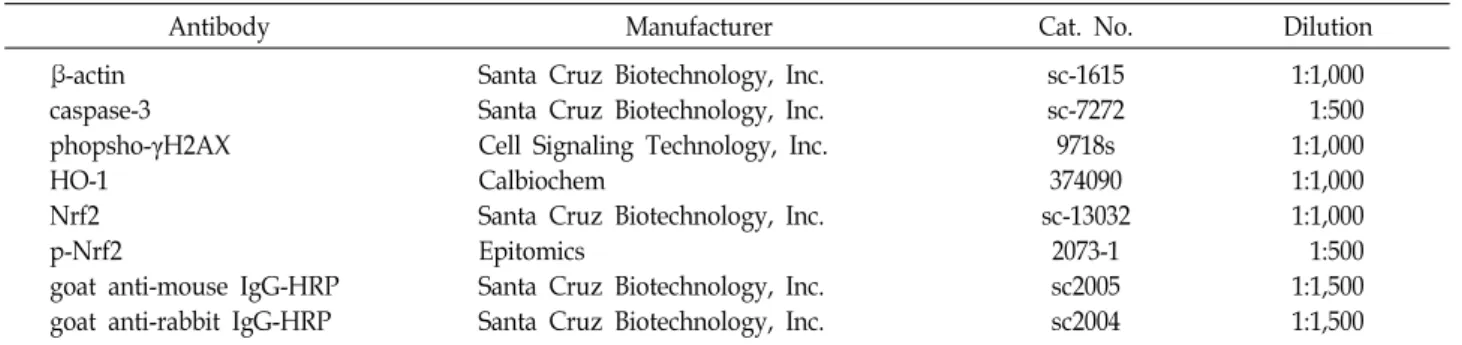
2를 동시 또는 단독 처리된 ARPE-19 세포에 lysis buffer [25 mM Tris-Cl (pH 7.5), 250 mM NaCl, 5 mM ethylenediaminetetraacetic acid, 1% Noni- det P-40, 1 mM phenymethylsulfonyl fluoride, 5 mM dithio- threitol]를 첨가하여 4℃에서 30분간 반응시킨 후 14,000 rpm, 4℃, 30분간 원심분리하여 상층액을 얻었다. 단백질 농도는 Bio-Rad 단백질 정량 시약(Bio-Rad Lab., Hercules, CA, USA) 을 이용하여 정량하였고, Laemilni sample buffer (Bio-Rad Lab.)를 혼합하였다. 동량의 단백질을 sodium dodecyl sul- phate (SDS)-polyacrylamide gel을 이용하여 전기영동을 실시 하였고, nitrocellulose membrane (Schleicher and Schuell, Keene, NH, USA)에 전이시켰다. 그 후 5% skim milk를 30분 처리하였고 1차 항체(Table 1)를 처리하여 4℃에서 over night 시킨 다음 1x phosphate buffered saline (PBS)-T (PBS with Tween 20)로 10분간 3번 세척하였다. 그 다음 적정 2차 항체 (Table 1)를 사용하여 상온에서 1시간 30분 반응시킨 후 1x PBS-T로 세척 후 Chemi-smart (Vilber Lourmat, France)를 이 용하여 분석하였다.
면역 형광 검사
다양한 조건에서 배양된 ARPE-19 세포에 100% 메탄올을 넣어 -20℃, 10분 동안 고정시킨 후 1X PBS로 3번 세척하였다.
Blocking buffer [5% bovine serum albumin (BSA) in 1X PBS-T (0.1% Triton X)]로 1시간 상온에서 반응시키고 2.5%
BSA로 희석한 p-γH2AX antibody (Table 1)를 첨가하여 4℃에 서 over night 시킨 다음 1X PBS-T로 2회 10분간 세척하였다.
그 후 2.5 μg/ml의 4‘,6’-diamidino-2-phenylindole (DAPI, Sigma-Aldrich Chemical Co., St. Louis, MO, USA) 용액을 처리하여 암실에서 20분 동안 반응시키고 1X PBS로 3회 세척 한 다음 형광현미경(Carl Zeiss, Oberkochen, Germany)을 이 용하여 분석하였다.
Mitochondrial membrane potential (MMP, Δψ
m)의 측정
H
2O
2처리에 따른 ARPE-19 세포의 미토콘드리아 기능 손
상에 미치는 감초 추출물의 효과를 조사하기 위하여 MMP
값의 변화를 측정하였다. 이를 위하여 H
2O
2를 처리하기 전에
감초 추출물을 1시간 전 처리하였다. 24시간 배양 후 10 μM의
Table 1. Primary and secondary antibodies used for immunoblotting
Antibody Manufacturer Cat. No. Dilution
β-actin caspase-3 phopsho-γH2AX HO-1
Nrf2 p-Nrf2
goat anti-mouse IgG-HRP goat anti-rabbit IgG-HRP
Santa Cruz Biotechnology, Inc.
Santa Cruz Biotechnology, Inc.
Cell Signaling Technology, Inc.
Calbiochem
Santa Cruz Biotechnology, Inc.
Epitomics
Santa Cruz Biotechnology, Inc.
Santa Cruz Biotechnology, Inc.
sc-1615 sc-7272 9718s 374090 sc-13032
2073-1 sc2005 sc2004
1:1,000 1:500 1:1,000 1:1,000 1:1,000 1:500 1:1,500 1:1,500
A
B
C
Fig. 1. Protection of H2O2-induced ROS generation and induc- tion of Nrf2 and HO-1 by GU (ethanol extract of G. ur- alensis Fischer) in human retinal pigmented epithelial ARPE-19 cells. ARPE-19 cells were pretreated with the indicated concentrations of GU or 10 μM NAC for 1 hr, and then stimulated with or without 300 μM H2O2 for 30 min (A and B) or 24 hr (C). (A) After staining with DCF-DA fluorescent dye, DCF fluorescence was moni- tored by a flow cytometer. (B) The data are shown as mean ± SD obtained from three independent experiments (*p<0.05 vs control group). (C) Cell lysates were subjected to gel electrophoresis, and then Western blot analysis was performed using the indicated antibodies. Actin was used as an internal control.
5,5‘,6,6’-tetrachloro-1,1‘,3,3’-tetraethyl-imidacarbocyanine io- dide (JC-1, Sigma-Aldrich Chemical Co.) 용액을 처리하여 37
℃, 20분 염색한 후 1x PBS로 2-3회 세척한 후 형광현미경을 이용하여 형광 강도를 관찰하였다. 또한 동일한 조건에서 배 양된 세포에 PBS를 첨가하여 세포를 부유시킨 다음 flow cy- tometer (Becton Dickinson, San Jose, CA, USA)에 적용시켜 MMP의 변화를 정량적으로 평가하였다.
세포사멸 유도의 정량적 평가를 위한 flow cytometry 분석 H
2O
2처리에 의한 세포사멸 유발 및 감초 추출물에 의한 억제 효과를 정량적으로 분석하기 위하여 준비된 세포들을 모은 다음 2,000 rpm으로 5분간 원심분리하여 상층액을 제거 하였다. 이들 세포에 1x binding buffer를 첨가 후 annexin V-fluorescein isothiocyanate (FITC, Becton Dickinson) 및 propidium iodide (PI, Becton Dickinson)를 처리하여 암실에 서 반응시켰다. 15분 후 1x binding buffer 200 μl을 추가하여 flow cytometer를 이용하여 세포사멸이 유발된 세포를 형광 반응에 따라 분석하였다.
통계처리
GraphPad Prism® version 5.0 (Graphpad Inc., San Diego, CA, USA)의 one-way ANOVA를 사용하여 통계분석을 실시 하였으며, Tukey's test로 사후 검정하여 p<0.05 값을 유의한 값으로 보고 통계 처리하였다.
결과 및 고찰
ARPE-19 세포에서 H
2O
2에 의한 ROS 생성에 미치는 감 초 추출물의 영향
산화적 스트레스 유발을 위한 H
2O
2의 처리 농도는 선행 연 구에 준하여 ARPE-19 세포에 24시간 처리 후 약 60% 정도의 생존율을 보이는 300 μM로 설정하였다[11, 24]. 그리고 감초 추출물의 농도는 세포독성을 나타내지 않는 150 μg/ml 이하 로 설정하였다(data not shown). Fig. 1A 및 Fig. 1B에 나타낸 결과는 다양한 농도의 감초 추출물을 1시간 전 처리 후, 300 μM의 H
2O
2에 30분간 노출된 ARPE-19 세포를 대상으로 ROS
의 생성 정도를 비교한 것이다. H
2O
2단독 처리된 세포는 정상
배지에서 배양된 세포에 비하여 ROS의 생성이 현저하게 증가
A
B
Fig. 2. Inhibition of H2O2-induced DNA damage by GU in ARPE- 19 cells. The cells were pretreated with 300 μg/ml GU and/or 10 μM NAC for 1 hr, and then stimulated with or without 300 μM H2O2 for 24 hr. (A) Cell lysates were subjected to gel electrophoresis, and then Western blot analysis was performed using anti-phospho (p)-γH2AX antibody. (B) Cell were stained with p-γH2AX antibody (red) and DAPI (nuclear stain; blue), then the cells were visualized using a fluorescence microscope (scale bar; 100 μm). Representative photographs are presented.
되었지만, 감초 추출물이 전 처리된 조건에서는 감초 추출물 처리 농도 의존적으로 유의하게 ROS의 생성이 감소하였다.
이러한 ROS 생성 억제 효과는 대조군으로 사용된 대표적인 ROS 소거제인 NAC 전 처리군에서도 유사하게 나타나 감초 추출물은 강력한 ROS 생성 억제 효능을 지니고 있음을 알 수 있었다.
Li et al. [22]에 의하여 ARPE-19 세포에서 산화적 스트레스 에 의한 ROS 생성 차단 효과에 Nrf2 신호계가 관여할 가능성 이 제기된 이후, 다양한 항산화 효능을 가지는 물질들의 ROS 생성 차단에 이 신호계의 활성이 관여됨이 보고되고 있다[1, 20, 26, 30, 31, 33, 36]. 아울러 최근 감초 추출물 또는 유효 성분들의 항산화 효능이 Nrf2 활성과 연관성이 있음이 알려졌 다[5, 10, 16, 28]. 따라서 감초 추출물에 의한 ROS 생성의 차단 에도 Nrf2 신호계의 활성이 관련이 있는지를 조사한 결과, H
2O
2단독 처리군에 비하여 감초 추출물과 H
2O
2가 동시 처리 된 세포에서 Nrf2의 생성이 증가되었으며, Nrf2의 대표적인 조절자인 HO-1의 발현도 매우 증가되었다(Fig. 1C). 아울러 Nrf2 의 발현 증가가 HO-1을 포함한 하위 유전자들의 발현을 증가시키기 위한 전사활성의 증가와 연관성이 있는지를 조사 하기 위하여 Nrf2의 인산화형(phosph-Nrf2, p-Nrf2)의 발현을 조사한 결과, 감초 추출물과 H
2O
2가 동시 처리된 세포에서 p-Nrf2의 발현이 증가되었음을 알 수 있었다. 비록 감초 추출 물과 H
2O
2의 동시 처리에 의한 HO-1의 발현 증가가 p-Nrf2 의존적인지의 여부와 증가된 HO-1의 기능에 대한 추가적인 연구가 요구되지만, 본 연구의 결과는 H
2O
2에 의한 산화적 스트레스를 억제하기 위해 Nrf2 신호계가 최소한 관여할 가능 성을 보여주었다.
H
2O
2에 의한 ARPE-19 세포의 DNA 손상에 미치는 감초 추출물의 영향
망막색소상피세포에서 과도한 ROS의 생성은 주요 세포 구 성 단백질의 변성뿐 만 아니라 심각한 DNA 손상을 유발하여 AMD의 발병 원인으로 작용하므로[14, 25] ROS의 생성 억제 는 망막색소상피세포의 항상성 유지에 필수적이다. 산화적 스 트레스에 의하여 DNA 이중 나선이 절단되면 히스톤 변이체 γH2AX 단백질의 인산화(serine 139)가 유발되며, 이는 DNA 손상을 평가하기 위한 바이오 마커로서 광범위하게 사용되고 있다[9]. 따라서 감초 추출물에 의한 ROS 생성의 억제 효과가 DNA 손상의 차단과 연관성이 있는지를 γH2AX의 인산화 여 부로 평가하였다. Fig. 2의 immunoblotting 및 면역 형광 분석 결과로 H
2O
2에 노출된 ARPE-19 세포에서 DNA 손상 지표인 γH2AX 단백질의 인산화가 현저히 증가되어 산화성 DNA 손 상이 유도되었음을 확인하였다. 그러나 H
2O
2의 이러한 DNA 손상 유발 효과는 감초 추출물이 전 처리된 조건에서는 유의 적으로 차단되었다. 이러한 현상은 다양한 항산화 효능을 가 지는 천연물들에 의한 ARPE-19 세포의 H
2O
2에 의한 DNA
손상 차단이 ROS 생성의 억제와 연관성이 있다는 선행 결과 들과 잘 일치되며[21, 27], 특히 이러한 효능이 Nrf2의 활성과 연관성이 있을 것이라는 결과와도 유사하였다[21]. 따라서 본 연구에 사용된 감초 추출물에 의한 ROS의 생성의 억제는 DNA 손상 보호와 직접 연관성이 있음을 알 수 있었다.
H
2O
2에 의한 ARPE-19 세포의 미토콘드리아 기능 손상 에 미치는 감초 추출물의 영향
세포 내 에너지 생산에 핵심적으로 관여하는 미토콘드리아
는 ROS 생성에 관여하는 주요 기관이면서 ROS에 손상을 받는
대표적인 기관이다. 미토콘드리아는 산화적 스트레스에 취약
하며, 과도한 ROS는 미토콘드리아의 기능 손상을 유발하여
에너지 항상성의 불균형을 야기한다[4, 6]. 특히 망막색소상피
세포에서도 ROS 및 자유 라디칼에 의한 산화적 스트레스는
미토콘드리아 손상을 일으키며, 이는 심각한 망막색소상피세
포의 기능 장애를 유발하여 AMD에서 맥락막 신생 혈관 형성
의 위축을 유발하는 원인이 된다[3, 13]. 따라서 감초 추출물의
항산화 효능이 H
2O
2에 의한 미토콘드리아 기능 손상을 차단
할 수 있는지의 여부를 조사하였다. 이를 위하여 미토콘드리
아 기능을 평가하는 대표적인 지표인 MMP의 소실에 미치는
감초 추출물의 영향을 조사하였다[3, 6]. Fig. 3A에 나타낸 JC-1
염색을 통한 면역 형광 분석 결과에 의하면, 선행 연구들과
유사하게 H
2O
2에 노출된 ARPE-19 세포에서 미토콘드리아 기
A
B
C
Fig. 3. Attenuation of H2O2-induced mitochondrial dysfunction by GU in ARPE-19 cells. The cells were pretreated with the indicated concentration of GU and/or 10 μM NAC for 1 hr, and then stimulated with or without 300 μM H2O2 for 24 hr. The cells were stained with JC-1 dye, and were then analyzed by a fluorescence microscope and a flow cytometer to evaluate the changes in MMP.
(A) Fluorescence images of MMP at a magnification of
×1,200. Representative photographs are presented. (B) Flow cytometry analysis was performed by JC-1 staining.
(C) The data of flow cytometry analysis are shown as mean ± SD obtained from three independent experi- ments (*p<0.05 vs control group, #p<0.05 vs H2O2-treated group).
A
B
C
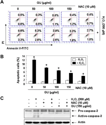
Fig. 4. Suppression of H2O2-induced apoptosis by GU in ARPE- 19 cells. The cells were treated with 300 μg/ml GU and/or 10 μM NAC for 1 hr, and then incubated with or without 300 μM H2O2 for 24 hr. (A and B) Flow cy- tometry analysis was performed by Annexin V and PI staining. The percentage of annexin V+/PI+ cells in the top and annexin V+/PI- cells in the bottom right quad- rant are indicated. Each point represents the mean of three independent experiments. (A) Representative pro- files. The percentages of apoptotic cells were determined by expressing the numbers of Annexin V+ cells as per- centages of all cells. (B) The data are shown as mean
± SD obtained from three independent experiments (*p<0.05 vs control group). (C) Cell lysates were subjected to gel electrophoresis, and then Western blot analysis was performed using anti-caspase-3 antibody. Actin was used as an internal control.
능의 손상을 의미하는 JC-1 단량체(monomer)의 발현이 증가 하였으며[27, 32], 감초 추출물이 전 처리된 조건에서 배양된 세포의 경우 대조군 수준을 유지하고 있었다. JC-1 다량체 (aggregate)의 형성의 감소에 의한 단량체의 증가는 MMP의 소실을 의미하며, Fig. 3B 및 C의 flow cytometry 분석 결과에 서도 이와 유사한 경향성을 보여 주었다. 흥미롭게도, Zhao et al. [35]은 Nrf2가 결손된 실험동물에서 AMD의 발병이 증 가하였으며, 이는 미토콘드리아 기능의 부재와 연관성이 있음 을 보고하였다[15, 20]. 비록 망막색소상피세포에서 항산화 효 능을 가지는 물질들에 의한 Nrf2의 발현 증가에 따른 HO-1의 기능에 대한 연구는 현재까지 자세히 이루어진 바 없으나, Nrf2가 HO-1의 전사활성에 핵심적인 역할을 하기 때문에
Nrf2의 부재는 산화적 스트레스에 의한 망막색소상피세포의 미토콘드리아 항상성 유지에 부정적인 영향을 줄 수 있을 것 으로 추정된다.
H
2O
2에 의한 ARPE-19 세포의 apoptosis 유도에 미치 는 감초 추출물의 영향
산화적 스트레스에 의한 미토콘드리아의 기능 손상은 내재
적 apoptosis 유도를 위한 신호로서 작용한다. 망막색소상피
세포에서 H
2O
2에 의한 ROS 생성의 증가는 DNA 손상 및 미토
콘드리아 기능 손상과 연계된 apoptosis 개시와 연관성이 있
다[21]. 또한 H
2O
2에 의한 망막색소상피세포의 apoptosis는 미
토콘드리아에서 세포질로의 cytochrome c 유리를 통한 cas-
pase cascade 활성화 촉진에 의해 유도된다[12, 26]. Caspase 의존적 apoptosis 유도를 위해서는 최종적으로 caspase-3이 활 성화 되어야 하며, H
2O
2에 의한 망막색소상피세포의 apopto- sis 유도에서도 caspase-3의 활성이 핵심적인 역할을 한다. 따 라서 ARPE-19 세포에서 H
2O
2에 의한 apoptosis에 미치는 감 초 추출물의 영향을 조사하였다. Fig. 4A 및 Fig. 4B에 나타낸 flow cytometry 결과에서 알 수 있듯이, 대조군에 비하여 H
2O
2에 노출된 ARPE-19 세포에서 apoptosis 유발이 유의적 으로 증가되었지만, 감초 추출물이 존재하는 조건에서는 처리 농도 의존적으로 H
2O
2에 의한 apoptosis 유도가 억제되었다.
아울러 H
2O
2가 단독으로 처리된 ARPE-19 세포에서 증가되었 던 caspase-3의 활성형 발현이 감초 추출물이 전 처리된 세포 에서는 감소되었다(Fig. 4C). 이는 H
2O
2에 의한 미토콘드리아 의 기능 손상이 감초 추출물에 의해서 억제됨으로서 나타난 결과로서, ROS 생성의 억제에 기인된 것으로 해석될 수 있다.
이상의 결과에서 감초 추출물은 H
2O
2에 의한 ROS의 생성 을 차단함으로써 미토콘드리아 기능 손상을 예방하고, 그에 따른 DNA 손상과 apoptosis로부터 ARPE-19 세포를 보호하 였음을 알 수 있다. 따라서 산화적 스트레스로부터 망막색소 상피세포를 보호 할 수 있는 항산화제로서 감초 추출물의 활 용 가능성을 제시하고자 한다. 비록 이 과정에서 Nrf2 의존적 HO-1 활성의 가능성도 제시하였으나, 이에 관한 추가적인 연 구와 감초 추출물에 함유된 생리활성 성분의 분석에 따른 추 가적인 기전 연구가 수행되어야 할 것이다.
감사의 글
본 연구는 BGN 밝은눈안과병원과 (주)비지엔케어의 연구 비 지원으로 수행되었다.
References
1. Arumugam, B., Palanisamy, U. D., Chua, K. H. and Kup- pusamy, U. R. 2019. Protective effect of myricetin deriva- tives from Syzygium malaccense against hydrogen per- oxide-induced stress in ARPE-19 cells. Mol. Vis. 25, 47-59.
2. Bao, F., Bai, H. Y., Wu, Z. R. and Yang, Z. G. 2019. Phenolic compounds from cultivated Glycyrrhiza uralensis and their PD-1/PD-L1 inhibitory activities. Nat. Prod. Res. 25, 1-8.
3. Brown, E. E., Lewin, A. S. and Ash, J. D. 2018. Mitochondria:
Potential targets for protection in age-related macular de- generation. Adv. Exp. Med. Biol. 1074, 11-17.
4. Caito, S. W. and Aschner, M. 2015. Mitochondrial redox dys- function and environmental exposures. Antioxid. Redox. Sig- nal. 23, 578-595.
5. Cao, L. J., Hou, Z. Y., Li, H. D., Zhang, B. K., Fang, P. F., Xiang, D. X., Li, Z. H., Gong, H., Deng, Y., Ma, Y. X., Tang, H. B. and Yan, M. 2017. The ethanol extract of Licorice (Glycyrrhiza uralensis) protects against triptolide-induced ox- idative stress through activation of Nrf2. Evid. Based Comple-
ment. Alternat. Med. 2017, 2752389.
6. Demine, S., Renard, P. and Arnould, T. 2019. Mitochondrial uncoupling: A key controller of biological processes in phys- iology and diseases. Cells 8, E795.
7. Deng, Y., Zhu, J., Mi, C., Xu, B., Jiao, C., Li, Y., Xu, D., Liu, W. and Xu, Z. 2015. Melatonin antagonizes Mn-induced oxi- dative injury through the activation of keap1-Nrf2-ARE sig- naling pathway in the striatum of mice. Neurotox. Res. 27, 156-171.
8. Dunaief, J. L., Dentchev, T., Ying, G. S. and Milam, A. H.
2002. The role of apoptosis in age-related macular degener- ation. Arch. Ophthalmol. 120, 1435-1442.
9. Gerić, M., Gajski, G. and Garaj-Vrhovac, V. 2014. γ-H2AX as a biomarker for DNA double-strand breaks in ecotoxicol- ogy. Ecotoxicol. Environ. Saf. 105, 13-21.
10. Gong, H., Zhang, B. K., Yan, M., Fang, P. F., Li, H. D., Hu, C. P., Yang, Y., Cao, P., Jiang, P. and Fan, X. R. 2015. A protective mechanism of licorice (Glycyrrhiza uralensis): iso- liquiritigenin stimulates detoxification system via Nrf2 acti- vation. J. Ethnopharmacol. 162, 134-139.
11. Hao, Y., Liu, J., Wang, Z., Yu, L. L. and Wang, J. 2019. Pice- atannol protects human retinal pigment epithelial cells against hydrogen peroxide induced oxidative stress and apoptosis through modulating PI3K/Akt signaling pathway.
Nutrients 11, E1515.
12. He Y., Leung K. W., Ren Y., Pei J., Ge, J. and Tombran-Tink, J. 2014. PEDF improves mitochondrial function in RPE cells during oxidative stress. Invest. Ophthalmol. Vis. Sci. 55, 6742- 6755.
13. Hoffman, D. L. and Brookes, P. S. 2009. Oxygen sensitivity of mitochondrial reactive oxygen species generation de- pends on metabolic conditions. J. Biol. Chem. 284, 16236- 16245.
14. Hsieh, F. C., Hung, C. T., Cheng, K. C., Wu, C. Y., Chen, Y. C., Wu, Y. J., Liu, W. and Chiu, C. C. 2018. Protective effects of Lycium barbarum extracts on UVB-induced damage in human retinal pigment epithelial cells accompanied by attenuating ROS and DNA damage. Oxid. Med. Cell. Longev.
2018, 4814928.
15. Hyttinen, J. M. T., Viiri, J., Kaarniranta, K. and Błasiak, J.
2018. Mitochondrial quality control in AMD: does mitoph- agy play a pivotal role? Cell. Mol. Life Sci. 75, 2991-3008.
16. Ji, S., Li, Z., Song, W., Wang, Y., Liang, W., Li, K., Tang, S., Wang, Q., Qiao, X., Zhou, D., Yu, S. and Ye, M. 2016.
Bioactive constituents of Glycyrrhiza uralensis (Licorice):
Discovery of the effective components of a traditional herbal medicine. J. Nat. Prod. 79, 281-292.
17. Kao, T. C., Wu, C. H. and Yen, G. C. 2014. Bioactivity and potential health benefits of licorice. J. Agric. Food Chem. 62, 542-553.
18. Kaspar, J. W., Niture, S. K. and Jaiswal, A. K. 2009. Nrf2:
INrf2 (Keap1) signaling in oxidative stress. Free Radic. Biol.
Med. 47, 1304-1309.
19. Léveillard, T. and Klipfel, L. 2019. Mechanisms underlying the visual benefit of cell transplantation for the treatment of retinal degenerations. Int. J. Mol. Sci. 20, E557.
20. Li, K. R., Yang, S. Q., Gong, Y. Q., Yang, H., Li, X. M., Zhao, Y. X., Yao, J., Jiang, Q. and Cao, C. 2016. 3H-1,2-dithiole-3- thione protects retinal pigment epithelium cells against Ultra- violet radiation via activation of Akt-mTORC1-dependent Nrf2-HO-1 signaling. Sci. Rep. 6, 25525.
21. Li, X. M., Huang, D., Yu, Q., Yang, J. and Yao, J. 2018. Neu- roligin-3 protects retinal cells from H2O2-induced cell death via activation of Nrf2 signaling. Biochem. Biophys. Res. Com- mun. 502, 166-172.
22. Li, Z., Dong, X., Liu, H., Chen, X., Shi, H., Fan, Y., Hou, D. and Zhang, X. 2013. Astaxanthin protects ARPE-19 cells from oxidative stress via upregulation of Nrf2-regulated phase II enzymes through activation of PI3K/Akt. Mol. Vis.
19, 1656-1666.
23. Liu, D., Huo, X., Gao, L., Zhang, J., Ni, H. and Cao, L. 2018.
NF-κB and Nrf2 pathways contribute to the protective effect of licochalcone A on dextran sulphate sodium-induced ul- cerative colitis in mice. Biomed. Pharmacother. 102, 922-929.
24. Liu, Y., Ren, Y., Wang, X., Liu, X., Xu, Y. and He, Y. 2019.
Down regulation of UCP2 expression in retinal pigment epi- thelium cells under oxidative stress: an in vitro study. Int.
J. Ophthalmol. 12, 1089-1094.
25. Marazita, M. C., Dugour, A., Marquioni-Ramella, M. D., Figueroa, J. M. and Suburo, A. M. 2016. Oxidative stress-in- duced premature senescence dysregulates VEGF and CFH expression in retinal pigment epithelial cells: Implications for age-related macular degeneration. Redox. Biol. 7, 78-87.
26. Ni, T., Yang, W. and Xing, Y. 2019. Protective effects of del- phinidin against H2O2-induced oxidative injuries in human retinal pigment epithelial cells. Biosci. Rep. 39, BSR20190689.
27. Park, C., Lee, H., Hong, S. H., Kim, J. H., Park, S. K., Jeong, J. W., Kim, G. Y., Hyun, J. W., Yun, S. J., Kim, B. W., Kim, W. J. and Choi, Y. H. 2019. Protective effect of diphlor- ethohydroxycarmalol against oxidative stress-induced DNA damage and apoptosis in retinal pigment epithelial cells.
Cutan. Ocul. Toxicol. 38, 298-308.
28. Rotstein, N. P, Miranda, G. E, Abrahan, C. E. and German, O. L. 2010. Regulating survival and development in the reti- na: key roles for simple sphingolipids. J. Lipid Res. 51, 1247- 1262.
29. van Lookeren Campagne, M., LeCouter, J., Yaspan, B. L.
and Ye, W. 2014. Mechanisms of age-related macular degen- eration and therapeutic opportunities. Pathology 232, 151- 164.
30. Wang, K., Jiang, Y., Wang, W., Ma, J. and Chen, M. 2015.
Escin activates AKT-Nrf2 signaling to protect retinal pig- ment epithelium cells from oxidative stress. Biochem.
Biophys. Res. Commun. 468, 541-547.
31. Wang, Z., Hao, Y., Wang, Y., Liu, J., Yuan, X., Sun, B. and Wang, J. 2019. Wheat alkylresorcinols protect human retinal pigment epithelial cells against H2O2-induced oxidative damage through Akt-dependent Nrf2/HO-1 signaling. Food Funct. 10, 2797-2804.
32. Yan, G., Yu, L., Jiang, S. and Zhu, J. 2018. Melatonin antago- nizes oxidative stress-induced mitochondrial dysfunction in retinal pigmented epithelium cells via melatonin receptor 1 (MT1). J. Toxicol. Sci. 43, 659-669.
33. Yang, P. M., Wu, Z. Z., Zhang, Y. Q. and Wung, B. S. 2016.
Lycopene inhibits ICAM-1 expression and NF-κB activation by Nrf2-regulated cell redox state in human retinal pigment epithelial cells. Life Sci. 155, 94-101.
34. Zhang, D. D. 2006. Mechanistic studies of the Nrf2-Keap1 signaling pathway. Drug Metab. Rev. 38, 769-789.
35. Zhao, Z., Chen, Y., Wang, J., Sternberg, P., Freeman, M. L., Grossniklaus, H. E. and Cai, J. 2011. Age-related retinopathy in NRF2-deficient mice. PLoS One 6, e19456.
36. Zhu, C., Dong, Y., Liu, H., Ren, H. and Cui, Z. 2017. Hesper- etin protects against H2O2-triggered oxidative damage via upregulation of the Keap1-Nrf2/HO-1 signal pathway in ARPE-19 cells. Biomed. Pharmacother. 88, 124-133.
초록:망막색소상피세포에서 감초 추출물의 산화적 스트레스에 의한 DNA 손상 및 apoptosis 유발의 차단 효과
김소영
1,2․김정환
3․김성옥
4․박세광
3․정지원
5․김미영
3․이혜숙
1,2․정재훈
6․최영현
1,2*
(1동의대학교 한의과대학 생화학교실, 2동의대학교 항노화연구소, 3㈜비지엔케어 연구개발전담부서, 4경성대학교
공과대학 식품영양학전공, 5BGN 밝은눈안과병원, 6부산대학교 자연과학대학 분자생물학과)