http://dx.doi.org/10.9721/KJFST.2016.48.6.597
597
©The Korean Society of Food Science and Technology
증숙 횟수에 따른 천마 추출물의 급성 위염 개선효과
이아름·권오준
1·노정숙
2·노성수*
대구한의대학교 한의과대학 본초약리학 교실, 1경북지역산업평가단, 2동명대학교 식품영양학과
Protective effects of Gastrodia elata extract by steaming time on
acute gastritis
Ah Reum Lee, O Jun Kwon1, Jeong Sook Noh2, and Seong-Soo Roh* College of Korean Medicine, Daegu Haany University
1Kyeoungbuk Institute for Regional Program Evaluation, Gyeongbuk TP 2Department of Food Science & Nutrition, Tongmyong University
Abstract This study aimed to explore the protective effect of Gastrodia elata (GE) in an HCl/ethanol induced acute gastritis model by differing the steaming time. The samples GE1 (GE by steaming for 1 time) and GE9 (GE by steaming for 9 times), were selected based on the results of HPLC analysis, free radical scavenging activities, and total phenol and flavonoid contents. To evaluate the anti-inflammatory effect of GE, ICR mice were divided into 5 groups; normal mice (Nor), gastritic mice with distilled water (Con), gastritic mice with 100 mg/kg GE1, gastritic mice with 100 mg/kg GE9 and gastritic mice with 10 mg/kg sucralfate (SC). HCl/ethanol-induced gastric mucosal injury was markedly improved by GE9 treatment as observed during histological evaluation. The increased reactive oxygen species levels in the serum were diminished by GE9 treatment. Furthermore, peroxynitrite levels of the stomach tissue were decreased in the GE9-treated group. The analyses of stomach proteins indicated that GE9 treatment effectively reduced inflammatory cytokine levels as compared to that by GE1 treatment. These results suggest that GE9 improves health during acute gastritis.
Keywords: Gastrodia elata, acute gastritis, anti-oxidant, anti-inflammation
서
론
위염은 위 점막 염증성 질환을 총칭하는 것으로 위 점막을 보 호하는 고유의 방어인자와 손상시키는 공격인자 사이의 균형이 깨져, 공격인자가 강해지거나 방어인자가 약해지면 발생하게 된 다(1). 히스타민(Histamine), 위산, 헬리코박터 필로리(Helicobacter pylori) 감염, 비스테로이드성 소염 진통제(non-steroidal anti-inflam-matory drugs, NSAIDs) 과다복용과 불규칙한 식습관, 음주, 스트 레스 등이 대표적인 공격인자이다(2). 이때, 위 점막 보호물질의 감소 또는 산화적 스트레스, 히스타민 분비량 증가 등은 위 손상 과 염증을 악화시킨다.
많은 연구에서 활성산소종(reactive oxygen species, ROS)은 위 염의 발병에서 중요한 원인이 된다고 보고되고 있다. 위에서 발 생된 자유라디칼은 여러 효소 반응을 통해 신속하게 제거되지만 과다한 활성산소종의 증가는 세포의 과산화지질 형성증가로 인 하여 세포벽의 손상을 일으켜 위 점막의 손상을 유발시킨다(3). 또한 활성산소종은 세포 내 고리산소화효소(cyclooxygenase-2), 인 터루킨-6 (interleukin-6), 종양괴사인자 알파(tumor necrosis factor
alpha), 유도성 산화질소 합성효소(inducible nitric oxide synthase) 와 같은 사이토카인의 증가를 유발하여 염증반응을 일어나게 한 다. 그러므로 세포 내의 산화방지 작용의 증가는 위염의 발병을 감소시킬 수 있으며 위 점막을 보호하는 역할을 할 수 있다(4).
위염의 치료는 위산분비를 억제하는데 중점을 두고 있으며, 치 료제는 수소수용체 길항물질(H2-receptor antagonist)와 양성자 펌
프 억제제(proton pump inhibitor)류의 약물이 대표적이다(5,6). 이 러한 약물들은 임상적으로 우수한 치료효과를 보이고 있지만 위 장 장애를 일으키거나, 발진, 가려움, 두드러기와 같은 피부과적 인 질환을 일으키는 부작용을 나타내고 있다(7-9). 따라서 약물만 으로는 치료의 한계가 있어, 위염 개선에 효능이 있으면서 동시 에 부작용을 줄이는 소재의 개발이 필요한 실정이다. 천마(Gastrodia elata)는 난초과 식물에 속하는 다년초로서 덩 이줄기를 약재와 식품소재로 사용하며 약리성분으로는 가스트로 딘(gastrodin), 바닐릴 알코올(vanillyl alcohol), 바닐린(vanillin), 벤 즈알데히드(benzaldehydes), 파라-하이드록시벤질 알코올(p-hydrox-ybenzyl alcohol) 등을 함유하고 있다(10). 천마는 단단하여 절단 하기가 어려워 예부터 다양한 가공 방법이 사용되었으나 현재는 조직의 연화와 유효성분의 증가를 목적으로 증숙하는 방법이 가 장 많이 사용되고 있다. 또한, 천마를 증숙하지 않고 씹어 먹을 경우 혀와 인후를 자극하는 부작용이 있어 최소 1회 증숙 과정 을 거친 천마가 유통되고 있다(11). 기존 연구에서는 천마의 산 화방지와 항암 활성(12) 항균활성(13), 혈청에서 중성지질을 저하 시키는 효과(14) 뿐만 아니라 발효천마의 암세포 증식 억제효과 (15)에 관한 연구가 보고되었다. 그러나, 증숙 천마의 위염 개선 *Corresponding author: Seong-Soo Roh, College of Korean
Medi-cine, Daegu Haany university, Daegu 42158, Korea Tel: +82-53-770-2351
Fax: +82-53-768-6340 E-mail: [email protected]
Received June 18, 2016; revised September 1, 2016; accepted September 13, 2016
파우더 25 g을 400 mL의 증류수에 넣어 100C에서 1시간 가열하 여 추출물을 얻었다. 이를 거름종이(Whatman No. 2, GE health-care, Arlington Heights, IL, USA)를 사용하여 여과한 뒤 여과액 을 미리 항량된 용기에 넣어 45-50oC의 수온에서 회전진공증발
기(rotary vacuum evaporator) (JP/N-1000X, Sunileyela Co., Ltd., Gyeonggido, Korea)를 사용하여 감압농축 후 냉동건조하고 −20oC 에 보관하여 사용하였다. 실험동물 ICR 마우스 6주령 수컷 30마리를 (주)오리엔트(Gyeonggido, Korea)에서 구입하여 1주일 동안 실험실 환경에 적응시킨 후 실 험에 사용하였다. 동물 사육실의 조건은 conventional system으로 온도 22±2oC, 습도 50±5% 명암주기(light: dark cycle)는 12시간
주기로 조절하였다. 사료는 고형사료(Samyang corporation, Seoul, Korea) (조단백질 22.1% 이상, 조지방 8.0% 이하, 조섬유 5.0% 이하, 조회분 8.0% 이하, 칼슘 0.6% 이상, 인 0.4% 이상, 항생제 무첨가)와 물을 충분히 공급하였다. 실험은 대구한의대학교 동물 실험 윤리위원회의 승인(DHU2016-011)을 얻어 시행하였으며 동 물관리 규정을 준수하였다. 성분 분석 HPLC 분석에 사용한 이동상은 A: 물(water) (0.2% 인산(phos-phoric acid))와 B: 아세토나이트릴(aceto-nitrile) (0.2% phosphoric acid)을 gradient로 사용하여 용매기울기로 유속 1.0 mL/min에서 column 온도 40oC를 유지하면서 검출기는 UV (225 nm)를 사용 하였고 주입량은 1 mL를 주입하였다. 천마 시료액의 1 mL에 함유된 gastrodin, gastrodigenin 성분을 분석하여 함량을 표로 나타내었다. 천마의 분석에 사용한 표준품 은 이화여자대학교 서은경 교수로부터 천마에서 분리한 gastrodin, gastrodigenin 2종을 제공받아 사용하였다. 산화방지 활성 측정 산화방지 활성은 DPPH와 ABTS 라디칼 소거법으로 측정되었 다. DPPH (Sigma Aldrich, St. Louis, MO, USA)는 시료 중에 포함된 산화방지 물질의 양을 측정하는데 사용된다. 시료 100 μL 과 60 μm DPPH 용액(dissolved in ethanol) 100 μL를 혼합한 후, 실온에서 30분간 반응시켰다. 이 반응액을 사용하여 540 nm에서 흡광도를 사용하여 측정한 후, 전자공여능은 계산하여 산출하였 다(16). ABTS 라디칼 소거활성은 Re 등(17)의 방법을 이용하여 측정하였다. 7 mM 2,2'-azino-bis(3-ethylbenzothiazoline-6-sulphonic acid), 2.45 mM 과황산포타슘(potassium persulfate) (Sigma Ald-rich)을 증류수에 녹인 다음 12시간 동안 차광하여 반응시켰다.
준 검량선을 구하고, 시료추출물의 총 페놀 함량을 산출하였다. 총 플라보노이드 함량은 Lister 등(19)의 방법에 의해 구하였다. 1 mL의 디에틸렌글리콜(diethylene glycol)과 시료추출물 100 μL와 1 N NaOH 10μL를 잘 혼합시켜 37oC의 물중탕(water bath)에서 1시간 동안 반응시킨 후 420 nm에서 흡광도를 측정하였다. 표준 물질로는 나린긴(naringin)를 사용하였으며, 표준 보정선을 구하여 추출물의 총 플라보노이드 함량을 구하였다. 급성 위염 유발과 동물 처치 실험동물은 군당 6마리씩 5그룹으로 구분하여 실험을 실시하 였다. 실험 전 날까지 고형사료와 물을 충분히 공급하였고 위 점 막 손상 유발 실험 24시간 전부터 절식하였다. 실험 전, 정상군 (Nor)은 아무런 처치를 하지 않았으며, 위 점막 손상 대조군(Con) 은 증류수를 경구 투여하였고, 1회 증숙 천마 추출물(100 mg/kg body wight, GE1), 9회 증숙 천마추출물(100 mg/kg body weight, GE9)과 양성대조군인 슈크랄페이트(sucralfate) (Sigma Aldrich, 10 mg/kg body weight, SC) 투여군은 각 농도에 맞게 경구 투여 한 후, 1시간 방치하였다. 그 다음 위 점막 손상을 유발하기 위 하여, 150 mM HCl/60% ethanol을 각 0.5 mL씩 경구 투여하였고 1시간 후 아이소플루레인(isoflurane) (Sigma Aldrich)으로 흡입 마 취 하여 개복 후 위 조직을 적출하였다(20).
조직학적 관찰
적출한 위 조직을 고정한 다음, 광학 디지털 카메라(DSC-HX50V, Sony, Tokyo, Japan)를 이용하여 촬영하였다. 손상된 위 점막 측정은 I-Solution lite (Innerview Co., Gyeonggido, Korea) 프로그램을 이용하여 실제 손상 부위의 면적을 측정한 후, 위 전 체 면적과 비교하여 비율로 표시하였다.
산화적 스트레스 바이오마커 측정
심장에서 채혈한 혈액을 4,000 rpm 10분 동안 원심 분리하여 혈청을 얻었다. ROS 측정은 혈청과 25 mM 2',7'-dichlorofluores-cein diacetate (DCFDA) (Molecular Probes, Eugene, OR, USA) 를 혼합한 후, 형광 광도계를 이용하여 0분부터 매 10분씩 방출 (emission) 파장 530 nm와 들뜸(excitation) 파장 485 nm를 이용하 여 30분간 측정한 산출 값을 계산하였다.
위 조직은 1 mM ethylenediaminetetraacetic acid (EDTA) (Wako Pure Chemical Industries, Ltd., Osaka. Japan)-50 mM 인산소듐완충용액(sodium phosphate buffer, pH 7.4)를 이용하여 분쇄한 후 ONOO−를 측정하기 위하여 DHR 123 buffer
(rodamin buffer, 5 mM DTPA, 10 mM DHR123)와 혼합하여 37oC에서 5분간 교반하였다. 그 다음 방출 파장 480 nm와 들뜸
파장 535 nm에서 측정한 값을 계산하여 나타내었다. ROS와 ONOO−의 실험방법은, 각각 Ali 등(21)과 Kooy 등(22)의 방법을 이용하여 시행하였다.
위 조직 웨스턴 블로팅(Western blotting)
위의 세포질을 얻기 위해 100 mM Tris-HCl (pH 7.4), 5 mM Tris-HCl (pH 7.5), 2 mM 염화마그네슘(MgCl2), 15 mM 염화칼슘 (CaCl2), 1.5 M sucrose, 0.1 M DTT, protease inhibitor cocktail을 첨가한 buffer A를 넣고 조직 분쇄기(tissue grinder) (Biospec Product, Bartlesville, OK, USA)로 분쇄한 후 10% NP-40 용액을 첨가하였다. 아이스 위에서 20분간 정치시킨 후 12,000 rpm으로 2분간 원심분리 하여 세포질을 포함하고 있는 상층액을 분리하 였다. 핵을 얻기 위해 10% NP-40가 더해진 buffer A에 두 번 헹 구고 100 mL의 buffer C (50 mM HEPES, 50 mM 염화포타슘 (KCl), 0.3 mM 염화소듐(NaCl), 0.1 mM EDTA, 1 mM DTT, 0.1 mM PMSF, 10% glycerol)를 첨가해 재부유시킨 뒤 10분마다 vortex을 3번 하였다. 4oC에서 12,000 rpm으로 10분간 원심 분리 한 후 핵을 포함하고 있는 상층액을 얻어 −80oC에서 각각 냉동
보관하였다. 위 조직의 세포질의 iNOS, COX-2, TNF-α, IL-6, 베 타액틴(β-actin) 단백질의 발현을 측정하기 위하여 10 μg의 단백 질을 8-15% SDS 폴리아크릴아미드겔(polyacrylamide gel)을 이용 하여 전기 연동 후, 아크릴아마이드겔(acrylamide gel)을 니트로셀 룰로스막(nitrocellulose membrane)으로 이동시켰다. 준비된 막(mem-brane)에 각각의 1차 antibody를 처리하여 4oC에서 overnight 시킨 다음 PBS-T로 6분마다 5회 세척하고, 각각 처리된 1차 항체에 사용되는 2차 항체(PBS-T로 1:3000로 희석해서 사용)를 사용하 여 상온에서 1시간 반응시킨 후, PBS-T로 6분마다 5회 세척하였 다. 그리고 membrane을 enhanced chemiluminescence (ECL) 용액 에 노출시킨 후, Sensi-Q2000 Chemidoc (Lugen Sci Co., Ltd., Seoul, Korea)에 감광시켜 단백질 발현을 확인한 후, 해당 band를
ATTO Densitograph Software (ATTO Corporation, Tokyo, Japan) 프로그램을 사용하여 정량하였다.
통계분석
In vitro의 수치는 평균과 표준편차로, in vivo의 수치는 평균과 표준오차로 표시하였으며, SPSS (Version 22.0, IBM, Armonk, NY, USA)을 사용하여 일원배치분산분석(one-way analysis of vari-ance, ANOVA) test를 실시한 후 Tukey multiple comparison test 로 사후검증을 실시하여 군 간의 유의성을 측정하였다. 각 군의 평균 차이에 대한 통계적 유의성을 5% 수준에서 검증하였다. Student’s paired t-test을 사용하여 위염 유발 대조군과 다른 군간 의 통계적 유의성을 측정하였다(p<0.05).
결과 및 고찰
성분 분석천마의 주요 생리활성 성분인 gastrodin은 간 섬유증 보호효과 (23)와 산화방지효과를 통한 알츠하이머 억제효과(24) 등이 보고 되었다. gastrodigenin 성분은 gastrodin의 아글리콘(aglycone)인 형 태로서 뇌손상 방지와 신경염증 치료효과(25)와 멜라닌합성 억제 효과(26)가 발표되었다. 이러한 천마의 주된 약리효능을 나타내 는 gastrodin은 건조하면 양이 감소하지만 증숙한 후 건조하면 gastrodin의 함량이 늘어나는데 이는 가열하면 gastrodin 분해효소 가 불활성화되어 gastrodin이 분해되지 않기 때문이다(27). 따라서 본 실험에서는 증숙 횟수를 달리하여 천마의 gastrodin과 gastro-digenin의 함량을 분석하였다. HPLC를 분석한 결과, gastrodin의 머무름시간(retention time, RT)은 23.053분이었고, gastrodigenin의 머무름시간은 24.979분으로 나타났다(Fig. 1). 천마의 gastrodin, gastrodigenin의 함량을 분석한 결과, 면적비를 이용한 검량선은 gastrodin, gastrodigenin 모두 R2=0.9999로 양호한 직진성을 나타 Fig. 1. HPLC chromatogram of gastrodin and gastrodigenin. Components of Gastrodia elata: 1): gastrodin, 2): gastrodigenin
내었다. Gastrodin의 경우 GE6와 GE9 모두 GE1에 비하여 높은 함량을 보였고, gastrodigenin 함량 또한 GE1에 비하여 GE6와 GE9이 높은 함량을 보였다(Table 1). 산화방지 활성 측정 결과 증숙 천마의 산화방지 활성을 평가하기 위하여, DPPH와 ABTS 라디칼 소거법을 활용하여 측정하였다. ABTS 라디칼의 경우, 극 성과 비극성 시료의 소거활성을 모두 측정할 수 있어 DPPH radical 소거법보다 적용범위가 넓다. 뿐만 아니라 DPPH 라디칼 은 자유 라디칼, ABTS 라디칼은 양이온 라디칼로서 기질의 특 징과 반응물과의 결합정도가 서로 달라, 추출물의 특성에 따라 소거능이 다르게 나타날 수 있다(28). 따라서 증숙 천마 추출물 의 산화방지 활성을 두 종류의 라디칼 소거능을 모두 활용하여 분석하였다. DPPH 라디칼 소거능 측정에서 1회 증숙한 천마 열 수추출물의 IC50값은 1426.841.73 μg/mL으로 나타났고 6회 증숙 하였을 때는 326.97±1.40 μg/mL, 9회 증숙하였을 때는 321.43± 0.42μg/mL의 IC50값을 나타내어 증숙 횟수에는 비례하지 않지만, 증숙 과정이 산화방지 효과를 크게 증가시키는 것을 확인할 수 있었다. ABTS 라디칼 소거 활성 효과 측정 실험에서는 천마를 6회 증숙 하였을 때는 249.96±0.03 μg/mL, 9회 증숙하였을 때는 226.68±0.43μg/mL으로 나타나 증숙 횟수가 증가 할수록 산화방 지 활성 또한 증가하는 것으로 나타났다(Table 2). 따라서 천마는 산화방지 효과를 가지며 1회 증숙한 천마와 비교하였을 때, 6회, 9회 증숙한 천마가 더 높은 산화방지효과를 나타낸 것으로 판단 된다. 결과적으로 천마는 1회 증숙하였을 때 보다 9회 증숙하였 을 때 DPPH 라디칼 소거능이 ABTS 라디칼 소거능에 비해 크 게 증가하였다, 이와 같은 결과는 증숙 천마의 산화방지 활성이 양이온 라디칼보다 자유 라디칼 소거능에서 더 큰 활성 증가를 나타내는 것으로 사료되며, 따라서 DPPH 라디칼과 ABTS 라디 칼 소거능에서 효능 증가 정도의 차이를 보이는 것으로 사료된다.
Total phenol 및 total flavonoid 함량 측정 결과
증숙 횟수를 달리한 천마의 total phenol 함량을 측정 한 결과, GE1은 9.07±0.51 mg/g, GE6는 13.24±0.39 mg/g, GE9는 15.30± 0.12 mg/g로 나타났다(Table 3). 천마의 페놀성 물질은 p-hydroxy-benzyl alcohol, p-hydroxybenzaldehyde, vanillyl alcohol, vanillin 등이 있으며 이러한 성분들의 증가로 증숙 횟수에 비례하게 total phenol함량이 증가하는 것으로 유추할 수 있다(29). 증숙 천마의 total flavonoid 함량은 각각 0.59±0.09, 3.14±0.13, 3.34±0.19 mg/g 으로 나타나 total phenol 함량에 비하여 적은 수치였지만 이 또 한 증숙 과정 중 증가하는 것을 확인할 수 있었다(Table 3). 식물 L-ascorbic acid 01.26±0.2 03.50±0.13 GE12) 1426.84±1.73a0 251.01±0.82a GE6 326.97±1.40b 0249.96±0.03ab GE9 321.43±0.42b 226.68±0.43b 1)All values are mean±SD of three replications.
2)GE1-GE9: Gastrodia elata by steaming-drying 1-9 times.
a-cResults were assessed by one-way ANOVA followed by Tukey
multiple comparison test (p<0.05).
Table 3. Total polyphenol and total flavonoid contents of steamed Gastrodia elata extract
Sample Total polyphenol (mg/g) Total flavonoid (mg/g)
0GE11) 009.07±0.512)c 0.59±0.09b GE6 13.24±0.39b 3.14±0.13a GE9 15.30±0.12a 3.34±0.19a 1)GE1-GE9: Gastrodia elata by steaming-drying 1-9 times. 2)All values are mean±SD of three replications.
a-cResults were assessed by one-way ANOVA followed by Tukey
multiple comparison test (p<0.05).
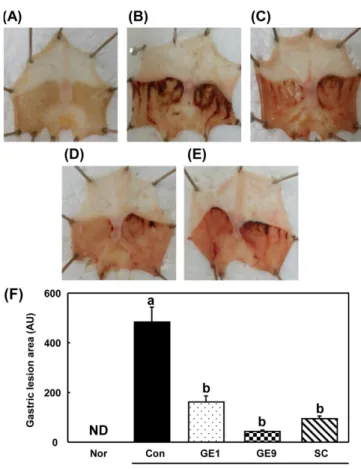
Fig. 2. Optical change stomach tissues of HCl/ethanol-induced acte gastritic mice. All values are mean±SE (n=6) (A) Nor, Normal mice, (B) Con, HCl/ethanol-induced acute gastritic mice with the administration of distilled water, (C) GE1, HCl/ethanol-induced acute gastritic mice with the administration of 100 mg/kg Gastrodia elata by steaming 1 time (D) GE9, HCl/ethanol-induced acute gastritic mice with the administration of 100 mg/kg Gastrodia elata by steaming 9 time (SE) SC, HCl/ethanol-induced acute gastritic mice with the administration of 10 mg/kg sucralfate, (F) gastric mucosal injury area. ND: Not detected. Different letters show a significant difference at p<0.05 as determined by one-way ANOVA followed by Tukey multiple comparison test.
에 존재하는 phenol과 flavonoid는 산화방지제로 작용하며, 여러 연구에서 phenol 성분은 hydroxy 라디칼, superoxide 라디칼과 lipid peroxy 라디칼 소거능과 연관이 있고, flavonoid는 alkyl per-oxyl 라디칼 소거능과 연관이 있다고 보고되었다(30). 증숙 천마 는 증숙 횟수에 따라 total phenol과 flavonoid가 증가하였고 이는 DPPH와 ABTS radical 소거능과 일치하는 결과로서 천마는 9회 증숙하였을 때 더 높은 산화방지 효과를 가지는 것으로 사료되 며 따라서 in vitro 실험 결과를 종합하여 볼 때, GE9의 위 손상 억제효과를 평가하기 위하여 GE1과 GE9의 위 손상 비교실험이 가능 할 것으로 판단되었다. 위점막 손상도 변화 HCl/ethanol을 이용한 급성 위염 유발실험에서, ethanol은 위 점 막을 자극하여 국소적인 출혈과 점막 손상을 일으키는 역할을 하 고 HCl은 위 운동을 증가시켜 위염 증상을 더욱 악화시키는 것 으로 보고되었다(31). 본 실험에서는 증숙 시간을 달리한 천마를 경구투여한 뒤 HCl/ethanol 급성위염을 유발하여 그 개선효과를 평가한 결과, 아무런 처치를 하지 않은 정상군인 마우스의 위 점 막은 손상이 발견되지 않았으나 수술 전 증류수를 처리한 대조 군은 위 점막이 HCl/ethanol에 의해 손상을 받아 출혈, 발적과 부 종이 발생하였고, 손상 부위 면적 비율은 485.07±58.82로 정상군 에 비하여 매우 높게 나타났다. 이는 Kim 등(32)의 연구결과와 일치하는 것으로 보아 HCl/ethanol에 의한 위 점막 손상이 정상 적으로 이루어 졌음을 의미한다. 하지만 양성대조군인 SC투여군 에서는 93.98±11.15의 위 손상을 보여 HCl/ethanol에 의한 손상을 유의하게 억제하는 결과를 확인하였다. 이는 scuralfate가 점액의 분비와 상피세포의 재생을 촉진하여 점막을 강화시키는 위 점막 보호제로서 역할을 하여 급성 위염의 개선효과를 보이는 것으로 사료된다(33). 이에 비하여 1회 증숙 천마 투여군은 161.19±25.83, 9회 증숙 천마 투여군은 42.60±6.35의 위 손상을 나타내었으며 특히 9회 증숙 천마 투여군은 SC투여군에 비교하였을 때, 더 높 은 개선효과를 보였다(Fig. 2). 산화적 스트레스 바이오마커 변화 산화적 스트레스는 정상적인 생리 상태에서는 일정한 수준으 로 생성되고, 과다 생성될 때는 산화방지적 방어 메커니즘에 의 해서 제거된다. 그러나, 병리적인 요인이 발생하였을 때는 산화 적 스트레스가 과다하게 증가하고 그 결과로 과산화지질을 형성 하여 세포 손상을 일으킨다(34). 에탄올의 경구투여는 초과산화 물제거효소(superoxide dismutase)(35), 카탈레이스(catalase)(36), 글 루타싸이온(glutatione)(37)과 같은 산화방지 관련 효소를 감소 시 켜 hydroxy 라디칼과 superoxide 라디칼 등의 ROS 증가를 돕고, 사이토카인과 같은 매개체를 통해 직간접적으로 심각한 위 점막 손상에 이르게 한다(34). 최근 연구에서는 HCl/ethanol로 유발된 위염에서 산화적 스트레스의 생성이 중요한 역할을 하고, 나아가 위궤양, 위암과 같은 소화기계통의 질환을 악화시키는 요인으로 보고되어지고 있다(38,39). 그러므로 추출물의 산화방지 활성은 위 조직의 손상을 보호와 치료하는데 중요한 역할을 한다. 본 연 구자는 in vitro 실험에서 DPPH, ABTS 라디칼 소거능, total phe-nol과 flavonoid 함량을 측정하여 천마의 산화방지효능에 관하여 평가하였고, in vivo 실험을 통하여 천마의 산화방지 효능을 검증 하기 위하여 급성 위염 유발 동물 모델의 혈청과 위 조직을 이 용하여 산화적 스트레스의 마커인 ROS, ONOO-를 측정하였다. 그 결과, 위 점막 손상 대조군(293.00±18.57 fluorescence/min)은 정상군(191.50±9.77)에 비해 수치가 증가하였고, 양성대조군인 SC 투여군(215.25±14.12)에서는 유의성 있게 감소하였으며 1회 증숙 천마 투여군(200.00±26.34)과 9회 증숙 천마 투여군(191.75±14.39) 에서 또한 유의하게 수치가 감소하였다. 위 조직을 분쇄하여 ONOO−를 측정한 결과, 대조군(370.20± 20.85 fluorescence)에 비하여 SC투여군(288.00±9.19)에서는 유의 성 있게 감소하였고, 1회 증숙 천마 투여군(303.67±16.24)과 9회 증숙 천마 투여군(272.60±6.68)에서 또한 유의성 있게 감소하였 다(Fig. 3). 증숙 천마는 혈청내의 ROS와 위조직 내의 ONOO−를
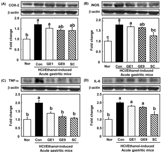
감소시킴으로서 in vitro에서의 결과와 in vivo 내에서의 결과가 유사한 양상을 보이는 것을 확인하였으며 이는 천마추출물의 경 구투여가 ROS와 ONOO−의 발생을 감소시켜 위 점막 보호효과 를 나타냈던 것으로 판단된다. 위 조직의 염증성 사이토카인 발현 산화적 스트레스가 증가함에 따라 염증반응이 일어나게 되고, 이로 인하여 COX-2, iNOS, TNF-α, IL-6와 같은 염증성 사이토 카인이 분비되어 세포손상이 발생하게 된다. TNF-α가 분비되면 각종 염증과 세포사멸 유도현상이 나타나며(40), IL-6는 TNF-α와 함께 T세포의 분화를 자극하고 염증반응을 촉진한다(41). COX-Fig. 3. Effect of steamed Gastrodia elata on rective oxygen species in serum peroxynitrite in stomach tissues. All values are mean±SE (n=6). Normal mice: Nor, HCl/ethanol-induced acute gastritic mice with the administration of distilled water: Con, HCl/ethanol-induced acute gastritic mice with the administration of 100 mg/kg Gastrodia elata by steaming 1 time: GE1, HCl/ethanol-induced acute gastritic mice with the administration of 100 mg/kg Gastrodia elata by steaming 9 times: GE9, HCl/ethanol-induced acute gastritic mice with the administration of 10 mg/kg sucralfate: SC. Different letters show a significant difference at p<0.05 as determined by one-way ANOVA followed by Tukey multiple comparison test.
2는 PGE2를 생성하여 염증반응을 지속하는 역할을 하며(42) iNOS 는 NO의 대량 생성에 의해 발현되어 세포 내에서 염증매개물질 로 작용한다(43). 따라서 본 연구에서는 염증성 매개인자인 COX-2, iNOS, TNF-α, IL-6의 발현을 확인하였다. COX-2의 발현은 정 상군(1.00±0.08)에 비해 대조군(1.69±0.11)은 유의성 있게 증가하 였으며 양성대조군인 SC투여군(1.41±0.14)에서 감소하였고, 1회 증숙 천마 투여군(1.51±0.10)보다 9회 증숙 천마 투여군(1.42±0.07) 에서 감소하였으나 통계적 유의성은 없었다. iNOS의 경우 정상군(1.00±0.06)에 비해 대조군(1.77±0.14)이 증 가하였으며 SC투여군(1.25±0.15)에서는 감소하였고, 1회 증숙 천 마 투여군(1.68±0.05)과 9회 증숙 천마 투여군(1.63다. 급성 위염 개선 효능 실험에서 증숙 천마 추출물의 섭취는 HCl/ethanol로 유 발된 위 점막 손상 마우스에서 위 조직의 육안적 손상을 감소시 켰다. 9회 증숙 천마 추출물의 섭취는 위 점막 손상마우스에서 혈청의 ROS와 조직의 ONOO−를 감소시켰고, 위 조직에서 염증 성 매개인자인 TNF-α 또한 감소시켰다. 결론적으로 9회 증숙한 천마의 경구투여는 1회 증숙한 천마에 비하여 효과적으로 위 점 막 손상을 억제하였다. 따라서 9회 증숙 천마 추출물의 투여가 급성 위염 유발 마우스 모델에서 위 점막 손상 억제에 효과가 있 다고 사료된다.
요
약
본 연구는 증숙 횟수에 따른 천마 추출물의 경구 투여가 산화 방지 효과를 통한 위 점막 손상 억제에 미치는 효과를 평가하는 실험이다. 증숙 횟수를 달리한 천마 추출물을 준비하여 DPPH, ABTS 라디칼 소거능, total phenol, flavonoid, gastrodin, gastrodi-genin 성분 분석 결과, 1회 증숙 천마와 9회 증숙 천마 추출물간 의 급성 위염 개선 효능 평가가 가능할 것으로 판단되었다. HCl/ ethanol로 유발된 급성 위염 동물 모델에 GE1과 GE9 (100 mg/kg body weight)과 sucralfate (10 mg/kg body weight)를 HCl/ethanol 처리 전 경구 투여하였다. 그리고 이를 정상군과 대조군과 비교 분석하였다. 급성 위염 개선 효능 실험에서 증숙 천마 추출물의 섭취는 HCl/ethanol로 유발된 위 점막 손상 마우스에서 위 조직 의 육안적 손상을 감소시켰다. 9회 증숙 천마 추출물의 섭취는 위 점막 손상마우스에서 혈청의 ROS와 조직의 ONOO−를 감소 시켰고, 위 조직에서 염증성 매개인자인 TNF-α 또한 감소시켰 다. 결론적으로 9회 증숙한 천마의 경구투여는 1회 증숙한 천마 에 비하여 효과적으로 위 점막 손상을 억제하였다. 따라서 9회 증숙 천마 추출물의 투여가 급성 위염 유발 마우스 모델에서 위 점막 손상 억제에 효과가 있다고 사료된다.Fig. 4. Effect of steamed Gastrodia elata on inflammatory factors of stomach tissues in HCl/ethanol induced acute gastritic mice. All values are mean±SE (n=6). Normal mice: Nor, HCl/ethanol-induced acute gastritic mice with the administration of distilled water: Con, HCl/ ethanol-induced acute gastritic mice with the administration of 100 mg/kg Gastrodia elata by steaming 1 time: GE1, HCl/ethanol-induced acute gastritic mice with the administration of 100 mg/kg Gastrodia elata by steaming 9 times: GE9, HCl/ethanol-induced acute gastritic mice with the administration of 10 mg/kg sucralfate: SC. Different letters show a significant difference at p<0.05 as determined by one-way ANOVA followed by Tukey multiple comparison test.
References
1. Banks WJ. Applied veterinary histology 2nd. pp.393-396. William & Wilkins Baltimore. Philadelphia, USA. (1986)
2. Shay J, Komarov SA, Fels SS, Merance D, Gruenstein M, Siplet H. A simple method for the uniform production of gastric ulcer-ation in the rats. Gastroenterology 5: 43-61 (1945)
3. Suzuki H, Nishizawa T, Tsugawa H, Mogami S, Hibi T. Role of oxidative stress in stomach disorders. J. Clin. Biochem. Nutr. 50: 35-39 (2012)
4. Thomson A, Hemphill D, Jeejeebhoy KN. Oxidative stress and anti-oxidants in intestinal disease. Dig. Dis. 16: 152-158 (1998) 5. Feldman M, Burton ME. Histamine2-receptor antagonists-standard
therapy for acid-peptic diseases. N. Engl. J. Med. 20: 1672-1680 (1990)
6. Bell NJ, Hunt RH. Progress with proton pump inhibition. Yale J. Biol. Med. 65: 649-657 (1992)
7. Berardi RR, Savitsky ME, Nostrant TT. Maintenance therapy for prevention of recurrent peptic ulcers. Drug Intell. Clin. Pharm. 21: 493-507 (1987)
8. Fullarton GM, McLauchlan G, Macdonald A, Crean GP, McColl KE. Rebound nocturnal hypersecretion after four weeks treatment with an H2 receptor antagonist. Gut 30: 449-454 (1989)
9. Szabo S, Bynum TE. Alternatives to the acid-oriented approach to ulcer disease: does ‘cytoprotection’ exist in man? A new clas-sification of antiulcer agents. Scand. J. Gastroenterol. 23: 1-6 (1988)
10. Taguchi H, Yosioka I, Yamasaki K, Kim IH. Studies on the con-stituents Gastrodia elata Blume. Chem. Pharm. Bull. 29: 55-62 (1981)
11. Ahn DK, Kim HC. Hanyakpojaehak. Iljung Publishing House, Seoul, Korea. pp. 285-287 (2000)
12. Heo JC, Park JY, An SM, Lee JM , Yun CY, Shin HM, Kwon TK, Lee SH. Anti-oxidant and anti-tumor activities of crude extracts by Gastrodia elata Blume. Korean J. Food Preserv. 13: 83-87 (2006) 13. Kang TS, Kong JY, Kwon HJ, Choi, Hong JG, Park YK. A
Stud-ies on the chemical composition and in vitro biological activitStud-ies of hot water extracts of Gastrodia elata. Korean J. Mycol. 30: 136-141 (2004)
14. Cho HE, Choi YH, Park SH, Park YS, Ahn BY. Effect of Gas-trodiae rhizoma powder on serum and liver lipid levels of rats with high fat diet. Korean J. Food Nutr. 21: 64-70 (2008)
15. Kim MH. Screening of biological activities of ethanol extracts from fermented Gastrodia elata Blume. Korean J. Food Nutr. 27: 837-844 (2014)
16. Blosis MS. Antioxidant determinations by the use of a stable free radical. Nature 26: 1199-1200 (1958)
17. Re R, Pellegrini N, Proteggente A, Pannala A, Yang M, Rice-Evans C. Antioxidant activity applying an improved ABTS radi-cal cation decolorization assay. Free Radic. Bio. Med. 26: 1231-1237 (1999)
18. Folin O, Denis W. On phosphotungastic-phosphomolybdic com-pounds as color reagent. J. Biol. Chem. 12: 239-243 (1912) 19. Lister CE, Lancaster JE, Sutton KH, Walker JR. Developmental
changes in the concentration and composition of flavonoids in skin of a red and a green apple cultivar. J. Sci. Food Agr. 64: 155-161 (1994)
20. Martins JLR, Rodrigues ORL, da Silva DM, Galdino PM, de Palula JR, Romao W, da Costa HB, Vaz BG, Ghedini PC, Costa EA. Mechanisms involved in the gastroprotective activity of Celtis iguanaea (Jacq.) Sargent on gastric lesions in mice. J. Eth-nopharmacol. 155: 1616-1624 (2014)
21. Ali SF, LeBel CP, Bondy SC. Reactive oxygen species formation as a biomarker of methylmercury and trimethyltin neurotoxicity. Neurotoxicology 13: 637-648 (1992)
22. Kooy NW, Royall JA, Ischiropoulos H, Beckman JS. Peroxyni-trite-mediated oxidation of dihydrohodamine 123. Free Radic. Biol. Med. 16:149-156 (1994)
23. Zhao S, Li N, Zhen Y, Ge M, Li Y, Yu B, Shao RG. Protective effect of gastrodin on bile duct ligation-induced hepatic fibrosis
in rats. Food Chem. Toxicol. 86: 202-207 (2015)
24. Zhang JS, Zhou SF, Wang Q, Guo JN, Liang HM, Deng JB, He WY. Gastrodin suppresses BACE1 expression under oxidative stress condition via inhibition of the PKR/eIF2α pathway in Alzheimer’s disease. Neuroscience 325: 1-9 (2016)
25. Kam KY, Yu SJ, Jeong N, Hong JH, Jalin AMA, Lee S, Kang SG. p-Hydroxybenzyl alcohol prevents brain injury and behav-ioral impairment by activating Nrf2, PDI, and neurotrophic factor genes in a rat model of brain ischemia. Mol. Cells. 31: 209-215 (2011)
26. Liu SH, Pan IH, Chu IM. Inhibitory effect of p-hydroxybenzyl alcohol on tyrosinase activity and melanogenesis. Biol. Pharm. Bull. 30: 1135-1139 (2007)
27. Yun-Choi HS, Pyo MK. Isolation of 4,4'-dihydroxybenzyl sulfox-ide from Gastrodia elata. Arch. Pharm. Res. 20: 91-92 (1997) 28. Shin SL, Lee CH. Antioxidant effects of the methanol extracts
obtained from aerial part and rhizomes of ferns native to Korea. Korean J. Plant Res. 23: 38-46 (2010)
29. Park MR, You C, Chang YN, Ahn BY. Change of total polyphe-nol content of fermented Gastrodia elata Blume and radical scav-enging. Korean J. Plant Res. 25: 379-386 (2012)
30. Jeon HY, Kim MH, Kim MR. Antioxidative and antimutagenin activity of ethanol extracts from Cuscutae semen. Korean J. Food Cook. Sci. 24: 46-51 (2008)
31. Ishii Y, Fujii Y, Homma M. Gastric acid stimulating action of cysteamine in the rat. Eur. J. Pharmacol. 36: 331-336 (1976) 32. Kim SJ, Baek JY, Park CK, Kim GW. Gastroprotective effect of
Korean rice-wine (Yakju). Korean J. Food Sci. Technol. 36: 818-822 (2004)
33. Jung KW. Momordicae cochichinensis seed extract as a therapeu-tic agent for acute & chronic gastritis and its constituents. PhD thesis, Seoul national University, Seoul, Korea (2012)
34. Kenter M, Demir H, Karakaya C, Ozbek H. Gastroprotective activity of Nigella sativa L oil and its constituent, thymoquinone against acute alcohol-induced gastric mucosal injury in rats. World J. Gastoenterol. 11: 6662-6666 (2005)
35. Alrashdi AS, Salama SM, Alkiyumi SS, Abdulla MA, Hadi AHA, Abdelwahab SI, Asykin N. Mechanisms of gastroprotective effects of ethanolic leaf extract of Jasminum sambac against HCl/ ethanol-induced gastric mucosal injury in rats. Evid.-Based Compl. Alt. 2012: 786426 (2012)
36. Boligon AA, de Freitas RB, de Brum TF, Waczuk EP, Klimacze-wski CV, de Avila DS, de Freitas Bauermann L. Antiulcerogenic activity of Scutia buxifolia on gastric ulcers induced by ethanol in rats. Acta Pharm. Sin. B. 4: 358-367 (2014)
37. Bilici D, Suleyman H, Banoglu ZN, Kizltunc A, Avci B, Clftcio-glu A, Bilici S. Melatonin prevents ethanol-induced gastric mucosal damage possibly due to its antioxidant effect. Dig. Dis. Sci. 47: 856-861 (2002)
38. Suzuki Y, Ishihara M, Segami T, Ito M. Anti-ulcer effects of antioxidants, quercetin, alpha-tocopherol, nifedipine and tetracy-cline in rats. Jpn. J. Pharmacol. 78: 435-441 (1998)
39. Atsushi O, Kenjirou O, Mamoru K, Hideaki H. Protective effects of a gastrointestinal agent containing Korean red ginseng on gas-tric ulcer models in mice. BMC Complem. Altern. M. 10: 10.1186/1472-6882-10-45 (2010)
40. Van Antwerp DJ, Martin SJ, Verma IM, Green DR. Inhibition of TNF-α induced apoptosis by NF-κB. Trends Cell Biol. 8: 107-111 (1998)
41. Opal SM, DePalo VA. Anti-inflammatory cytokines. Chest 117: 1162-1172 (2000)
42. Lee SW, Shin SM, Kim HY, Han SH, Kim KH, Kwon JH, Kwak JH, Lee CK, Yim DS, Kim KJ. Anti-inflammatory function of arctiin by inhibiting COX-2 expression via NF-κB pathways. J. Inflamm. 8: 10.1186/1476-9255-8-16 (2011)
43. Ekmekcioglu S, Ellerhorst J, Smid CM, Prieto VG, Munsell M, Buzaid AC, Grimm EA. Inducible nitric oxide synthase and nitrotyrosine in human metastatic melanoma tumors correlate with poor survival. Clin. Cancer Res. 6: 4768-4775 (2000)