INTRODUCTION
Interstitial lung disease (ILD) relatively often develops in patients with connective tissue diseases, such as rheumatoid arthritis (RA), polymyositis-dermatomyositis, progressive systemic sclerosis, and mixed connective tissue disease. The prevalence of ILD in patients with RA has been variably reported according to the method of assessment. In radio- graphic studies, the reported incidence of ILD has varied from about 1.6 to 4.5% (1). However, up to 40% of patients who were evaluated with pulmonary function tests demonstrated abnormalities of diffusing capacity (2).
ILD associated with RA may be slowly progressive or relent- lessly fatal. Traditionally, corticosteroids have been the main- stay in the treatment of these patients, but their effect is gen- erally limited and may be temporary. Although there have been some anecdotal reports on the efficacies of cytotoxic agents such as methotrexate, D-penicillamine, azathioprine, and cyclophosphamide (3-6), no convincing study has been established for these agents. So far, a limited information has been available for cyclosporine trial in ILD associated with RA. We describe a 49-yr-old female with RA and progres- sive ILD refractory to corticosteroid and cyclophosphamide who was effectively treated with cyclosporine.
CASE REPORT
In March 1999, a 49-yr-old woman presented with a 1-yr history of polyarthralgia and shortness of breath on exertion.
She did not smoke and had no past history of environmental toxin exposure. There was no medical history of photosensi- tivity, oral ulcer, alopecia, and Raynaud phenomenon. On physical examination, inspiratory crackles were heard on the both lower lung fields, and heart sounds were normal. The joint swelling and tenderness were noted on the small joints of both hands, right elbow, right first metatarsophalangeal joint, and left third metatarsophalangeal joint. Laboratory studies at this time showed a hemoglobin level of 11.3 g/dL and a white blood cell count of 5,200/ L. The erythrocyte sedimentation rate was 50 mm/hr. The C-reactive protein was not increased. The rheumatoid factor was 340 IU/mL and the antinuclear antibody titer was 1:640 with a speckled pattern. The anti-extractable nuclear antigen and anti-dou- ble-stranded DNA antibodies were absent. The complement level was normal. There were no specific abnormalities on other chemistry profiles.
A chest radiograph revealed bibasilar interstitial infiltrates (Fig. 1A). High resolution computerized tomography (HRCT) of the chest showed interstitial fibrosis, and ground glass
Hyun Kyu Chang*, Wann Park, Dae Sik Ryu�
Department of Internal Medicine, Diagnostic Radiology�, University of Ulsan Asan-Kangnung Hospital, Kangnung, Korea
Address for correspondence Hyun Kyu Chang, M.D.
Department of Internal Medicine, College of Medicine, Dankook University, 16-5 Anseo-dong, Choeonan 330-715, Korea
Tel : +82-33-610-3122, Fax : +82-33-641-8130 E-mail: [email protected]
270 J Korean Med Sci 2002; 17: 270-3
ISSN 1011-8934
Copyright � The Korean Academy of Medical Sciences
Successful Treatment of Progressive Rheumatoid Interstitial Lung Disease With Cyclosporine
: A Case Report
Treatment of interstitial lung disease (ILD) in rheumatoid arthritis (RA) has been controversial. Although there have been several anecdotal reports on the effica- cies of corticosteroids or cytotoxic agents such as methotrexate, cyclophos- phamide, azathioprine, and D-penicillamine for the treatment of ILD associated with RA, no controlled studies have been performed. To date, corticosteroids have been a central agent for the treatment of this disease, but their effects are partial and temporary in most cases. In addition, the adverse effects of these agents are considerable. On the other hand, limited information is available on the cyclosporine use in ILD associated with RA. We describe a 49-yr old female patient with RA and ILD that had initially responded to high dose prednisolone and cyclophosphamide intravenous pulse therapy, and the lung disease was aggravated with the tapering of prednisolone. After 10 months of follow-up loss, the patient was successfully treated with low dose cyclosporine without high dose corticosteroids.
Key Words : Lung Disease, Interstitial; Arthritis, Rheumatoid; Cyclosporine
*Present address: See address for correspondence Received : 8 February 2001
Accepted : 14 May 2001
Cyclosporine in Progressive Rheumatoid Interstitial Lung Disease 271
opacities suggesting active inflammation on the both lower lung fields. A pulmonary function test revealed a mild restric- tive pattern with forced vital capacity (FVC) of 78% of pre- dicted. The carbon monoxide diffusing capacity was 46% of predicted. Although juxta-articular osteoporosis in the plain radiographs was noted around proximal interphalangeal joints of both hands, right wrist, and right elbow, bony erosions were absent.
A diagnosis of RA with active ILD was established. Her initial medications included prednisolone 1 mg/kg daily, hy- droxychloroquine 200 mg b.i.d. and nabumetone 500 mg b.i.d. Monthly cyclophosphamide intravenous (IV) pulse ther-
apy with a dose of 600 mg/m2was started. Three weeks later, dyspnea and polyarthralgia improved. The pulmonary func- tion test was also improved with FVC of 85% of predicted and diffusing capacity of 56% of predicted. The dosage of prednisolone was gradually tapered to 7.5 mg over the next 4 months. Monthly cyclophosphamide IV pulse therapy was continued.
In September 1999, she complained of a shortness of breath of a greater severity. At that time, pulmonary function was aggravated with FVC 72% of predicted and diffusing capac- ity of 50%. HRCT showed progressive basal interstitial infil- trates and increased ground glass opacities compared with
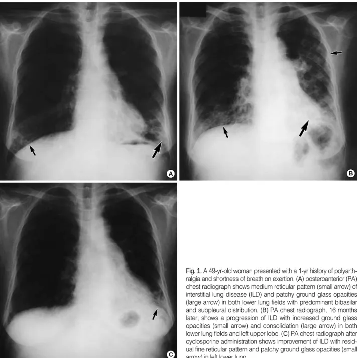
Fig. 1.A 49-yr-old woman presented with a 1-yr history of polyarth- ralgia and shortness of breath on exertion. (A) posteroanterior (PA) chest radiograph shows medium reticular pattern (small arrow) of interstitial lung disease (ILD) and patchy ground glass opacities (large arrow) in both lower lung fields with predominant bibasilar and subpleural distribution. (B) PA chest radiograph, 16 months later, shows a progression of ILD with increased ground glass opacities (small arrow) and consolidation (large arrow) in both lower lung fields and left upper lobe. (C) PA chest radiograph after cyclosporine administration shows improvement of ILD with resid- ual fine reticular pattern and patchy ground glass opacities (small arrow) in left lower lung.
A B
C
272 H.K. Chang, W. Park, D.S. Ryu
the findings in March 1999. Cyclosporine 3.6 mg/kg (200 mg) daily was prescribed, and prednisolone was increased to 20 mg daily, but she did not take this medication and was lost from follow-up. During this period she had not taken any medical treatment except herb medications.
In July 2000, she was admitted again with complaints of shortness of breath on minimal exertion and severe polyarthral- gia. Physical examination revealed inspiratory crackles and coarse breath sounds on both lower lung fields. There were swelling and tenderness on the multiple joints such as small joints of both hands, both elbows, both knee joints, and right ankle. On laboratory studies, a white blood cell count was 10,000/ L and a hemoglobin level was 11 g/dL. Arterial blood gas analysis without O2supplementation showed a PaO2of 54.1 mmHg, PaCO2of 27.6 mmHg, and O2satura- tion of 90.1%. The lung infiltrates on the chest radiograph (Fig. 1B) and HRCT (Fig. 2) were more aggravated with an extension to left upper lobe. The pulmonary function was also aggravated with FVC of 54% of predicted and diffusing capacity of 37% of predicted. A transbronchial lung biopsy showed interstitial fibrosis and mononuclear cell infiltrates, which were compatible with interstitial pneumonitis. Bac- terial cultures grew normal flora of the upper respiratory tract. Cultures for fungus and mycobacteria were negative.
She was considered to have as advanced ILD with active inflammation in association with RA. Daily cyclosporine 3.6 mg/kg (200 mg), prednisolone 5 mg, and celecoxib 200 mg were started. Within 4 weeks, her breathlessness and joint symptoms markedly improved. The pulmonary function test revealed FVC of 69% of predicted and diffusing capaci- ty of 39% of predicted. Serum trough level of cyclosporine was 119 ng/mL. In December 2000, her shortness of breath further improved and stabilized. Chest radiograph (Fig. 1C) and HRCT at this time showed markedly reduced pulmonary
infiltrates. The dosage of cyclosporine could be reduced to 2.7 mg/kg (150 mg) daily. Mild hypertension which was the only side effect of cyclosporine, was controlled with amlodip- ine. In January 2001, she was free of joint symptoms, and only had mild dyspnea on exertion.
DISCUSSION
The clinical course of ILD associated with RA is variable.
Whereas some patients take gradually progressive clinical course (7), other patients run a rapidly progressive course that can be fatal within several months (2). Spontaneous remission for this disease has been rarely reported (8). The clinical presentation and the pathologic findings of ILD in RA are known to be very similar to those of idiopathic pul- monary fibrosis, and the therapeutic modalities for these two diseases are not much different. Although some RA patients with progressive ILD may respond to aggressive immuno- suppressive therapy, treatment for this disease still remains controversial.
To date, the corticosteroids have been the central agent for the treatment of progressive ILD in RA. However, the favor- able response rate to these agents has been reported less than 40%, and documentation of sustained effect is even rare (2, 9, 10). Although there have been several anecdotal reports for the efficacies of cytotoxic agents such as methotrexate, azathioprine, D-penicillamine, and cyclophosphamide in patients with RA and progressive ILD, few controlled studies have demonstrated the sustained effects of these agents (2).
The efficacy of cyclosporine for the treatment of RA has been proven (11-13). Even if there have been a few reports for the successful cyclosporine use in progressive ILD or acute interstitial pneumonitis in association with RA, the available
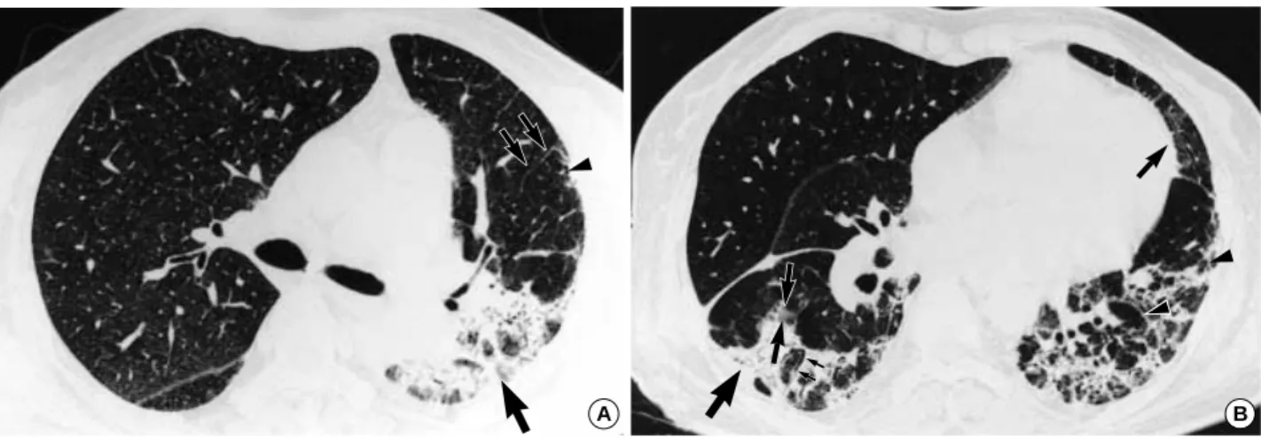
Fig. 2. (A) HRCT at the level of the carina shows consolidation (large arrow) in the posterior segment of left upper lobe and superior seg- ment of left lower lobe. Interlobular septal thickening (small arrows) and subpleural traction bronchiolectasis (arrowhead) are noted in anterior segment of left upper lobe. (B) HRCT at left atrium level shows ground glass opacity (small arrows) and consolidation (large arrow). Traction bronchiectasis (thin arrow), thickening of major fissure, and irregular interlobular septal thickening (arrowhead) are pre- sent in the both lower lobe.
A B
Cyclosporine in Progressive Rheumatoid Interstitial Lung Disease 273
information is still limited (14-17). Alegre et al. first described the successful treatment of aggressive pulmonary fibrosis in RA with cyclosporine (14). In contrast to our case, that patient had symptoms for less than one month before treatment, and had not been treated with any other immunosuppressive agents. In addition they used high dose prednisone and high- er dose cyclosporine than in our case. Puttick et al. reported the effect of cyclosporine in a patient with long-standing RA and progressive ILD that had been refractory to corticosteroids and cytotoxic agents such as chlorambucil and oral cyclophos- phamide. They used similar dose of cyclosporine and higher dose of prednisolone, compared with our case (15).
Our patient had initially responded to high dose pred- nisolone and cyclophophamide IV pulse therapy. However, her lung disease was aggravated with the tapering of pred- nisolone for 6 months. After 10 months of follow-up loss, ILD was much more aggravated. At that time, she was treat- ed with low doses of cyclosporine and prednisolone (5 mg/day).
Despite subjective symptoms, her pulmonary function such as forced vital capacity, and radiographic pulmonary infiltrates have much improved with these agents, the diffusing capacity did not change. This may reflect the ventilation-perfusion mismatch in irreversibly damaged lung lesions. Most patients in the literature who showed the effectiveness of cyclosporine, including our case, the improvement exhibited within one month. Because other patients than ours had used cyclosporine together with higher dose of corticosteroids, it is not certain whether the therapeutic improvement was due to cyclosporine or to corticosteroids. On the other hand, as the spontaneous remission is extremely rare in progressive ILD associated with RA, the improvement of the lung disease in the present case was mostly considered to be the effect of cyclosporine.
Cyclosporine, a complex fungal decapeptide, has highly selective immunosuppressive properties. This agent appears to reduce interleukin-2 synthesis by activated T-cell (18) and to disrupt cytokine-dependent T lymphocyte-macrophage interaction, and thus prevents fibroblast-mediated fibrosis that is thought to be important in the pathogenesis of ILD (2). Recently, Sekigawa et al. described 4 cases of steroid-resis- tant autoimmune diseases who were effectively treated with an extremely low dose of cyclosporine (1 mg/kg/day) (19). Due to the limited information of cyclosporine in the treatment of RA with ILD, the controlled studies are needed to clarify whether cyclosporine is effective agent for the treatment of ILD associated with RA, whether high dose corticosteroids are required together with cyclosporine, and which dosage of cyclosporine is optimal.
REFERENCES
1. Hunninghake GW, Fauci AS. Pulmonary involvement in the colla-
gen vascular disease. Am Rev Respir Dis 1979; 119: 471-503.
2. Roschmann RA, Rothenberg RJ. Pulmonary fibrosis in rheumatoid arthritis: a review of clinical features and therapy. Semin Arthritis Rheum 1987; 16: 174-85.
3. Scott DGI, Bacon PA. Response to methotrexate in fibrosing alve- olitis associated with connective tissue disease. Thorax 1980; 35:
725-32.
4. Lorber A. Penicillamine therapy for rheumatoid lung disease: effects on protein suphydryl groups. Nature 1966; 210: 1235-7.
5. Cohen JM, Miller A, Spiera H. Interstitial pneumonitis complicat- ing rheumatoid arthritis. Chest 1977; 72: 521-4.
6. Brown CH, Turner-Warwick M. The treatment of cryptogenic fibrosing alveolitis with immunosuppressant drugs. Q J Med 1971;
158: 289-302.
7. Patterson CD, Harvill WE, Pierce JA. Rheumatoid lung disease.
Ann Intern Med 1965; 62: 685-97.
8. Scadding JG. The lungs in rheumatoid arthritis. Proc R Soc Med 1980; 62: 227-38.
9. Walker WC, Wright V. Pulmonary lesions and rheumatoid arthri- tis. Medicine 1968; 47: 501-20.
10. Turner-Warwick M, Evans RC. Pulmonary manifestations of rheumatic disease. Clin Rheum Dis 1977; 3: 549-64.
11. Bombardier C, Buchbinder R, Tugwell P. Efficacy of cyclosporin A in rheumatoid arthritis: long-term follow-up data and the effect on quality of life. Scand J Rheumatol Suppl 1992; 95: 29-33.
12. Tugwell P. Cyclosporine in rheumatoid arthritis: docomented effi- cacy and safety. Semin Arthritis Rheum 1992; 21(Suppl 3): 30-8.
13. Tugwell P, Pincus T, Yocum D, Stein M, Gluck O, Kraag G, McK- endry R, Tesser J, Baker P, Wells G. Combination therapy with cyclosporine and methotrexate in severe rheumatoid arthritis. The Methotrexate-Cyclosporine Combination Study Group. N Engl J Med 1995; 333: 137-41.
14. Alegre J, Teran J, Alvarez B, Viejo JL. Successful use of cyclosporine for the treatment of aggressive pulmonary fibrosis in a patient with rheumatoid arthritis. Arthritis Rheum 1990; 33: 1594-6.
15. Puttick MP, Klinkhoff AV, Chalmers A, Ostrow DN. Treatment of progressive rheumatoid interstitial lung disease with cyclosporine.
J Rheumatol 1995; 22: 2163-5.
16. Miyazawa S, Hotta O, Kitamura H, Sudou K, Horigome I, Chiba S, Tokoi T, Taguma Y. Successful treatment of interstitial pneumonitis with cyclosporin A in a patient with rheumatoid arthritis accompa- nied by acute interstitial nephritis. Nippon Jinzo Gakkai Shi 1996;
38: 33-9.
17. Ogawa D, Hashimoto H, Wada J, Ueno A, Yamasaki Y, Yamamu- ra M, Makino H. Successful use of cyclosporin A for the treatment of acute interstitial pneumonitis associated with rheumatoid arthri- tis. Rheumatology (Oxford) 2000; 39: 1422-4.
18. Kahan BD. Cyclosporine. N Engl J Med 1989; 321: 1725-38.
19. Sekigawa I, Ogasawara H, Sugiyama M, Kaneko H, Hishikawa T, Tokano Y, Iida N, Hahimoto H, Hirose S. Extremely low dose treat- ment of cyclosporine for autoimmune diseases. Clin Exp Rheumatol 1998; 16: 352.