INTRODUCTION
Although occlusive lesions of the arch vessels are not uncom- mon among patients with atherosclerosis, these lesions are encountered less frequently than carotid bifurcation lesions and account for fewer than 5% of the surgical procedures performed on the cerebral vasculature. Most patients with arch vessel lesions are asymptomatic, because of the rich col- lateral circulation of the head and shoulder girdle. When symptoms occur, they are usually flow-related, and are quite variable. Neurological symptoms are most common and are related to the degree of posterior circulation insufficiency in patients with a subclavian or innominate artery disease.
Localized in vivo proton magnetic resonance spectroscopy (1H-MRS) has been used to measure the metabolic status of the human brain in a non-invasive manner; thus, it is often called “a non-invasive biochemical assay” (1). MRS is more sensitive than magnetic resonance imaging (MRI) in detect- ing ischemic damage by measuring the metabolic changes that occur prior to the anatomic changes. Therefore, 1H-MRS can aid the diagnosis and management of cerebral hypoper- fusion at a much earlier stage.
This report describes a patient with innominate artery occlu- sion and symptoms of posterior circulation insufficiency who showed metabolic changes by 1H-MRS after revascularization.
CASE REPORT
A 61-yr-old male was admitted to our department because of intermittent dizziness, blurred vision, and right arm clau- dication of 6 yr duration. He had never suffered from a cere- bral event. MRI, carotid duplex scan (ATL HDI 3000: Ad- vanced Technology Laboratories High Definition Imaging 3000, Bothell, Washington, U.S.A.), and four-vessel cerebral angiography studies were undertaken to assess the posterior cerebral circulation. The MRI showed no visible lesion. Carotid duplex scan showed a damped waveform and markedly de- creased volume flow of the right common carotid artery (60 mL/min) and reversed flow of the ipsilateral vertebral artery (Fig. 1). Four-vessel cerebral angiography exhibited complete occlusion of the innominate artery and a retrograde flow from the right vertebral artery into the common carotid and sub- clavian artery (Fig. 2). Duplex and angiographic findings were consistent with the subclavian steal syndrome caused by complete occlusion of the innominate artery. 1H-MRS was performed on a GE 1.5T SIGNA system equipped with shielded gradients and proton brain examination (PROBE) package (General Electric Medical System, Milwaukee, U.S.A., Version 5.4) in a resting state, after informed consent. Image- guided stimulated echo acquisition mode (STEAM)-spectra were obtained from 7-10 mL voxels in the middle cerebral artery territory and cerebellum of both hemispheres (Fig. 3A).
Yong Pil Cho, Jung Hee Lee*, Geun Eun Kim�
Department of Surgery, University of Ulsan College of Medicine, Gangneung Asan Hospital, Gangneung;
NMR Laboratory*, Asan Institute for Life Sciences, and Department of Vascular Surgery�, Asan Medical Center, University of Ulsan College of Medicine, Seoul, Korea
Address for correspondence Yong Pil Cho, M.D.
Department of Surgery, Gangneung Asan Hospital, 415 Bangdong-ri, Sacheon-myeon, Gangneung 210-711, Korea
Tel : +82.33-610-3229, Fax : +82.33-641-8120 E-mail : [email protected]
267
Metabolic Changes after Revascularization in a Patient with
Innominate Artery Occlusion by Localized in vivo Proton Magnetic Resonance Spectroscopy
Localized in vivo proton magnetic resonance spectroscopy (1H-MRS) has been used to measure the metabolic status of the human brain in a non-invasive man- ner; thus, it is often called “a non-invasive biochemical assay”. MRS is more sen- sitive than magnetic resonance imaging (MRI) in detecting ischemic damage by measuring the metabolic changes that occur prior to the anatomic changes. We report a patient who presented with innominate artery occlusion and symptoms of posterior circulation insufficiency and showed favorable metabolic changes by
1H-MRS after revascularization. He showed no visible lesion in brain MRI, but in
1H-MRS, decreased N-acetylaspartate (NAA) signal was noted in a resting state.
After revascularization, both symptomatic improvement and recovery of NAA signal were observed. 1H-MRS may provide valuable clinical information in diag- nosis and management of cerebral hypoperfusion at a much earlier stage prior to the anatomic changes.
Key Words : Magnetic Resonance Spectroscopy; Brachiocephalic trunk; Arterial Occlusive Diseases;
Reperfusion
Received : 29 March 2002 Accepted : 17 May 2002
Fig. 3B demonstrates the 1H-MR spectrum acquired from the right cerebellum. The spectrum showed a decreased N- acetylaspartate (NAA) signal as compared to the contralat- eral side (Table 1). Axillary-to-axillary bypass with 8-mm externally supported polytetrafluoroethylene (PTFE) graft was performed without complications. At 10 days after surgery, carotid duplex scan and 1H-MRS were done for the evalua- tion of hemodynamic changes and the metabolic status of the
right cerebellum. Postoperative carotid duplex scan showed a normal arterial waveform of the right common carotid artery with a markedly increased volume flow (400 mL/min) and a to-and-fro waveform of the ipsilateral vertebral artery (Fig. 4).
Postoperative 1H-MR spectra were acquired from the same region of the middle cerebral artery territory and cerebellum of both hemispheres. The spectra showed increased NAA values in the right middle cerebral artery territory and cerebel-
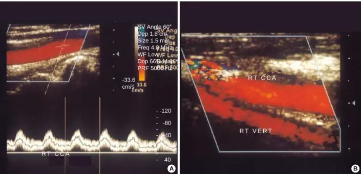
Fig. 1.Preoperative carotid duplex scan shows (A) a damped waveform and markedly decreased volume flow of the right common carotid artery (RT CCA), and (B) reversed flow of the ipsilateral vertebral artery (RT VERT).
A B
R T C C A
R T C C A
+19
-19 cm/
R T V E R T
R T S U B C - -100
-- -80 -- -60 -- -40 -- -20 -- cm/s -- 20 +24.0
SV Angle 60。
Dep 1.9 cm Size 1.5 mm Freq 4.0 MHz WF Low Dop 70% Map PRF 3731 Hz -24.0
cm/s
Fig. 2.Transfemoral four-vessel cerebral angiography shows (A) complete occlusion of the innominate artery and (B) retrograde flow from the right vertebral artery into the common carotid and subclavian artery.
A B
27.8 32.7
44.2
lum as compared to the preoperative values (Fig. 3C, Table 1).
DISCUSSION
Neurospectroscopy is defined as the field of study resulting from MRS examination of the human brain (1). It measures neuronal markers, energy and redox states, specific fuels of tissue respiration, maturation, and possibly myelination. It provides diagnostic patterns of altered neurochemistry. Positron emission tomography (PET), and to a lesser extent single photon emission computed tomography (SPECT), magnet- ic resonance angiography (MRA), functional magnetic reso- nance imaging (fMRI), and “diffusion-imaging” address
blood flow, glucose turnover, and oxygen consumption. How- ever, until the advent of nuclear magnetic resonance, no direct noninvasive assay of the cerebral metabolites was available.
There was no neuronal marker, no astrocyte marker, and no technique to directly determine the energy metabolism. These gaps are now filled by neurospectroscopy. With increased clinical experience of neurospectroscopy, an increasingly use- ful metabolic information emerges. Single-voxel 1H-MRS can measure the cerebral metabolites for patients with vari- ous diseases (1-3).
Of the many expected and new resonances now identified in the clinical practice of neuro-MRS, none has created more diagnostic information than NAA. Multiple lines of evidence suggest that NAA can be used as a neuronal marker as it is
Fig. 3.(A) 1H-MR spectrum was acquired from the right cerebellum in T2-weighted MRI. 1H-MR spectra shows a markedly increased NAA signal as compared to the preoperative signal: before (B) and after (C) revascularization.
C A100
P100 A B
Cr
cho
NAA
4 3 2 1 0 ppm
4 3 2 1 0 ppm
A100
P100
found exclusively in neurons (4-6), and their processes in the mature brain were well previously investigated (7). In human brain spectra, in vivo NAA is reduced in situations known to be associated with neuronal loss (8, 9). It can begin to fall as early as two hours after the onset of focal infarction (10). The continued NAA decline over the first two weeks after stroke does not merely reflect clearing of debris from neurons killed at the initial outset, but it is an evidence of continuing neu- ronal loss after the acute period, possibly, in the ischemic pe- numbra (4). Reversible decreases in NAA emphasize that neuronal dysfunction or transient relative volume changes can also lead to a decreased level of NAA (3, 4, 11). Although we do not think that the increased NAA means the creation of new neurons, the postoperative NAA/Creatine (Cr) ratio showed a significant increase than the preoperative value, endorsing the authors’ contention that the improved cerebral perfusion after revascularization will positively affect the
hemodynamically compromised brain, e. g., better survival of neuronal cells in the ischemic penumbra. Total Cr concentra- tion is known to be relatively constant throughout the brain and tends to be relatively resistant to changes for many metabolic diseases. Therefore, it may be used as an internal reference for the other metabolic peaks of the 1H-MR spec- trum (1). The concentration of myo-Inositol (mI) fluctuates more than any of the other major compounds detected in the proton spectrum. While mI has been recognized as a cerebral osmolyte and its cellular specificity is believed to be that of an astrocyte marker, evidence is incomplete. The resonance intensity of mI has been labeled as a breakdown product of myelin, but the evidence is particularly indirect and rather weak on this point (1).
The term subclavian steal syndrome has been used to describe any condition in which there is reversal of vertebral blood flow distal to a subclavian artery lesion, and may be applied when cerebral symptoms are produced by exercise of the affected upper extremity. It is presumed that under these circumstances, the collateral circulation to the arm becomes insufficient. The decrease in the blood pressure in the vertebral system results in a “steal” of the blood from the intracranial circulation via the basilar artery. The decreased perfusion pressure in the intracranial circulation causes the transient neurological symp- toms. Cessation of the arm exercise stops or lessens the “steal”
from the intracranial circulation.
We described 1H-MRS findings in a patient with innomi- nate artery occlusion and symptoms of subclavian steal syn- drome, before and after revascularization. He showed no visi- ble lesion in brain MRI, but in 1H-MRS, a decreased NAA signal was noted in a resting state. After revascularization, both
Fig. 4.Postoperative carotid duplex scan shows (A) a normal arterial waveform and a markedly increased volume flow of the right com- mon carotid artery (RT CCA) and (B) a to-and-fro waveform of the ipsilateral vertebral artery (RT VERT).
A B
R T C C A
R T C C A
R T V E R T - -120
- - -80 - - -40 -- - cm/s - - 40 SV Angle 60。
Dep 1.8 cm Size 1.5 mm Freq 4.0 MHz WF Low Dop 66% Map PRF 5000 Hz -33.6
cm/s
NAA/Cr, pre- 1.22 1.31 0.71 0.93
post- 1.32 1.31 0.97 0.98
Cho/Cr, pre- 0.69 0.75 0.49 0.64
post- 0.59 0.70 0.63 0.60
mI/Cr, pre- 0.58 0.57 0.32 0.39
post- 0.46 0.41 0.55 0.35
Metabolite ratio Rt MCA Lt MCA Rt Cbll Lt Cbll Table 1. Metabolic changes in the middle cerebral artery ter- ritory and cerebellum of both hemispheres before and after revascularization procedure
MCA; middle cerebral artery territory, Cbll; cerebellum, pre-; pre-revas- cularization, post-; post-revascularization, NAA; N-acetylaspartate, Cho;
Choline-containing compounds, Cr; Creatine, mI; myo-Inositol.
symptomatic improvement and recovery of NAA signal were observed. 1H-MRS may provide valuable clinical information in diagnosis and management of cerebral hypoperfusion at a much earlier stage prior to the anatomic changes.
REFERENCES
1. Ross B, Michaelis T (eds). Clinical applications of magnetic reso- nance spectroscopy. Magn Reson Q 1994; 10: 191-247.
2. Kreis R, Ross BD, Farrow NA, Ackerman Z. Metabolic disorders of the brain in chronic hepatic encephalopathy detected with 1H-MR spectroscopy. Radiology 1992; 182: 19-27.
3. Roski R, Spetzler RF, Owen M, Chander K, Sholl JG, Nulsen FE.
Reversal of seven-year-old visual field defect with EC-IC arterial anastomosis. Surg Neurol 1978; 10: 267-8.
4. Simmons ML, Frondoza CG, Coyle JT. Immunocytochemical local- ization of N-acetylaspartate with monoclonal antibodies. Neuro- science 1991; 45: 37-45.
5. Moffett JR, Namboodiri MAA, Cangro CB, Neale JH. Immunohis- tochemical localization of N-acetylaspartate in rat brain. Neuro Report 1991; 2: 131-4.
6. Birken DL, Oldendorf WH. N-acetyl-L-aspartic acid: a literature review of a compound prominent in 1H-NMR spectroscopic studies of brain. Neurosci Biobehav Rev 1989; 13: 23-31.
7. Jacobs LA, Ganji S, Shirley JG, Morrell RM, Brinkman SD. Cogni- tive improvement after extracranial reconstruction for the low flow- endangered brain. Surgery 1983; 93: 683-7.
8. van der Knaap MS, van der Grond J, Luyten PR, den Hollander JA, Nauta JJP, Valk J. 1H and 31P magnetic resonance spectroscopy of the brain in degenerative cerebral disorders. Ann Neurol 1992; 31:
202-11.
9. Duijn JH, Matson GB, Maudsley AA, Hugg JW, Weiner MW.
Human brain infarction: proton MR spectroscopy. Radiology 1992;
183: 711-8.
10. Petroff OAC. 1H spectroscopic imaging of stroke in man: histopatho- logy correlates of spectral changes. In Proceedings, 10th Society of Magnetic Resonance in Medicine. San Francisco, Society of Mag- netic Resonance in Medicine, 1991; 227.
11. Kim GE, Lee JH, Cho YP, Kim ST. Metabolic changes in the ischemic penumbra after carotid endarterectomy in stroke patients by localized in vivo proton magnetic resonance spectroscopy (1H- MRS). Cardiovasc Surg 2001; 9: 345-55.