ISSN 2288-0356(Online)
Original Article
Antioxidant and Oxidative DNA Damage Protection Potential of Methanol Extract of Red Tea Stem
Anil Kumar Yadav, Sun Chul Kang
*Department of Biotechnology, Daegu University, Kyoungsan, Kyoungbook 712-714, Republic of Korea
Abstract
This study was undertaken to determine free radical scavenging capacity and oxidative DNA damage protecting activity of methanol extract of red tea stem. The extract was subjected to assess their antioxidant potential using various in vitro systems such as DPPH
•, ABTS
•+, super oxide and nitric oxide free radicals and it exhibited IC
50values of 68.88 ± 1.1, 12.08 ± 0.65, 404.38 ± 1.6, 93.6 ± 2.7, µg/mL respectively. Red tea extract also showed ferric reducing ability (FRAP) with 2606.85 mmol Fe (II)/g of extract. Furthermore, Methanol extract of red tea stem showed significant DNA damage protecting activity in concentration dependent manner against H
2O
2+UV induced photolysis on pUC19 plasmid DNA. Results of this study showed that the methanol extract of Red Tea stem has strong antioxidant potential along oxidative DNA damage protecting capacity that would be the significant sources of natural antioxidants, which might be helpful in preventing the progress of various oxidative stress generated diseases.
Further study is necessary for isolation and characterization of the active antioxidants, which may serve as a potential source of natural antioxidant.
Keywords:Antioxidant, Nitric oxide, Oxidative DNA damage, Red tea stem, Superoxide radicals
Introduction
1)In living beings, free radicals like superoxide radical (O
2∙) hydroxyl radicals (
∙OH) and singlet oxygen (
1O
2) are constantly generated as endogenous metabolism process (Porteret al. 1995) as well as by exogenous sources such as ionizing radiations, UV light, pesticides (Briviba and Sies 1994). Fortunately, living beings possess an innate antioxidant enzymes defense mechanism including catalase, superoxidedismutase (SOD), glutathione (GSH) and peroxidase, to protect the functional and structural integrity of the biological molecules such as DNA, proteins, and lipids from detrimental effects of ROS(Ames et al. 1993; Tvazziet al. 2000). However, during oxidative stress, which results from imbalance between formation and neutralization of pro-oxidants, antioxidant defence system become compromised and this oxidative state become sole culprit for the physiological and pathological conditions such as cancer, aging, rheumatoid arthritis, atherosclerosis and neurodegenerative diseases(Florence 1995; Martinez-Cayuela 1995; Schoneich 1999; Young and Woodside 2001;
Pardo-Andreuaet al. 2006).
The extent of damage caused by free radicals might be mitigated via supplementation with one or more antioxidants. Therefore, in recent years research has been more focused towards to dugout
non-toxic and more effective bioactive phyto-chemicals having antioxidant property, to substitute synthetic preservative antioxidants like butylatedhydroxyanisole (BHA), butylatedhydroxytoluene (BHT), whose applications are circumscribed because of their toxic and/or mutagenic concerns(Ito et al. 1983).
The tea plant Camellia sinensis (L.) Kuntze (family Theaceae) is grown in about 30 countries worldwide. Research on the effects of tea on human health has been fuelled by the growing need to provide naturally healthy diets that include plant-derived polyphenols. There is already growing evidence that tea polyphenols mitigate the risk of cancer and heart diseases inhumans (Vanessa and Williamson 2004). Moreover some studies have reported about anti-allergic (Yamamoto et al. 2004) and antimicrobial properties (Paola et al. 2005) properties of tea. Therefore, tea appears to be an effective chemo-preventive agent for toxic chemicals and carcinogens. The major antioxidant compounds in teas are catechins, then theaflavins, thearubigins, oxyaromatic acids, flavonols, such as kaempferol, myricetin, quercetin; flavones, such as apigenin; derivatives of gallic acid, such as tannins, etc.
Red tea also known as black tea is more oxidized than oolong, green and white teas(Reeves et al. 1987). These all types of tea are prepared from leaves of Camellia sinensis . Red tea is
Received: June 11, 2013 / Revised: June 20, 2013 / Accept: June 30, 2013
*Corresponding Author: Sun Chul Kang, Tel. 82-53-850-6553, Fax. 82-53-850-6559, Email. [email protected]
©2012 College of Agricultural and Life Science, Kyungpook National University
generally stronger in flavor than the less oxidized teas. However antioxidant property of green tea, as measured by different methods, is usually higher than the antioxidant activity of Red tea or black tea (Cao et al. 1996). But it was also evaluated that theaflavins in black tea and catechins in green tea are equally effective antioxidants (Stewart et al. 2005; Leung et al. 2001).
Despite the upsurge of interest in the therapeutic potential of red tea as sources of natural antioxidants, there is no study was carried out by using stem part because of no utility in preparation of tea. However it might contain bioactive compounds and might become the basis for identification and isolation of novel bioactive compounds.
Therefore, this study was undertaken to evaluate antioxidant and oxidative DNA damage protecting property of red tea stem.
2)
2. Materials and Methods 2.1 Chemicals
DPPH (1,1-diphenyl-2picrylhydrazyl), ABTS (2,2-azinobis- (3ethylthiazoline-6-sulphonic acid), ferric chloride, Griess reagent, nitro blue tetrazolium (NBT), xanthine, xanthine oxidase, 2,4,6-Tris(2-pyridyl)-s-triazine (TPTZ), quercetin, sodium nitroprusside (SNP), hydrogen peroxide, were purchased from Sigma-Aldrich (St. Louis, MO, USA). We used pUC19 plasmid DNA obtained from (New England Biolabs, UK). Methanol used for preparation of extract was of highest analytical grade.
2.2 Plant material and extraction
Red tea stem( Cameliasinensis ) was bought from a special tea store at Hawang market, Kyoungsan city, Republic of Korea in 2013. A total of 200 g of grinded stem of red tea was extracted 3 times for 24 h with the use of 95% ethanol in a laboratory shaker at ambient conditions. After filtration, solvent was evaporated under reduce pressure by using a rotary vacuum- evaporator (Laborota 4000, Heidolph, Germany) at 50℃. Thus after freeze-drying red tea extract (RTSE) was obtainedand dissolved in the respective solvent and used for assessment of antioxidant capacitythrough various assaysand oxidative DNA damage protecting potential.
2.3 In vitro antioxidant activity
2.3.1 Determination of DPPH
∙radical scavenging activity
DPPH
∙free radical scavenging activity of RTSE was estimated using the slightly modified method of Liyana-Pathirana, and Shahid (2005). 100 µL of different concentrations (5, 10, 25,
50, 100 and 200 µg/mL) of RTSE was added in different test tubes. A solution of 0.1 mM DPPH in methanol was prepared and 900 µl of this solution was added to these tubes, and then shaken vigorously. The tubes were allowed to stand for 20 min at 37℃ in dark. The control was prepared as above without any sample. The absorbance of the samples was measured at 517 nm. Radical scavenging activity was expressed as the inhibition percentage of free radicals by RTSE was calculated by using the following formula:
% radical scavenging activity = [(absorbance of control–
absorbance of sample)/absorbance of control] ×100. Quercetin was used as a standard antioxidant. All the concentrations were used in triplicate and the graph was plotted with the mean values.
2.3.2 Determination of ABTS
∙+radical scavenging activity
The ABTS
∙+radical scavenging activity of different concentration of RTSE was determined according to the procedure described by Re et al.(1999) with slight modifications.
ABTS
∙+was dissolved in water to a 7 mM concentration.
ABTS
∙+radical cation was produced by reacting ABTS stock solution with 2.45 mM potassium persulfate (final concentration) and allowing the mixture to stand in the dark at room temperature for 12-16 h before use. Prior to assay, the solution was diluted with ethanol (about 1:89 v/v) and equilibrated at 30℃ to give an absorbance of 0.700±0.02 in a 1 cm cuvette at 734 nm.
100 µL of different concentrations (1.25, 2.5, 5, 10, and 20 µg/mL) of RTSE wasadded to 900 µl ABTS
∙+free radical solution and incubated for 30 min. OD was taken at exactly 30 min after the initial mixing. Appropriate solvent blanks were also run in each assay. Radical scavenging activity was expressed as the inhibition percentage of free radicals by the RTSE and was calculated by using the following formula:
% radical scavenging activity = [(absorbance of control–
absorbance of sample)/absorbance of control] ×100. Quercetin was used as a standard antioxidant. All the concentrations were used in triplicate and the graph was plotted with the mean values.
2.3.3 Nitric oxide free radical scavenging activity
At physiologicalpH, SNP in aqueous solution spontaneously
generates nitric oxide (Marcocciet al. 1994), which interacts
with oxygen to produce nitric ions that can be determined by
using Greiss reagent. Scavengers of nitric oxide compete with
oxygen to minimize the production of nitric oxide. SNP (10
mM) in 0.1 mM phosphate buffer saline (PBS) was mixed with
different concentrations (25, 50, 100, 200, and 400 μg/mL) of RTSE and incubated at 25℃ for 150 min. After incubation, Greiss reagent A (2% sulphanilamide in 4% H
3PO
4and Greiss reagent B (0.2% napthylethylenediaminedihydrochloride) were added and incubated for 10 min. The OD of the chromaphore formed during the diazotization of nitrite with sulphanilamide and subsequent coupling with napthylethylenediamine was measured at 540 nm.
3)Radical scavenging activity was expressed as the inhibition percentage of free radicals by RTSE and was calculated by using the following formula:
%radical scavenging activity = [(absorbance of control–
absorbance of sample)/absorbance of control] ×100. Quercetin was used as a standard antioxidant. All the concentrations were used in triplicate and the graph was plotted with the mean values.
2.3.4 Super oxide free radical scavenging activity The superoxide anion radical protecting activity of RTSE was estimated according to the method Kuthan et al ., (1986) with slight modification. The reaction mixture contained 0.25 mL of 0.8 mM xanthine in 0.1 mM potassium phosphate (pH 7.8), 0.15 mL of 0.5 mM nitro-blue tetrazolium (NBT) in 0.1 mM potassium phosphate (pH 7.8) and 0.09 mL of different concentrations (25, 50, 100, 200 and 400 µg/mL) of RTSE.
After incubation at 25℃ for 15 min, the reaction was started by adding 0.5 U/ml xanthine oxidase and reaction mixtures were kept at 25℃ for 30 min and after that, by adding 0.5 mL of 1 N HCl, reaction of samples was stopped. The absorbance was measured at 560 nm.
Radical scavenging activity was expressed as the inhibition percentage of free radicals by RSTE and was calculated by using the following formula:
% radical scavenging activity = [(absorbance of control – absorbance of sample)/absorbance of control] ×100. Quercetin was used as a standard antioxidant. All the concentrations were used in triplicate and the graph was plotted with the mean values.
2.3.5 Ferric-reducing/antioxidant power (FRAP) assay The antioxidant capacity of RTSE was estimated according to the method described previously by Pulido et al. (2000). The absorbance of the reaction mixture was read at 593 nm. The values are expressed as mmol Fe (II)/g extract.
2.4. In vitro DNA damage protection efficiency of RTSE.
DNA damage protective activity of RTSE was checked on
pUC19 plasmid DNA ( E. coli ER2272, New England Bio labs, UK). Plasmid DNA was oxidized with H
2O
2+UV treatment in presence of different concentrations of RTSE and checked on 1% agarose according to Russo et al. (2000) after slight modifi cations.
In brief, the experiments were performed in a volume of 10 µL in a microfuge tube containing 186 ng of pUC19 plasmid DNA in 1×TE buffer (10 mMTris–Cl and 1 mM EDTA);
pH 8.0, H
2O
2was added at afinal concentration of 33 mM with and without 3 µL of different concentrations of RTSE.
The reactions were initiated by UV-C irradiation and continued for 35 seconds with 22500 µW/cm
2at 280 nm under room temperature on the surface of Trans-illuminator. After irradiation the reaction mixture (10 µL) along with gel loading dye (6X) was placed on 1% agarose gel for electrophoresis in 1 X TBE at 50 V, Untreated pUC19 plasmid DNA was used as a control.
Gel was stained with aqueous solution of ethidium bromide and visualized under UV Tranilluminator. Quercetin(50 µg/ml) was used as positive control.
Gel was scanned on Gel documentation system (Gel Doc- XR, Bio-rad, CA, USA). Bands were quantified by using Gel-Pro Analyzer, Media Cybernetics, USA.
2.5. Statistical analysis
The data were subjected to a one-way analysis of variance (ANOVA) and the significance of the difference between means was determined by Duncan’s multiple range test (P<0.05) using statistica (Statsoft Inc., Tulsa, USA). Values expressed are means of three replicatedeterminations±standard deviation.
3. Results and discussion 3.1. DPPH
∙radical scavenging activity
The DPPH
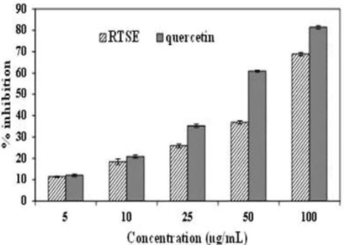
∙radical is a stable radical with a maximum absorbance at 517 nm that can readily undergo reduction by the antioxidant molecule. The radical scavenging capacity RTSE is showed in Figure. 1 expressed as a percentage reduction of the initial DPPH radical absorption by different concentration.
In this assay, RTSE at 100 μg/mL showed 68.84 % DPPH radical scavenging activity in a concentration dependent manner.
Consequently, the more rapidly the absorbance decreases, the
more potent antioxidant capacity of RTSE observed in terms
of hydrogen atom or electron donating ability. At same
concentrations quercetin used as a reference compound which
showed 81.4% inhibitory effect against DPPH
∙radicals at 100
μ g/mL in a dose dependent manner. Therefore, due to presence
of bioactive active molecules, RTSE could donate hydrogen
to free radicals, particularly to the lipid peroxides or hydroperoxide radicals that are the major propagators of the chain autoxidation of lipids, and to form non-radical species, resulting in the inhibition of propagating phase of lipid peroxidation (Bamforth et al. 1993).
4)Figure 1. DPPH
∙radical scavenging activity of RTSE and standard compound quercetin.
Values are mean of three replicate determinations (n = 3) ± standard deviation.
3.2. ABTS
∙+radical scavenging activity
Furthermore, the antioxidant capacity of RTSE was confirmed by ABTS
∙+radical scavenging activity at different concentrations.
As demonstrated in Figure. 2, RTSE showed 78.83 % inhibition of free radicalsat 25 µg/mL. RTSE showed a potent scavenging activity toward ABTS radical cations in a concentration dependent manner, showing a direct role in quenching of free radicals as at 25 µg/mL concentration standard compound
Figure 2. ABTS
∙+radical scavenging activity of RTSE and standard compound quercetin.
Values are mean of three replicate determinations (n = 3) ± standard deviation.
quercetin exhibited 72.53 % inhibition with 12.08 ± 0.65 IC
50value (Table2). There by, RTSE could play a crucial role in scavenging free radicals via a chain breaking reaction, and may survey aspotentialnutraceutical.
3.3. Nitric oxide free radical scavenging activity
Nitric oxide radical is key molecule that involved in several physiological processessuch as neural signal transmission, vasodilatation and regulation of blood pressure (Afanasev 2007).
Due to emergence in level of nitric oxide radical during oxidative stress, it becomes the major culprit for numerous carcinomas and inflammatory conditions including arthritis, ulcerative colitis juvenile diabetes, and multiple sclerosis (Rabkin and Klassen 2007). During aerobic metabolism, nitric oxide molecule reacts with oxygen molecule and generates intermediates such as NO
2, N
2O
4and N
3O
4. These products are highly genotoxic; causing deamination of purines, pyrimidines and denaturation of enzymes such as DNA ligase and DNA alkyltransferase (Lundberg et al. 2008).
Figure 3. Nitric oxide free radical scavenging activity of RTSE and standard compound quercetin.
Values are mean of three replicate determinations (n = 3) ± standard deviation.
Table 1. FRAP activity of RTSE and standard compound quercetin.
Sample FRAP
mmol Fe(Ⅱ)/mg extract
RTSE 2606.85±18.7
Quercetin 8326±115.2
Values are means of three replicate determinations (n = 3) ± standard deviation.
FRAP-Ferric reducing antioxidant power (Concentration of substance having
ferric-TPTZ reducing ability as equivalent to 1 µmol Fe (II).
In this study, RTSE reduced the levels of nitrite by competing with oxygen to react with nitric oxide radical, a possible protective effect against oxidative damage. As shown in Figure.
3, extract showed most efficient inhibition of nitric oxide radicals with 47.92% at 400 μg/mL in concentration-dependent manner.
RTSE showed IC
50value of 404.38± 1.6 (Table 2). Standard compound, quercetinshowed 90.98 % of inhibition at 400 µg/mL.
5)
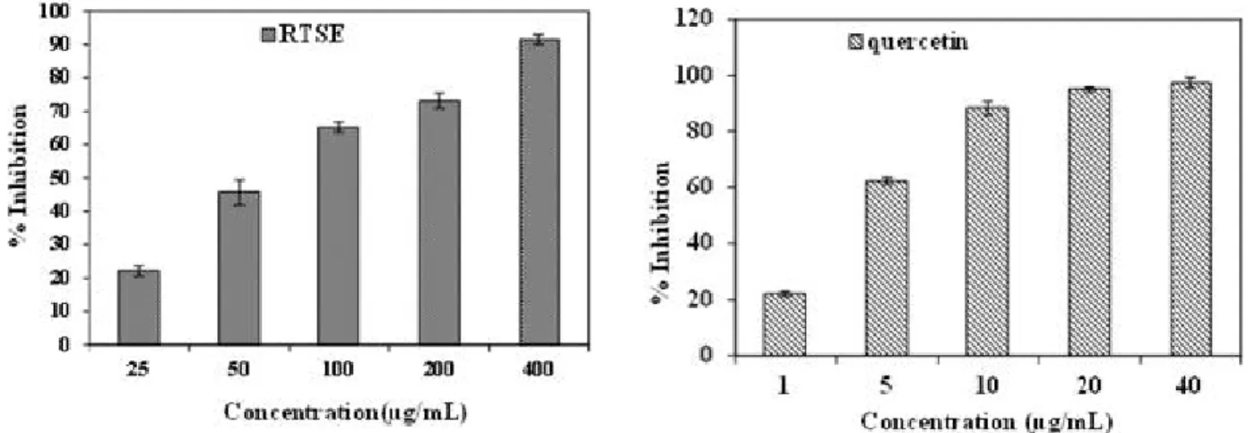
3.4. Super oxide free radical scavenging activity
In living beings superoxide radicals, a highly toxic species, are continuously produced in metabolic and physiological processes. Superoxide free radicals can generate more toxic and reactive free radicals i.e. hydroxyl radicals or singlet oxygen.
Biological molecules such as DNA, lipids and proteins are prominent targets of these oxygen species and cause metabolic and cellular disturbances (Martinez-Cayuela, 1995). Superoxide radical protecting activity of RTSE was measured by the NBT which generates superoxide radicals. In Figure. 4, RTSE showed superoxide radical scavenging activity in dose dependent manner with 91.57% inhibition at 400 µg/mL. RTSE exhibited 93.67
Figure 4. Superoxide free radical scavenging activity of RTSE and standard compound quercetin.
Values are mean of three replicate determinations (n = 3) ± standard deviation.
Table 2. IC
50values of DPPH, ABTS, Nitric oxide and Superoxideradicals scavenging potential of RTSE and standard compound quercetin.
Sample IC
50(μg/mL)
DPPH ABTS Nitric oxide Super oxide RTSE 68.88±1.1 12.08±0.65 404.38±1.6 93.6±2.7 Quercetin 48.41±0.7 13.62±0.4 29.34±1.3 4.32±0.8 Values are means of three replicate determinations (n = 3) ± standard deviation.
± 2.7 µg/mL IC
50value as compare to standard compound quercetin 4.32±0.8 µg/mL (Table 2).
On the basis of this assay, we can evidence that RTSE contains bioactive compounds which quench superoxide free radicals and can terminate radical chain reaction (Hamid et. al. 2010).
3.5. Ferric reducing antioxidant power (FRAP) assay Antioxidants can be explained as reductants, and in-activators of pro-oxidants (Siddhuraju and Becker, 2007). Some previous studies have also reported that the reducing power may serve as a significant indicator of potential antioxidant activity.
Anti-oxidative activity has been proposed to be related to reducing power. Therefore, the antioxidant potential of RTSE was estimated for their ability to reduce TPTZ–Fe (III) complex to TPTZ–Fe (II). The ferric reducing ability of the RTSE showed thatit has significant FRAP activity with 2606. 85 mmol Fe (II)/g extract (Table 1.) FRAP assay was used by several authors for the assessment of antioxidant activity of various food product samples (Pellegrini et al. 2003). Halvorsen et al.
(2006) suggested most of the secondary metabolites are
redox-active compounds that will be picked up by the FRAP assay.
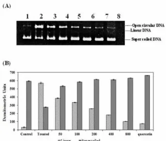
3.6. In vitro DNA damage protection efficiency of RTSE against hydroxyl radical
Hydroxyl radicals can be produced inside cells by various
reactions as UV-induced decomposition of H
2O
2used in the
present study and can damage macromolecules such as lipids,
proteins and DNA resulting in the onset of several skin diseases
including skin cancer (Halliwell and Gutteridge 1990).
Therefore, in this study, we check the DNA damage protection capacity of RTSE on pUC19 plasmid DNA against hydroxyl radicals generated by UV photolysis of H
2O
2. Figure. 5 shows electrophoretic pattern of pUC19 plasmid DNA in the presence or absence of different concentration (50, 100, 200, 400 and 800 µg/mL) of RTSE. On gel electrophoresis, pUC19 plasmid DNA shows two bands as in lane 1, fast moving prominent band corresponded to native supercoiled circular (Sc) DNA and slow moving, very fade band represents open circular DNA (Oc). After the exposure of UV irradiations in the presence of H
2O
2, supercoiled DNA converted into prominent band of open circular DNA, due to scission by
∙OH free radicals, generated by UV photolysis of H
2O
2(Lane 2). In biological system,
∙OH radicals, generated by the reaction between O
2•-and H
2O
2are major culprit for oxidative damage (Guitteridge, 1984).
6)