Comparison of survival outcomes after anatomical resection and non-anatomical resection in patients with hepatocellular carcinoma
Seheon Kim, Seokwhan Kim, Insang Song, and Kwangsik Chun
Department of surgery, Chungnam National University Hospital, Daejeon, Korea
Backgrounds/Aims: Liver resection is a curative procedure performed worldwide for hepatocellular carcinoma (HCC).
Deciding on the appropriate resection range for postoperative hepatic function preservation is an important surgical consideration. This study compares survival outcomes of HCC patients who underwent anatomical or non-anatomical resection, to determine which offers the best clinical survival benefit. Methods: One hundred and thirty-one patients underwent liver resection with HCC, between January 2007 and February 2015, and were divided into two groups:
those who underwent anatomical liver resection (n=88) and those who underwent non-anatomical liver resection (n=43).
Kaplan-Meier survival analysis and Cox regressions were used to compare the disease-free survival (DFS) and overall survival (OS) rates between the groups. Results: The mean follow-up periods were 27 and 40 months in the anatomical and non-anatomical groups, respectively (p=0.229). The 3- and 5-year DFS rates were 70% and 60% in the anatomical group and 62% and 48% in the non-anatomical group, respectively. The 3 and 5-year OS rates were 94% and 78%
in the anatomical group, and 86% and 80% in the non-anatomical group, respectively. The anatomical group tended to show better outcomes, but the findings were not significant. However, a relative risk of OS between the anatomical and non-anatomical group was 0.234 (95% CI, 0.061-0.896; p=0.034), which is statistically significant. Conclusions:
Although statistical significance was not detected in survival curves, anatomical resection showed better results. In this respect, anatomical resection is more likely to perform in HCC patients with preserve liver function than non-ana- tomical resection. (Korean J Hepatobiliary Pancreat Surg 2015;19:161-166)
Key Words: Hepatocellular carcinoma; Liver resection; Anatomical resection; Non-anatomical resection
Received: October 1, 2015; Revised: November 4, 2015; Accepted: November 8, 2015 Corresponding author: Kwangsik Chun
Deparment of surgery, Chungnam National University Hospital, 282 Munwha-ro, Jung-gu, Daejeon 35015, Korea Tel: +82-42-280-7185, Fax: +82-42-257-8024, E-mail: [email protected]
Copyright Ⓒ 2015 by The Korean Association of Hepato-Biliary-Pancreatic Surgery
This is an Open Access article distributed under the terms of the Creative Commons Attribution Non-Commercial License (http://creativecommons.org/
licenses/by-nc/4.0) which permits unrestricted non-commercial use, distribution, and reproduction in any medium, provided the original work is properly cited.
Korean Journal of Hepato-Biliary-Pancreatic Surgery ∙ pISSN: 1738-6349ㆍeISSN: 2288-9213
INTRODUCTION
Liver resection is a curative procedure performed world- wide for hepatocellular carcinoma (HCC).1 Anatomical re- section is an especially effective curative treatment; how- ever, massive anatomical liver resection can be difficult to execute in patients with existing liver conditions, such as hepatitis or hepatic cirrhosis. In the Korean population, one study reports that 74.6% of HCC patients were positive of Hepatitis B virus (HBV) and 9.3% were positive of hep- atitis C virus (HCV).2 Further, it is predicted that one-third of cirrhotic liver patients will develop HCC.3 Because of this, to preserve liver function, there are cases in which limited liver resection is performed, irrespective of the ana- tomical structure of the liver.4,5
Because recurrence is common after HCC surgery, hep-
atic function at the time of recurrence is critical to de- termine the survival rate after recurrence. In particular, mortality after liver surgery can result from not only re- currence, but also decreased hepatic function and ex- acerbation of existing liver disease.6 Therefore, deciding on the appropriate resection range for preservation of postoperative hepatic function is important to make in- formed surgical treatment decision.
From oncological and anatomical perspectives, anatom- ical liver resection is theoretically superior to non-anatom- ical liver resection, however clinical studies have failed to show any differences in survival benefit between them.7-9
The present study examined 131 cases of liver resection performed by the authors with the aim of comparing dis- ease-free survival (DFS) and overall survival (OS) be-
tween anatomical and non-anatomical resection, in order to determine which offers the best survival benefits.
MATERIALS AND METHODS
Patients
The diagnosis of HCC was based on imaging modality, including enhanced computed tomography (CT) or mag- netic resonance imaging (MRI) and tumor markers.
Considering cancer cell seeding during liver biopsy, pre- operative liver biopsy was not suggested for all patients initially. Diagnosis of HCC mainly depended on typical findings: early-phase enhancement or late-phase contrast washout. Elevation of alpha-fetoprotein (AFP) and history of hepatic viral infection or heavy alcohol drinking were also considered. Before treatment, all patients underwent basal laboratory tests including bilirubin, albumin and prothrombin activity. To predict postoperative liver fail- ure, an ICG test and fibroscan were performed.
Two hundred and twenty-seven patients diagnosed with HCC between January 2007 and February 2015 were evaluated. Thirty-four patients had undergone previous ra- diofrequency ablation (RFA), transcatheter arterial chemo- embolization, or surgery after HCC diagnosis were excluded. Eighteen patients who had other cancers, such as cholangiocarcinoma or stomach cancer at the time of the operation were also excluded. An additional 44 pa- tients who underwent concomitant RFA during liver re- section were excluded. The remaining 131 patients were divided into two groups, those who underwent anatomical liver resection (n=88) and those who underwent non-ana- tomical liver resection (n=43).
Liver resection
Anatomical liver resection involved segment-oriented resection from the areas that included the tumor and where the hepatic portal veins and hepatic veins divided, follow- ing the terminology proposed by Strasberg.10 Non-anatom- ical liver resection involved resection of the lesion area regardless of anatomical segment or lobe. For a single HCC with well-preserved liver function and ≤15% of ICG R15, anatomical major liver resection was initially considered for curative treatment. For multiple tumors, anatomical major liver resection was considered when all tumors located in single lobe with good liver function.
Non-anatomical liver resection was considered for a single tumor with suboptimal liver function or >15% of ICG R15 and for multiple tumors located in different lobes. In this study, a single segmental resection, such as S5 seg- mentectomy were not considered anatomical resection.
Anatomical resection included right or left hemi- hepatectomy, right posterior or anterior sectionectomy, left lateral sectionectomy and central bisectionectomy.
Perioperative factors
Preoperative albumin, total bilirubin, prothrombin time international normalization ratio (PT [INR]), platelet count, tumor markers, and the presence of underlying liver dis- eases, such as hepatitis or hepatic cirrhosis, were compared between the groups. HCC staging was performed according to the American Joint Committee on Cancer/Union for International Cancer Control TNM staging guidelines.11 Histology was compared using Edmondson-Steiner grading.12
Statistical analyses
Clinical characteristics and perioperative factors be- tween the anatomical and non-anatomical groups were an- alyzed using an independent sample t-test. DFS and OS were analyzed using the Kaplan-Meier method and the relative ratio was analyzed using the Cox regression model. Statistical analyses were conducted using SPSS software (version 19, SPSS Inc., Chicago, IL).
RESULTS
Patient characteristics are shown in Table 1. There were no significant differences in age, sex, total bilirubin, tu- mor number or PT-INR between the groups. In addition, platelet counts and serum albumin levels were statistically different; however, there was no difference in liver func- tion of between the results of the two groups. Nevertheless, tumor size in the anatomical group was significantly larg- er than that in the non-anatomical group (p=0.004). Most patients had chronic hepatitis or cirrhosis as an underlying disease and most were hepatitis B virus positive. Histologic grades in all patients were mostly grade II or III. The ma- jority of patients had TNM stage I or II disease. Alfa-feto- protein levels were not evaluated in all patients, although there appeared to be no significant differences in patients whose levels were measured between the groups.
Table 1. Clinical characteristics and perioperative factors in patients with HCC
Anatomical resection (n=88)
Non-anatomical resection (n=43) p-value Age (range)
Sex Male Female
Platelet count (103/l)*
Albumin (g/dl)*
PT (INR)†
Total bilirubin (mg/dl)*
Background liver Normal
Chronic hepatitis Cirrhosis Hepatic infection HBV
HCV
HBV and HCV None
Tumor size (cm)*
Tumor number 1
2
Histology grade I
II III IV Stage I II III IV AFP† (range)
58.5 (20-80) 65 23 175.1 4.02 1.09 0.91
11 22 55 59 4 0 0 3.67
82 6 4 24 56 4 63 20 5 0 26.7 (1-20,000)
(n=62)
59.5 (42-77) 34
9 139.5 4.30 1.05 0.80
5 6 32 25 1 0 0 2.48
42 1 2 18 22 1 35 6 1 1 9.85 (0.22-16,460)
(n=42)
0.583 0.519
0.020 0.021 0.478 0.849 0.281
0.177
0.004 0.283
0.151
0.445
0.840
PT, prothrombin time; INR, international normalized ratio HBV, hepatitis B virus; HCV, hepatitis C virus; G, grade; AFP, alfa-fetoprotein. *Values are presented as average; †Values are presented as medians with range
Fig. 1. Disease-free and overall survival of the anatomical and non-anatomical groups by the Kaplan-Meier Method.
The mean follow-up periods were 27 and 40 months in the anatomical and non-anatomical groups, respectively (p=0.229). As shown in Fig. 1, the 3- and 5-year DFS rates were 70% and 60% in the anatomical group and 62% and 48% in the non-anatomical group, respectively.
The 3 and 5-year OS rates were 94% and 78% in the ana- tomical group and 86% and 80% in the non-anatomical group, respectively. Although, the anatomical group tend- ed to have better DFS and OS outcomes, the differences between the groups were not significant.
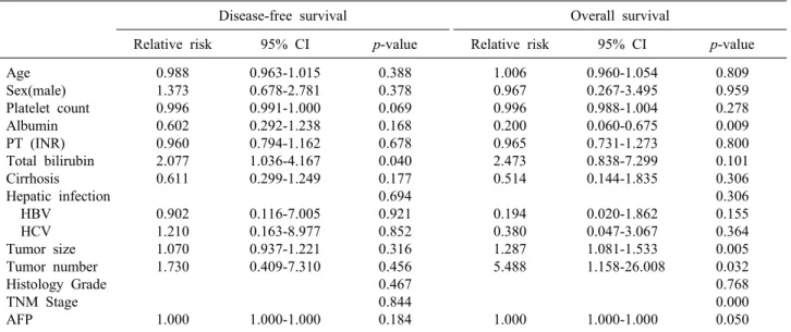
Univariate analysis of relative risk was performed on the perioperative factors analyzed (Table 2). For DFS, to- tal bilirubin and platelet counts were significant predictors. However, a multivariate analysis showed that total bilirubin was a significant predictor of DFS (Table 3). In addition, a relative risk (RR) of DFS between the anatomical and non-anatomical groups was 0.804 (95%
confidence interval [CI], 0.423-1.529; p=0.506), which is not statistically significant (Fig. 2). Serum albumin, tumor number, tumor size, stage and alfa-fetoprotein (AFP) were significant predictors of OS in univariate analysis.
Furthermore, a multivariate analysis showed that serum albumin and stage were also significant predictors. RR of OS between the anatomical and non-anatomical groups was 0.234 (95% CI, 0.061-0.896; p=0.034), which is stat- istically significant (Fig. 2).
In subjects with a single tumor, anatomical resection groups showed better overall survival in the graph, how- ever, there was no statistical difference of two groups (Fig. 3).
Table 3. Multivariate analysis of factors related to the disease-free and overall survival
Disease-free survival Overall survival
Relative risk 95% CI p-value Relative risk 95% CI p-value
Total bilirubin Platelet count Albumin Tumor size Tumor number TNM Stage AFP
2.149 0.997
- - - - -
1.055-4.376 0.992-1.002
- - - - -
0.035 0.195
- - - - -
- - 0.272 1.235 5.003 3.376 1.000
- - 0.071-1.043 0.868-1.485 0.793-23.657
1.478-7.713 1.000-1.000
- - 0.045 0.333 0.123 0.024 0.612 AFP, alfa-fetoprotein
Table 2. Univariate analysis of factors related to the disease-free and overall survival
Disease-free survival Overall survival
Relative risk 95% CI p-value Relative risk 95% CI p-value
Age Sex(male) Platelet count Albumin PT (INR) Total bilirubin Cirrhosis Hepatic infection HBV
HCV Tumor size Tumor number Histology Grade TNM Stage AFP
0.988 1.373 0.996 0.602 0.960 2.077 0.611 0.902 1.210 1.070 1.730
1.000
0.963-1.015 0.678-2.781 0.991-1.000 0.292-1.238 0.794-1.162 1.036-4.167 0.299-1.249 0.116-7.005 0.163-8.977 0.937-1.221 0.409-7.310
1.000-1.000
0.388 0.378 0.069 0.168 0.678 0.040 0.177 0.694 0.921 0.852 0.316 0.456 0.467 0.844 0.184
1.006 0.967 0.996 0.200 0.965 2.473 0.514 0.194 0.380 1.287 5.488
1.000
0.960-1.054 0.267-3.495 0.988-1.004 0.060-0.675 0.731-1.273 0.838-7.299 0.144-1.835 0.020-1.862 0.047-3.067 1.081-1.533 1.158-26.008
1.000-1.000
0.809 0.959 0.278 0.009 0.800 0.101 0.306 0.306 0.155 0.364 0.005 0.032 0.768 0.000 0.050 PT, prothrombin time; INR, international normalized ratio; AFP, alfa-fetoprotein; HBV, hepatitis B virus; HCV, hepatitis C virus
Fig. 2. Disease-free and overall survival of the anatomical and non-anatomical groups by the Cox regression model.
DISCUSSION
Liver resection is a curative procedure for HCC. In partic-
ular, eradication of intrahepatic metastasis occurring via vascular invasion is one of the most important considerations. Therefore, the non-anatomic approach is dis-
Fig. 3. Overall survival of the anatomical and non-anatomical group by the Cox regression model in patients with single tumor.
advantageous when considered from the standpoint of intra- hepatic metastasis eradication.13 Retrospective studies report that a three year survival rate with anatomical resection is better than with limited non-anatomical resection.14
However, only a few patients with HCC undergo liver resection because of poor liver function such as cirrhosis, chronic liver disease, and the difficulty in predicting post- operative liver failure.4,5 Although anatomical resection for HCC treatment is being recommended more often,13,15 recently conducted studies fail to demonstrate any distinct differences in DFS or OS between anatomical resection and non-anatomical resection.7-9 Furthermore, some sur- geons prefer to leave a greater portion of parenchyma of functional unit, such as in non-anatomic resection because most tumors arise in cirrhotic livers.16 In other studies, non-anatomical resection is equal to anatomical resection and in some cases, non-anatomical hepatic resection is recommended over anatomical resection.17,18
Some studies indicate that very restrictive patient se- lection is required to perform anatomical resection.
Although it is especially recommended for Child-Pugh class A patients19 or non-cirrhotic liver20 patient groups, it is reported that there is no difference in the improve- ment of survival or recurrence rates compared with those after non-anatomical resection in these patients.15
From an operative results perspective, although secur- ing the resection margin is more difficult in non-anatomi- cal resection compared with anatomical resection,7 the re- section volume of the normal liver parenchyma may be bigger in some anatomical resection cases. This suggests
that a smaller liver volume can be removed to achieve the same margin status in cases of non-anatomical re- section, which may be more favorable for postoperative liver function.21 Takano et al.7 report that partial resection is associated with a reduced frequency of operative com- plications, more so than anatomical resection, although there were no differences in morbidity rates between them. In addition, they demonstrated that non-anatomical resection offered more benefits in terms of hepatic paren- chyma preservation, which allows for additional surgery or other treatments in cases of recurrence. From a liver function perspective, Shirabe et al.22 report that patients with better postoperative liver function lived >10 years longer compared to those with poor liver function. They emphasized the importance of preserving hepatic function at the time of operation because survival was affected by underlying liver conditions.
Various studies have published the benefit of anatomi- cal resection or non-anatomical resection for the patients with preserved liver function. Nagasue et al.23 analyzed the outcome of anatomical liver resection and partial liver resection for patients with HCCs with preserved liver function (Child-Pugh A). Moreover, there were no sig- nificant differences in DFS and survival between the oper- ation methods. On the other hand, Hasegawa et al.13 ana- lyzed the outcomes of anatomical and non-anatomical re- section for HCC in Child-Pugh A and B groups. The five-year survival and DFS rates were better in the ana- tomical resection (Survival, 66% vs. 35%; DFS, 34% vs.
16%).
In the present study, the anatomical resection group showed better DFS and OS outcomes than the non-ana- tomical resection group, although the results of DFS and OS were not statistically significant. Further, the anatomi- cal resection group showed results that are more benefi- cial in relative risk of OS. The reason there is no stat- istical difference may be due to the difference between the follow-up periods of the two groups. However, liver function of our patients was well preserved after the pa- tients underwent liver resection. Furthermore, there were no major differences in clinical characteristics and peri- operative factors between the two groups. Therefore, if there was no difference in the follow-up period, we can expect that anatomical resection can provide better surviv- al outcomes when there are no major differences in under-
lying liver conditions and well preserved liver function.
The tumor number and TNM stage in the relative risk of overall survival showed significant results in the uni- variate study. However, tumor number in the multivariate analysis was not statistically significant. If the number of tumors is more diverse, it is expected to have a sig- nificantly better result.
As described above, our study had limitations. First, there was a large discrepancy in the mean follow-up dura- tion between the groups. Second, the sample size was small. Nevertheless, we believe that significant results can be derived in longer-term studies with a greater number of patients.
In conclusion, although statistical significance was not detected in survival curves, anatomical resection showed better results. In this respect, anatomical resection should be considered before non-anatomical resection for HCC patients with well-preserved liver function.
REFERENCES
1. Lai EC, Fan ST, Lo CM, Chu KM, Liu CL, Wong J. Hepatic resection for hepatocellular carcinoma. An audit of 343 patients.
Ann Surg 1995;221:291-298.
2. Kwak HW, Park JW, Nam BH, Yu A, Woo SM, Kim TH, et al. Clinical outcomes of a cohort series of patients with hep- atocellular carcinoma in a hepatitis B virus-endemic area. J Gastroenterol Hepatol 2014;29:820-829.
3. Sangiovanni A, Prati GM, Fasani P, Ronchi G, Romeo R, Manini M, et al. The natural history of compensated cirrhosis due to hepatitis C virus: a 17-year cohort study of 214 patients.
Hepatology 2006;43:1303-1310.
4. Kanematsu T, Takenaka K, Matsumata T, Furuta T, Sugimachi K, Inokuchi K. Limited hepatic resection effective for selected cirrhotic patients with primary liver cancer. Ann Surg 1984;199:
51-56.
5. Bismuth H, Houssin D, Ornowski J, Meriggi F. Liver resections in cirrhotic patients: a Western experience. World J Surg 1986;
10:311-317.
6. Castells A, Bruix J, Bru C, Fuster J, Vilana R, Navasa M, et al. Treatment of small hepatocellular carcinoma in cirrhotic pa- tients: a cohort study comparing surgical resection and percuta- neous ethanol injection. Hepatology 1993;18:1121-1126.
7. Takano S, Oishi H, Kono S, Kawakami S, Nakamura M, Kubota N, et al. Retrospective analysis of type of hepatic resection for hepatocellular carcinoma. Br J Surg 2000;87:65-70.
8. Ercolani G, Grazi GL, Ravaioli M, Del Gaudio M, Gardini A,
Cescon M, et al. Liver resection for hepatocellular carcinoma on cirrhosis: univariate and multivariate analysis of risk factors for intrahepatic recurrence. Ann Surg 2003;237:536-543.
9. Kondo K, Chijiiwa K, Makino I, Kai M, Maehara N, Ohuchida J, et al. Risk factors for early death after liver resection in pa- tients with solitary hepatocellular carcinoma. J Hepatobiliary Pancreat Surg 2005;12:399-404.
10. Strasberg SM, Belghiti J, Clavien PA, Gadzijev E, Garden Jo, Lau WY, et al. The Brisbane 2000 terminology of liver anatomy and resections. Terminology Committee of the International Hepato-Pancreato-Biliary Association. HPB 2000;2:333-339.
11. Edge S, Byrd DR, Compton CC, Fritz AG, Greene FL, Trotti A. AJCC cancer staging manual. 7th ed. New York: Springer, 2010:175.
12. Edmondson HA, Steiner PE. Primary carcinoma of the liver: a study of 100 cases among 48,900 necropsies. Cancer 1954;7:462-503.
13. Hasegawa K, Kokudo N, Imamura H, Matsuyama Y, Aoki T, Minagawa M, et al. Prognostic impact of anatomic resection for hepatocellular carcinoma. Ann Surg 2005;242:252-259.
14. Nagao T, Inoue S, Goto S, Mizuta T, Omori Y, Kawano N, et al. Hepatic resection for hepatocellular carcinoma. Clinical fea- tures and long-term prognosis. Ann Surg 1987;205:33-40.
15. Fuster J, García-Valdecasas JC, Grande L, Tabet J, Bruix J, Anglada T, et al. Hepatocellular carcinoma and cirrhosis. Results of surgical treatment in a European series. Ann Surg 1996;223:
297-302.
16. Cucchetti A, Cescon M, Ercolani G, Bigonzi E, Torzilli G, Pinna AD. A comprehensive meta-regression analysis on outcome of anatomic resection versus nonanatomic resection for hep- atocellular carcinoma. Ann Surg Oncol 2012;19:3697-3705.
17. Marubashi S, Gotoh K, Akita H, Takahashi H, Ito Y, Yano M, et al. Anatomical versus non-anatomical resection for hep- atocellular carcinoma. Br J Surg 2015;102:776-784.
18. Tomimaru Y, Eguchi H, Marubashi S, Wada H, Kobayashi S, Tanemura M, et al. Equivalent outcomes after anatomical and non-anatomical resection of small hepatocellular carcinoma in pa- tients with preserved liverfunction. Dig Dis Sci 2012;57:1942-1948.
19. Regimbeau JM, Kianmanesh R, Farges O, Dondero F, Sauvanet A, Belghiti J. Extent of liver resection influences the outcome in patients with cirrhosis and small hepatocellular carcinoma.
Surgery 2002;131:311-317.
20. Kosuge T, Makuuchi M, Takayama T, Yamamoto J, Shimada K, Yamasaki S. Long-term results after resection of hepatocellular carcinoma: experience of 480 cases. Hepatogastroenterology 1993;40:328-332.
21. Tanaka K, Shimada H, Matsumoto C, Matsuo K, Nagano Y, Endo I, et al. Anatomic versus limited nonanatomic resection for solitary hepatocellular carcinoma. Surgery 2008;143:607-615.
22. Shirabe K, Shimada M, Kajiyama K, Gion T, Ikeda Y, Hasegawa H, et al. Clinicopathologic features of patients with hepatocellular carcinoma surviving >10 years after hepatic resection. Cancer 1998;83:2312-2316.
23. Nagasue N, Yamanoi A, el-Assal ON, Ohmori H, Tachibana M, Kimoto T, et al. Major compared with limited hepatic resection for hepatocellular carcinoma without underlying cirrhosis: a ret- rospective analysis. Eur J Surg 1999;165:638-646.