• 검색 결과가 없습니다.

Malignant Schwannoma of the Esophagus: A Rare Case Report Biswajit Mishra, M.D.

N/A
N/A
Protected

Academic year: 2021

Share "Malignant Schwannoma of the Esophagus: A Rare Case Report Biswajit Mishra, M.D."

Copied!
4
0
0

로드 중.... (전체 텍스트 보기)

전체 글

(1)

ISSN: 2233-601X (Print) ISSN: 2093-6516 (Online)

− 63 −

Departments of

1

Radiodiagnosis,

2

Gastrointestinal Surgery, and

3

Pathology, All India Institute of Medical Sciences Received: July 6, 2015, Revised: August 16, 2015, Accepted: August 25, 2015, Published online: February 5, 2016

Corresponding author: Kumble Seetharama Madhusudhan, Department of Radiodiagnosis, All India Institute of Medical Sciences, Ansari Nagar, New Delhi 110029, India

(Tel) 91-11-26593326 (Fax) 91-11-26588663 (E-mail) drmadhuks@gmail.com

C

The Korean Society for Thoracic and Cardiovascular Surgery. 2016. All right reserved.

CC
This is an open access article distributed under the terms of the Creative Commons Attribution Non-Commercial License (http://creative- commons.org/licenses/by-nc/4.0) which permits unrestricted non-commercial use, distribution, and reproduction in any medium, provided the original work is properly cited.

Malignant Schwannoma of the Esophagus: A Rare Case Report

Biswajit Mishra, M.D.

1

, Kumble Seetharama Madhusudhan, M.D., FRCR

1

, Ragini Kilambi, M.Ch.

2

, Prasenjit Das, M.D.

3

, Sujoy Pal, M.Ch.

2

, Deep Narayan Srivastava, M.D.

1

Neurogenic tumors are the most prevalent tumors of the mediastinum, and schwannomas are the most common type of neurogenic tumor. Primary neurogenic neoplasm of the esophagus is uncommon and malignant schwanno- ma of the esophagus is extremely rare. We report a case of a 27-year-old female presenting with dysphagia and palpitations who was found to have a lobulated tumor in the mediastinum that was compressing the esophageal lumen. The tumor was successfully treated surgically without recurrence. The final diagnosis, on histopathological examination of the specimen, was malignant schwannoma.

Key words: 1. Schwannoma 2. Neurilemmoma

3. X-ray computed tomography 4. Esophagogastric junction

CASE REPORT

The most common benign tumor of the esophagus is leio- myoma, and primary esophageal schwannoma is extremely rare. To date, about 27 reports of esophageal schwannomas have been published in the literature, most of which are be- nign lesions. Malignant esophageal schwannoma is much more rare, with only three cases previously reported in the literature [1]. Esophageal schwannoma is more common in females and is mostly found in the upper and middle esoph- agus [2]. As the imaging appearance is similar to that of leio- myoma, preoperative diagnosis of schwannoma is difficult, and definitive diagnosis is mostly established after resection [3]. Here we present an unusual case of malignant schwanno- ma of the esophagus in a young female that was treated suc- cessfully with surgery.

A 27-year-old female presented to our hospital with com-

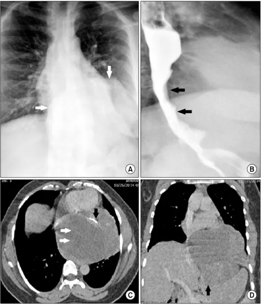
plaints of gradual onset of dysphagia for five years associated with intermittent episodes of palpitations, and weight loss without loss of appetite. Her symptoms had worsened over the preceding two months. Her medical history was otherwise unremarkable. A chest radiograph (Fig. 1A) revealed a smooth well-defined mass in the posterior mediastinum with a loss of the normal silhouette of the cardiac borders. A barium swal- low was performed to evaluate the dysphagia, and a smooth extrinsic impression was seen on the distal esophagus and gastro-esophageal junction (Fig. 1B). Endoscopy showed an extrinsic bulge 30 cm distal to the incisors at the gastro- esophageal junction, with normal overlying mucosa. Subse- quently, thoracic contrast-enhanced computed tomography (CT) was performed. This revealed a large lobulated low-density mass (Fig. 1C, D) in the middle mediastinum arising from the anterior and left lateral walls of the lower thoracic esoph- agus and containing areas of necrosis and calcification. The

Korean J Thorac Cardiovasc Surg 2016;49:63-66 Case Report

http://dx.doi.org/10.5090/kjtcs.2016.49.1.63

(2)

Biswajit Mishra, et al

− 64 −

Fig. 1. (A) Chest radiograph show- ing a well-defined retrocardiac soft tissue mass (white arrows) with in- tra-abdominal extension. (B) Barium swallow showing smooth luminal nar- rowing of the distal esophagus and gastroesophageal junction due to ex- trinsic compression by the mass (black arrows). (C) Axial contrast-enhanced CT image showing a large lobulated hy- podense well-defined mass (white ar- rows) containing areas of calcification (black arrow). (D) Coronal reformat of CT scan showing the mass ex- tending through the esophageal hia- tus and compressing the fundus of the stomach (black arrow). CT, com- puted tomography.

lesion extended into the abdomen through the esophageal hia- tus and compressed the fundus of the stomach. Based on the imaging studies a diagnosis of leiomyoma of the esophagus was made. Laboratory parameters, including pulmonary func- tion tests, were unremarkable.

The patient was prepared for surgery. The esophagus was approached through a left thoraco-abdominal incision. A large firm to hard mass was seen arising from the mid- and distal thoracic esophagus and adherent to the diaphragm, pericar- dium, and aorta. Esophagectomy was performed and a gastric conduit was brought into the neck through the posterior me- diastinal route. A tumor measuring 12×10×10 cm was seen adherent to the serosal aspect of the resected specimen of

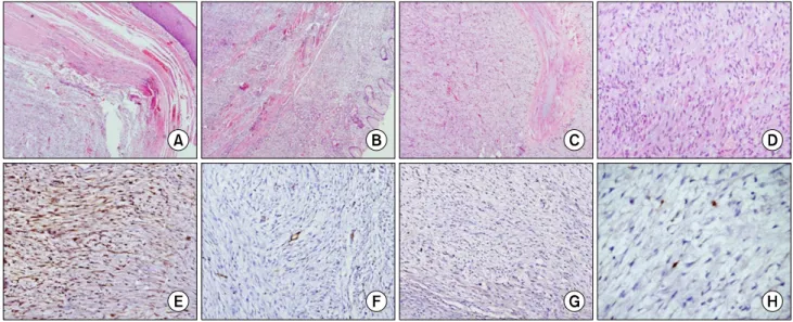
esophagus. A cut section of the tumor was solid and homo- geneous with areas of whorling. Histopathology showed a lo- bulated spindle cell tumor with a focally infiltrative peripheral margin and atypical mitoses in 4/10 high power fields (Fig. 2).

Spindle cells were immunopositive for S100 and negative for Dog1, CD117, CD34, and SMA. The Ki-67 labeling index was 2%–3% (Fig. 2). The dissected lymph node showed re- active features. Based on the above features, a diagnosis of low-grade malignant schwannoma of the esophagus was made.

Postoperatively, the patient had right recurrent laryngeal

nerve palsy, possibly due to injury during dissection of the

cervical esophagus. This was managed conservatively and the

(3)

Malignant Schwannoma of the Esophagus

− 65 −

Fig. 2. Photomicrographs show (A) a well-circumscribed spindle cell lesion arising from the lower end of the esophagus (H&E, ×40) and (B) reaching up to the gastric fundus (H&E, ×40). (C) The mass is lobulated (H&E, ×40) and (D) shows fascicles with spindle cells show- ing hyperchromasia and nuclear buckling on a myxoid background (H&E, ×100). On immunohistochemistry, (E) these cells were positive for S100 protein (S100, ×40) and (F, G) negative for CD34 and DOG 1 stains (CD34 and DOG-1, ×100). (H) The Ki67 labeling index is low (Ki67, ×100).

patient was discharged on the 17th postoperative day. A fol- low-up CT scan one year after surgery showed no evidence of tumor recurrence. The patient remained clinically asympto- matic 18 months after surgery.

DISCUSSION

Malignant schwannoma of the esophagus is a rare neoplasm. Patients usually present with dysphagia and dysp- nea; however, a wide range of presentations have been re- ported, including chest pain, cough, and palpitations, with symptoms becoming worse as the size of the tumor increases [4]. Usually located in the proximal third of the esophagus, schwannomas are often described as hypo- or iso-attenuating with a homogeneous texture on CT scan and may contain areas of calcification that differentiate them from leiomyomas.

Malignant schwannoma appears heterogeneous in non-contrast and contrast-enhanced CT scans and may have an ill-defined infiltrating border. A striking feature of gastrointestinal stro- mal tumors, in comparison with schwannomas, is the pres- ence of hemorrhage, necrosis, and cystic changes, which give them a more heterogeneous appearance on CT. On magnetic resonance imaging, benign schwannomas are iso- or slightly

hyperintense relative to muscles in T1-weighted images, and have a target appearance in T2-weighted images with a pe- ripheral hyperintense rim and central low intensity [5].

Intense contrast enhancement is usually seen in most cases of schwannoma. The typical target appearance may not be pres- ent in malignant schwannomas.

Endoscopic ultrasonography with sufficient resolution can

determine the tissue layer of origin of the tumor and a guid-

ed biopsy is helpful for diagnosis and management. A

18-fluoro-deoxyglucose uptake is seen in normal Schwann

cells due to normal glucose uptake for axon repolarization,

and is thus less useful for differentiating benign and malig-

nant schwannomas [1]. On immunohistochemistry, the diag-

nosis of schwannoma is based on immunopositivitity for

S100 protein and negative staining for CD-34, CD-117, and

muscle specific actin (MSA) [6]. CD-34 and CD-117 are pos-

itive in gastrointestinal stromal tumors and desmin and MSA

are positive in leiomyomas. Surgery is the only treatment op-

tion in schwannomas, as chemotherapy and radiotherapy are

ineffective. Enucleation with video-assisted thoracoscopic sur-

gery is ideal for small schwannomas less than 2 cm in size

[3]. However, for larger tumors, esophagectomy with esoph-

ago-gastrostomy is usually performed [7,8].

(4)

Biswajit Mishra, et al

− 66 − In conclusion, preoperative diagnosis of primary esophageal neurogenic tumors is challenging, as their imaging appearance mimics leiomyoma. A high index of clinical suspicion is nec- essary because of the rarity of these tumors, their non-specif- ic imaging appearance and vague clinical features, and the difficulty in sampling the tumor due to its submucosal position.

CONFLICT OF INTEREST

No potential conflict of interest relevant to this article was reported.

REFERENCES

1. Wang S, Zheng J, Ruan Z, Huang H, Yang Z, Zheng J.

Long-term survival in a rare case of malignant esophageal schwannoma cured by surgical excision. Ann Thorac Surg

2011;92:357-8.

2. Saito R, Kitamura M, Suzuki H, Ogawa J, Sageshima M.

Esophageal schwannoma. Ann Thorac Surg 2000;69:1947-9.

3. Park BJ, Carrasquillo J, Bains MS, Flores RM. Giant benign esophageal schwannoma requiring esophagectomy. Ann Thorac Surg 2006;82:340-2.

4. Byun JH, Park SD. Benign schwannoma of the esophagus:

surgical experience of two cases. Korean J Thorac Cardiovasc Surg 2005;38:589-93.

5. Anil G, Tan TY. Imaging characteristics of schwannoma of the cervical sympathetic chain: a review of 12 cases. AJNR Am J Neuroradiol 2010;31:1408-12.

6. Kobayashi N, Kikuchi S, Shimao H, et al. Benign esoph- ageal schwannoma: report of a case. Surg Today 2000;30:

526-9.

7. Davis EA, Heitmiller RF. Esophagectomy for benign disease:

trends in surgical results and management. Ann Thorac Surg 1996;62:369-72.

8. Dutta R, Kumar A, Jindal T, Tanveer N. Concurrent benign

schwannoma of oesophagus and posterior mediastinum. Interact

Cardiovasc Thorac Surg 2009;9:1032-4.

수치

Fig. 1. (A) Chest radiograph show- show-ing a well-defined retrocardiac soft  tissue mass (white arrows) with  in-tra-abdominal extension
Fig. 2. Photomicrographs show (A) a well-circumscribed spindle cell lesion arising from the lower end of the esophagus (H&E, ×40) and  (B) reaching up to the gastric fundus (H&E, ×40)

참조

관련 문서

44 글의 첫 번째 문장인 The most important thing in the Boat Race is harmony and teamwork.을 통해 Boat Race에서 가장 중요한 것은 조 화와 팀워크임을

Now that you have the right bike for you, it is important to learn the right riding position.. A poor riding position can lead to injuries

44 글의 첫 번째 문장인 The most important thing in the Boat Race is harmony and teamwork.을 통해 Boat Race에서 가장 중요한 것은 조 화와 팀워크임을

12 that절 내의 주어를 문장의 주어로 쓰고 that절 내의 동사를 to부정사로 써 서 수동태 문장을 만들 수 있다... 13 반복되는 the reason을 관계대명사 which로 바꿔주고

Now that you have the right bike for you, it is important to learn the right riding position.. A poor riding position can lead to

(The Office of International Affairs arranges a dormitory application for the new students only in the first semester.). 2)

Images that are required to be recorded include the upper esophagus, middle esophagus, lower esophagus, GEJ, front and reverse images of the fundus, front and reverse images

웹 표준을 지원하는 플랫폼에서 큰 수정없이 실행 가능함 패키징을 통해 다양한 기기를 위한 앱을 작성할 수 있음 네이티브 앱과