대한소아소화기영양학회지:제 11 권 제 1 호 2008
◇ 증 례 ◇
56
접수:2008년 1월 29일, 승인:2008년 2월 29일 책임저자:최연호, 135-710, 서울시 강남구 일원동 50번지
성균관대학교 의과대학 삼성서울병원 소아청소년과 Tel: 02-3410-3539, Fax: 02-3410-0043
E-mail: [email protected]
거대 간 종괴와 심방 중격 결손을 동반한 Abernethy 기형 2형 1예
성균관대학교 의과대학 삼성서울병원 소아과학교실, *진단병리과학교실, †진단방사선과학교실
이해정ㆍ이지현ㆍ허 준ㆍ강이석ㆍ이흥재ㆍ서연림*ㆍ유소영
†ㆍ최연호
A Case of Congenital Extra Hepatic Portocaval Shunt (Abernethy Malformation Type 2) with a very Large
Liver Mass and an Atrial Septal Defect
Hae Jeong Lee, M.D., Jee Hyun Lee, M.D., June Huh, M.D., I Seok Kang, M.D., Heung Jae Lee, M.D., Yeon-Lim Suh, M.D.*, So Young Yoo, M.D.† and Yon Ho Choe, M.D.
Departments of Pediatrics, *Pathology and †Radiology, Samsung Medical Center, Sungkyunkwan University School of Medicine, Seoul, Korea
Extrahepatic portosystemic shunts, known as Abernethy malformations, were first reported by John Abernethy in 1793. They are classified into two types: Type I refers to a congenital absence of the portal vein and Type II refers to a shunt involving a side-to-side anastomosis with reduced portal blood flow into the liver parenchyma. This malformation is so rare that less than 100 cases have been reported in the medical literature. We report the case of a 13-month-old boy who had a congenital extrahepatic portocaval shunt with a hypoplastic portal vein. This case was complicated with an atrial septal defect and a large hyperplastic nodule in the liver. The patient was diagnosed with a Type II Abernethy malformation. We planned on surgical occlusion of the extrahepatic portocaval shunt. However, six months later, the patient had a sudden onset of a fever of unknown origin and developed hepatic encephalopathy.
Although he underwent a liver transplantation, he died of acute hepatic failure. (Korean J Pediatr Gastroenterol Nutr 2008; 11: 56∼59)
Key Words: Abernethy malformation, Focal nodular hyperplasia, Hepatic encephalopathy
서 론
Abernethy 기형은 선천성 간외 문체정맥 단락(extra- hepatic portocaval shunt)으로 알려진 것으로 1793년 John Abernethy가 원인 불명으로 사망한 10개월 된 여 아의 부검을 통해 문맥의 기형을 처음으로 보고했다1).
이해정 외:거대 간 종괴와 심방 중격 결손을 동반한 Abernethy 기형 2형 1예ㆍ57
Fig. 2. (A) Portosystemic shunting vessel is shown in a contrast-enhanced CT scan of the abdomen. An arrow shows anastomosis between the portal vein and inferior vena cava. (B) Contrast-enhanced CT scan of the abdomen shows dilated left portal vein (arrow) and absence of the right portal vein.
Fig. 1. Contrast-enhanced CT scan of the abdomen shows large hepatic mass (arrow) in the posterior segment of the right hepatic lobe with centripetal enhancement pattern.
이것은 두 형태로 나누어 1형은 문맥이 없는 경우로, 2형은 저형성된 문맥 혈류가 간의 실질로 들어가면서 side-to-side 연결을 형성하는 경우로 분류된다2,3). 이러 한 기형은 매우 드문 질환으로 전 세계적으로 100예 미 만으로 보고되고 있으며4), 국내에서는 보고된 적이 없 다. 저자들은 간의 거대 종괴 및 심방 중격 결손으로 추적 관찰 중 복부 전산화단층촬영(computer tomo- graphy, CT) 및 조직 검사를 통해 진단된 Abernethy 기 형 2형 1예를 경험하였기에 문헌 고찰과 함께 보고하는 바이다.
증 례
과거력 상 생후 1개월에 황달, 간비대 및 심잡음이 관찰되어 시행한 복부 초음파 및 복부 CT, 심장 초음파 상 간의 거대 종괴(Fig. 1) 및 심방 중격 결손으로 진단 받고, 생후 2개월에 간 우엽 절제술을 시행 후 조직 검 사 상 국소 결절성 과증식(focal nodular hyperplasia, FNH)으로 확진되어 추적 관찰 중이던 13개월 된 남아 로 약 10 mm 크기의 심방 중격 결손 지속되어 수술을 위해 본원에 내원하였다.
상기 환아는 내원 당시 신체 검진 상 2도의 수축기성 심잡음이 좌측 흉부에서 청진되었으며, 황달이나 간비 대는 없었다. 혈액 검사 상 혈색소 12.9 g/dL, 총 단백/
알부민 5.7/3.7 g/dL, 암모니아 43.1μmol/dL로 정상 범 위였으며 AST 77 IU/L, ALT 44 IU/L, 총 빌리루빈 1.6 mg/dL, PT/PTT 22초/45초, PT (INR) 1.75로 약간 증가 되어있었다. 심방 중격 결손 수술 후 실시한 복부 도플 러 초음파 및 간 전산화단층촬영 상 우측 간문맥이 보 이지 않았고 전반적으로 커진 좌측 간문맥이 하대정맥 과 연결되는 문체정맥 단락이 발견되었다. 이에 생후 1개월 경 촬영하였던 복부 CT를 재검토한 결과 동일 소견이 확인되었다(Fig. 2). 생후 2개월 경 시행한 우측 간 절제술 후의 조직 검사를 재검토한 결과 상 간문맥 의 저형성(hypoplasia) 소견을 확인할 수 있었다(Fig. 3).
58ㆍ대한소아소화기영양학회지:제 11 권 제 1 호 2008
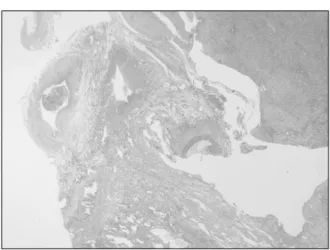
Fig. 3. The resected portion of the liver reveals a relatively smaller portal vein than the hepatic artery at the hilar portion (H&E, ×10).
이에 FNH와 심방 중격 결손을 동반한 Abernethy 기형 2형으로 진단하였다. 이후 문체정맥 단락 폐쇄 수술 예 정으로 정기적 추적 관찰하던 중, 생후 19개월 경 4일 간 지속되는 원인 불명의 발열, 황달, 처지는 증상으로 본원 내원하여 시행한 신체 검사 상 우상복부에서 간이 3 cm 정도 촉지되었으며, 좌상복부에서 비장이 5 cm 정도 촉지되었다. 환아는 지속적으로 심한 유해 자극을 주어야만 잠시 깨어나는 혼미(stupor) 상태였으며, 당시 시행한 혈액 검사 상 혈색소 6.3 g/dL, 총 단백/알부민 4.5/2.3 g/dL로 감소되어 있었으며, 총 빌리루빈/직접 빌 리루빈 12.6/8.1 mg/dL, 암모니아 100.4μmol/dL, AST 1285 IU/L, ALT 322 IU/L, PT/PTT 61초/117초, PT (INR) 7.36, C-반응 단백14.29 mg/dL로 증가된 소견 관찰되어 간성 혼수 진단 하에 간이식하였으나 사망하였다.
고 찰
선천성 간 단락(congenital hepatic shunts)은 아주 드 문 질환으로 증상이 있는 영아 또는 무증상의 어린이에 서 우연히 초음파를 통해 발견할 수 있다5). 선천성 간 단락은 하대정맥과 난황 정맥(vitelline veins)의 발생 과 정에서 생기는 것으로 생각된다. 정맥계는 재태 약 5주 경에 형성되기 시작된다6,7). 한 쌍의 난황 정맥이 난황 자루(yolk stalk)로 들어가서 십이지장 주위에서 서로 연결되고, 가로 중격(septum transversum)을 통해 정맥 동(sinus venosus)으로 지나간다. 좌측 난황 정맥의 근위
부는 복잡하게 얽히게 되고, 간의 좌측에서 오는 혈류 는 우측 난황 정맥으로 재분포된다. 전 우측 제대 정맥 과 좌측 제대 정맥의 일부, 그리고 좌측 정맥동이 퇴화 되며, 좌측 제대 정맥으로부터 모든 산소를 태아에게 운반하게 된다. 정맥관(ductus venosus)이 상대정맥과 함께 제대 정맥과 연결되고, 문맥은 십이지장 주변의 난황 정맥 연결부로부터 형성된다6,7). 이와 같이 난황 정맥에서 문맥으로 발달되는 과정에서 매우 다양한 형 태의 문체정맥 단락이 형성될 수 있다8).
간외 문체정맥 단락은 Abernethy에 의해 처음으로 보 고되었으며, Morgan과 Superina가 간외 문체정맥 단락 을 두 가지 형태로 분류하였다3). 1형은 간이 문맥 혈류 를 통해 관류되지 않는 경우로, 선천적으로 문맥혈류가 없는 경우이다. 이것은 다시 상부 장간막 정맥과 비장 정맥이 연결되지 않는 형태와 이 두 정맥이 전신순환으 로 들어가기 전에 서로 만나는 형태로 분류된다. 이러 한 1형 단락은 주로 여아에서 발생하며, 선천성 기형이 흔히 동반된다2). 동반 기형 중 심실 중격 결손, 심방 중 격 결손, 대동맥 축착과 같은 심혈관계 기형이 가장 흔 하며, 담도 폐쇄, 다비증, 내장 역위와 장 회전 이상 등 을 보였던 보고도 있다2). 그리고 아주 드물게 다낭포 이형성 신, Goldenhar 증후군, 총 담관낭, 골격 이상, 피 하 혈관종이 동반될 수 있다9). 2형은 문맥은 있으나 문 맥혈류가 하대정맥으로 간외 단락을 통해 연결되는 것 으로, 남아에서 주로 발생하며, 동반 기형은 1형에 비해 드물다. 폐동맥판 폐쇄증, 동맥관 개존, Goldenhar 증후 군, 다비증과 하대 정맥 기형, Cornelia de Lange 증후군 과 연관된 경우가 보고되었다9). 두 가지 형 모두 무증 상일 수 있으나, 간세포암, 선종, 국소 결절성 과증식 등의 간내 종양과 간성 혼수로 진행될 수 있다10∼12). 그동안 조직 검사를 통해 문맥이 없음을 확인함으로 써 Abernethy 기형 1형으로 확진되었던 여러 보고들이 있었다10,11,13,14)
. 본 증례는 복부 CT를 통해 문체정맥 단 락이 확인되었고 조직 검사 상 간문맥의 저형성이 확인 되어 Abernethy 기형 2형으로 진단되었다. Abernethy 기형 2형의 경우에서는 조직 검사 상 문맥이 정상이거 나 저형성을 보일 수 있으며15), 다른 기형과의 동반이 드문 것으로 알려져 있으나 본 증례에서는 FNH와 심 방 중격 결손이 동반된 매우 드문 경우였다. 본 증례와 같은 간내 종양의 발생은 간으로의 과도한 동맥혈의 공
이해정 외:거대 간 종괴와 심방 중격 결손을 동반한 Abernethy 기형 2형 1예ㆍ59
급으로 인해 문맥혈류가 줄어들고, 간 성장 인자 (hepatic growth factors)의 순환 수준이 증가되어 발생하 는 것으로 생각된다15). 환자는 생후 1개월 경 시행했던 복부 CT 상에서 거대 간 종괴와 문체정맥 단락이 있었 으나 진단이 늦어졌던 경우로 신생아 시기에 발견되는 간내 종양이 의심될 경우 문체정맥 단락이 동반될 수 있음을 간과해서는 안될 것이다.
본 증례에서는 문체정맥 단락 폐쇄술을 시행할 예정 으로 추적 관찰 중 원인 불명의 발열이 동반되면서 갑 작스럽게 간부전 및 간성 혼수 진행하여 간이식을 시행 했으나 사망하였다. 간성 혼수가 문체정맥 단락과의 연 관성이 있는지에 대해서는 밝히지 못했다. 이전의 문헌 에 간외 문체정맥 단락으로 인해 소아에서 간성 혼수로 진행된 예는 없었으나, 성인에서 간성 혼수로 진행된 예에 대한 보고는 있었다2,16). 선천성 문체정맥 단락에 서 발생할 수 있는 간성 혼수의 원인으로는 간에서 대 사되는 순환 독소가 뇌에 영향을 줌으로 인해 생기는 것으로 생각된다12). 문체정맥 단락에 의해 갈락토오스, 담즙산, 암모니아, 그리고 다른 혈장 내 질소 물질이 뇌 에 영향을 줌으로써 생길 수 있다. 간성 혼수의 발생 나이는 매우 다양하며 단락의 양, 기간, 그리고 동반되 는 간 질환 유무와 관련이 있다.
본 증례에서 발생한 간성 혼수가 문체정맥 단락과 연 관되어 발생한 것인지는 알 수 없으나 간성 혼수의 발 생 시기를 예측할 수는 없으므로 문체정맥 단락이 있는 환아들은 특별한 증상이 나타나지 않더라도 조기에 문 체정맥 단락 폐쇄술이 필요할 것으로 생각된다.
요 약
저자들은 간의 거대 종괴와 심방 중격 결손으로 수술 을 받았던 환아에서 복부 CT와 간 조직 검사를 통해 확인된 Abernethy 기형 2형 1예를 경험하였기에 문헌 고찰과 함께 보고하는 바이다.
참 고 문 헌
1) Abernethy J. Account of two instances of uncommon formation in the viscera of the human body. Philos Trans R Soc 1793;83:59-66.
2) Howard ER, Davenport M. Congenital extrahepatic porto- caval shunts-the Abernethy malformation. J Pediatr Surg 1997;32:494-7.
3) Morgan G, Superina R. Congenital absence of the portal vein: two cases and a proposed classification system for portasystemic vascular anomalies. J Pediatr Surg 1994;29:
1239-41.
4) Ratnasamy C, Kurbegov A, Swaminathan S. Cardiac anomalies in the setting of the Abernethy malformation of the portal vein. Cardiol Young 2007;17:212-4.
5) Gallego C, Miralles M, Marin C, Muyor P, Gonzalez G, Garcia-Hidalgo E. Congenital hepatic shunts. Radiogra- phics 2004;24:755-72.
6) Collins P, Gray H. Gray's anatomy. In: Collins P, editor.
Embryology and development. London, England: Chur- chill Livingstone, 1999:321-6.
7) Fasouliotis SJ, Achiron R, Kivilevitch Z, Yagel S. The human fetal venous system: normal embryologic, anato- mic, and physiologic characteristics and developmental abnormalities. J Ultrasound Med 2002;21:1145-58.
8) Bellah RD, Hayek J, Teele RL. Anomalous portal venous connection to the suprahepatic vena cava: sonographic demonstration. Pediatr Radiol 1989;20:115-7.
9) Stringer MD. The clinical anatomy of congenital porto- systemic venous shunts. Clin Anat 2007.
10) Arana E, Marti-Bonmati L, Martinez V, Hoyos M, Montes H. Portal vein absence and nodular regenerative hyperplasia of the liver with giant inferior mesenteric vein. Abdom Imaging 1997;22:506-8.
11) Grazioli L, Alberti D, Olivetti L, Rigamonti W, Codazzi F, Matricardi L, et al. Congenital absence of portal vein with nodular regenerative hyperplasia of the liver. Eur Radiol 2000;10:820-5.
12) Uchino T, Matsuda I, Endo F. The long-term prognosis of congenital portosystemic venous shunt. J Pediatr 1999;
135:254-6.
13) Murray CP, Yoo SJ, Babyn PS. Congenital extrahepatic portosystemic shunts. Pediatr Radiol 2003;33:614-20.
14) Yonemitsu H, Mori H, Kimura T, Kagawa K, Tsuda T, Yamada Y, et al. Congenital extrahepatic portocaval shunt associated with hepatic hyperplastic nodules in a patient with Dubin-Johnson syndrome. Abdom Imaging 2000;25:
572-5.
15) Kanamori Y, Hashizume K, Kitano Y, Sugiyama M, Motoi T, Tange T. Congenital extrahepatic portocaval shunt (Abernethy type 2), huge liver mass, and patent ductus arteriosus-a case report of its rare clinical presen- tation in a young girl. J Pediatr Surg 2003;38:E15.
16) Watanabe A. Portal-systemic encephalopathy in non- cirrhotic patients: classification of clinical types, diagnosis and treatment. J Gastroenterol Hepatol 2000;15:969-79.