J Korean Soc Coloproctol Vol. 22, No. 4, 2006
241
A Pilot Study as the Biochip Based Gene Expression Profiling in Patients with Hy- perplastic Colonic Polyp
Ung Chae Park, M.D., Kyong Rae Kim, M.D., Moo Kyung Seong, M.D., Joon Ho Wang, M.D.
1, Jae DongLee, M.D.
1, Sang Yoon Kim, M.D.2, Seung Hwa Park, Ph.D.3, Dong Kug Choi, Ph.D.4, Chan Gil Kim, Ph.D.4 Departments of Surgery, 1Internal Medicine, 2Pathology, 3Anatomy, College of Medicine, 4Department of Biotechnology, College of Biomedical and Health Science, Konkuk University, Chungju, KoreaPurpose: A microarray-based gene expression analysis may
offer a rapid and efficient means for assessing. However, the molecular genetic change in nonneoplastic colonic polyp is still poorly understood. To elucidate the molecular ge- netic basis, We now report the results of our initial microarray data to analyze the genom pattern in patients with hyperplastic polyps of colon. Methods: 36 samples (18 pairs of colonic polyps and normal colonic mucosa were) harvested from colonoscopic biopsy. 3 of 18 colonic polyps were pathologically identified as the serrated type of hyperplastic polyp. We used the oligonucleotide mi- croarray technique for analysis of the expression profiles of serrated polyps and normal mucosa. For the identi- fication of differentially expressed genes, SAM (Significance Analysis of Microarray) package method was used. The result was analysed by using global normalization, intensity dependent normalization and block-wise normalization.Results: Polypectomy specimens microscopically showed
the pathologically characteristic serration with a saw-teeth like luminal border (branching of the crypts). 8 genes including RHEB (Ras homolog enriched in brain), WASF2 (WAS protein family, member 2), TYRP1 (Tyrosinase-related protein 1), VSX1 (Visual system homeobox 1 homolog),ROS1 (V-ros UR2 sarcoma virus oncogene homolog 1), WEE1 (WEE1 homolog), TEC (Tec protein tyrosine kinase), TNFRSF10A (Tumor necrosis factor receptor superfamily, member 10a) in serrated polyp were up-regulated by more than 10 times as compared with normal colonic mucosa.
On the other hand, 6 genes including SIAT7D (Sialyl- transferase 7D), DRD1 (Dopamine receptor D1), SIAT1 (Sialyltransferase 1), ITSN1 (Intersectin 1), TNFSF12 (Tumor necrosis factor superfamily, member 12), CHES1 (Check- point suppressor 1) were down-regulated by less than a tenth of the expression as compared with normal colonic mucosa. Conclusions: Serrated polyps as a subset of hyperplastic colonic polyps were analyzed with the oli- gonucleotide microarray technique. We authors could identify 14 genes (8 up-regulated and 6 down-regulated genes) that showed the significant change of expression as compared with normal colonic mucosa. Specifically, we believe that current study will serve as a fundamental base to offer a bioinformative characteristics of the serrated colonic polyp in future clinical applications. J Korean Soc
Coloproctol 2006;22:241-249
Key Words: Hyperplastic colonic polyp, Serrated polyp, Gene expression
대장 증식성 용종, 거치상 용종, 유전자 발현 ꠏꠏꠏꠏꠏꠏꠏꠏꠏꠏꠏꠏꠏꠏꠏꠏꠏꠏꠏꠏꠏꠏꠏꠏꠏꠏꠏꠏꠏꠏꠏꠏꠏꠏꠏꠏꠏꠏꠏꠏꠏꠏꠏꠏꠏꠏꠏꠏꠏ
서 론
유전자의 발현은 특정 유전자의 mRNA 양을 측정함 으로써 간접적으로 알 수 있으며 이제까지의 분자생 물학 기술은 어떤 질환이나 자극에 대해 한개 내지는 몇 개 유전자들의 표현을 연구할 수 있었다. 이에 반해
대장의 증식성 용종 환자에서 바이오칩을 이용한 유전자 발현의 초기 선험적 연구
건국대학교 의과대학 외과학교실, 1내과학교실, 2병리학교실, 3해부학교실, 4의료생명대학 생명공학과
박웅채․김경래․성무경․왕준호1․이재동1․김상윤2․박승화3․최동국4․김찬길4
접수: 2006년 1월 10일, 승인: 2006년 8월 11일
책임저자: 박웅채, 380-704, 충북 충주시 교현동 620-5번지 건국대학교 의료원 충주병원 외과 대장항문클리닉 Tel: 043-840-8824, Fax: 043-848-0865
E-mail: [email protected] 상기 논문은 온라인으로 접수된 논문임.
본 논문은 2004년도 건국대학교 학술진흥연구비 지원에 의한 논문임.
Received January 10, 2006, Accepted August 11, 2006
Correspondence to: Ung Chae Park, Department of Surgery, College of Medicine, Konkuk University, 620-5, Gyohyeon-dong, Chungju 380-704, Korea.
Tel: +82-43-840-8824, Fax: +82-43-848-0865 E-mail: [email protected]
microarray 법은 수백 내지는 수천 종류의 유전자들의 표현이 증가되었는지 또는 감소되었는지를 한번의 반 응으로 알 수 있다. 이것은 종래의 방법들과 비교해 볼 때, 처리할 수 있는 양이 많고 시간을 절약할 수 있다 는 측면에서 매우 획기적인 기법이라 할 수 있다.1 1995년 Schena 등2의 연구진은 유전자의 발현을 양적 으로 측정하기 위하여 바이오칩을 사용한 microarray 법을 사이언스 지에 처음 발표하였다. 이 기법을 이용 한 종양 연구는 그동안 계속되어 왔으나 인간 게놈 프 로젝트가 완성됨에 따라 유전체 수준에서 대량으로 유전자 발현을 연구하려는 필요성이 대두되면서 더욱 주목을 받게 되었다.
대장에서 발견되는 대부분의 용종은 비종양성인 단순 증식에 의한 돌출로 이루어진 증식성 용종(hyperplastic polyp)이다. 과형성 용종이라 불리어 왔던 이것은 최근 에 용어 사용의 개선이 필요하다는 의견에 따라 증식 성 용종으로 부르기를 권장하고 있는데, 비신생물 용 종으로 악성의 잠재성이 없어서 임상적 중요성이 없다 는 것이 정설로 받아들여져 왔다.3 그러나 1980년대 후 반부터는 하부 대장의 증식성 용종이 상부 대장의 선 종성 용종의 표지자라는 연구 결과들이 보고되었으며 일부 연구 보고들에서는 대장 선종과 유전 역학적으로 뿐만 아니라 해부학적 분포의 특성을 공유하고 있어서 선종성 용종 이전 단계의 전구 질환으로 여겨지고 있 다.4 이것은 종래에 매우 잘 알려진 “선종-암 연결현상”
에 추가적으로 “증식상피 - 이형성 - 선종 - 암 연결현 상(hyperplastic epithelium - dysplasia - adenoma - carci- noma sequence)”이 있다는 것을 의미하며, 여기에는 중 간 단계에서 거치상 용종이 나타나는 소위 “거치상 경 로(serrated pathway)”가 관여한다는 주장이 있다.5 본 연구를 착안하게 된 배경은 발달된 분자 생물학 적의 기법을 증식성 용종에 적용하고자 함에 다름 아 니다. 국내에는 기존의 분자생물학 기술(Northern blot, RT-PCR, RNase protection assay, nuclear run-on assay)을 이용하여 밝혀진 용종에서의 유전자 변이들이 있기는 하지만 증식성 용종에 대한 microarray 연구 결과는 잘 알려져 있지 않다.
이에 저자들은 대장 내시경을 통해 얻어진 대장의 증 식성 용종 중에서 조직학 소견이 거치상 용종(serrated polyp)으로 나타나는 검체의 유전자 발현 분포를 알기 위해 바이오칩을 이용한 분자 표지자 또는 표적을 찾 고자 하였다.
방 법
본 연구에서는 병리 조직학적으로 거치상 증식성 용종으로 확인된 18쌍(18명 환자의 대장 용종 및 정상 점막 조직)의 검체를 시료 분리하여 사용하였다. 대장 내시경 검사에서 발견된 용종은 올가미로 절제하는 것을 원칙으로 하였으며 생검 겸자로 최소 75% 이상 을 절제하였다. 용종 조직의 절반은 진단을 위해 병리 조직 검사를 시행하고 나머지 절반은 RNA 파손을 막 기 위한 방법으로 RNA later (Ambion Inc. Austin, USA) 에 담근 후 4oC 이하에서 냉장 보관한 다음날 액체 질 소 용기에 급속 냉동한 후 실험실로 운반하였다. 이중 에서 Agilent 2100 Bioanalyzer (Agilent Technologies, CA, USA)분석을 통하여 RNA 상태가 양호한 3쌍의 검 체를 대상으로 microarray 분석 및 GeneSpring 프로그 램을 이용한 클러스터링 분석을 수행하였다.
사용된 바이오칩은 Genocheck Platinum Biochip Human cancer 3.0 K (Genocheck, Ansan, Korea) oligochip이었 다. 이것은 Qiagen Operon에서 제공하는 Array-Ready Oligo set의 human cancer oligo set을 기본으로 이미 알 려진 유전자(2,959개)와 기능이 알려지지 않은 EST 유 전자(81개)가 점적되어 있으며, 바이오칩의 정도관리 와 표준화를 위하여 3가지 종류의 spike control sample (24개)과 negative control 3X SSC (6개)들이 블록에 분 산하여 점적되어 있다.
1) 형광 DNA 표지와 혼성화
전체 RNA는 Trizol 시약(Gibco RBL, USA)을 이용하 여 병리조직학적으로 확인된 증식성 용종 검체 5예에 서 추출하였다. 전체 RNA 30μg에 역전사 혼합물인 400U Superscript RNase H-reverse transcriptase (Gibco RBL, USA), oligo (dT)18, 0.5 mM dATP, dTTP, 0.2 mM dCTP, 0.1 mM Cy3 또는 Cy5로 표지된 dCTP(NEN Life Science, USA)를 첨가하여 총 30μl의 형광 표지된 cDNA 탐색자를 합성하였다. 역전사 반응 후 5μl의 반 응 정지액(0.5M NaOH/50 mM EDTA)을 넣고, 65oC에 서 10분간 처리하였다. cy3, cy5로 형광 표지된 cDNA 는 에탄올 침전 을 통해 농축한 후 12μl의 혼성화 용 액(2X Denhardt's solution, 4.5% SDS, 1X SSC, 1 mM EDTA, 0.25 M Na2HPO4, 0.05 mg/ml yeast tRNA)을 이 용하여 각각 녹여준다. 합성된 두 개의 cDNA를 똑같 은 양으로 섞어서 95oC에서 2분간 변성시킨 후 45oC에 서 20분간 처리하였다. cDNA 혼합물을 3000개의 DNA
가 집적되어 있는 슬라이드 글라스(GenoCheck, Korea) 로 넣어준 후, cover 슬립(22×22 mm)을 덮고 혼합물이 글라스에 잘 분포되도록 하였다. 슬라이드를 반응
chamber에 넣어준 후 62oC에 14시간 동안 혼성화 시켰 다, 상온에서 2X SSC, 0.1% SDS용액을 이용하여 2분 간 먼저 세척한 후, 1X SSC 용액에서 3분간, 그리고, 0.2X SSC 용액에서 2분간 세척하였다.
2) 스캐닝과 이미지 분석
혼성화가 끝난 후 슬라이드의 스캐닝은 GenePix 4000 B scanner (Axon, CA, USA)를 사용하여 수행하였 다. 이미지 분석은 GenePix Pro 3.0 (Axon, CA, USA)와 GeneSpring (Silicon genetics, CA, USA)의 분석 프로그 램를 사용하였으며 개별 spot의 분석은 GenePix에 내 장된 알고리듬을 이용하여 수행하였다.
DNA chip의 data를 실험하는 sample 및 labeling dye 의 특성, 실험에서의 variation을 보정하고 data의 신뢰성 을 위해 normalization을 실시하였으며 본 실험에서는 intensity 및 localization-dependent normalization을 시행하 였다. 결과의 표준화를 위해 양성 대조 유전자(A. thaliana genes and Amp genes)를 각 슬라이드에 점적하여 그 신 호를 사용하였다. 데이터의 정확성을 높이기 위해 signal/
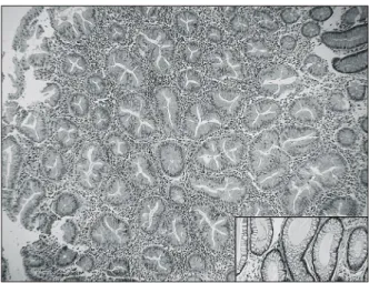
noise의 값이 100 이하가 되는 값은 데이터에서 제외시켰 다. 실험 결과의 통계학덕 정확성을 확인하기 위해 SAM Fig. 1. Serrated type of hyperplastic polyp. Polypectomy spec-
imen shows increased number and size of the glands. Serration and saw-teeth like luminal border (branching of the crypts) are characteristic. Nuclei of the glandular cells are not dysplastic.
Compare the smooth luminal border of usual hyperplastic glands without serration in the other case (right lower box)(H&E, ×100).
Fig. 2. Scanning and image of expression profiles in patients with serrated polyps. SAM (significance analysis of microarray) package method for identification of dif- ferentially expressed genes. Green colored section corresponds to down-regulated gene expression.
Red colored section corresponds to up-regulated gene expression.
Total image
(Stanford Univ., CA, USA) 프로그램을 이용하였으며 실험 결과는 원격차단(distant cutoff)이 1.5 배수의 변화를 보일 때 통계적으로 유의하였고 cutoff 상관 계수는 0.95였다.
3) 통계분석
t-test는 실험군과 대조군 사이의 통계적 차이를 결 정하기 위해 사용되었으며 다면 비교의 경우, 유의수 준은 Bonferroni 조정법을 따랐다.
결 과
병리 조직 소견에서 증식성 용종의 톱니형 형태를
보이는 거치상 용종(Fig. 1)으로 확인된 3명의 환자는 모두 남자였으며 평균 연령은 평균 64 (60∼68)세였다.
용종의 크기는 모두 1 cm 이하였으며 평균 크기는 0.6 (0.5∼0.9) cm였다. 용종의 유전자 분석을 위해 단계적 무리 연산법(hierarchial clustering algorithm)을 사용한 스캔 영상 (Fig. 2)에서 과발현된 유전자들은 붉은 색 깔로, 저발현된 유전자들은 녹색으로 나타났다.
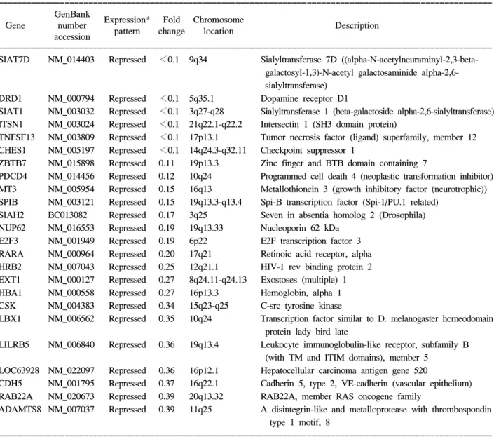
Oligonucleotide microarray의 배열막에 점적된 3,096 개의 유전자 중 67개(2.2%)의 유전자들에서 정상 점막 과 비교하였을 때 유의한 증가 혹은 감소를 보였다. 발 현의 차이를 보인 유전자들 중 34개는 정상 점막의 것 들과 비교하였을 때 과발현된 양상을 보였고 33개는
Table 1. Expression profiles of serrated polyps
ꠚꠚꠚꠚꠚꠚꠚꠚꠚꠚꠚꠚꠚꠚꠚꠚꠚꠚꠚꠚꠚꠚꠚꠚꠚꠚꠚꠚꠚꠚꠚꠚꠚꠚꠚꠚꠚꠚꠚꠚꠚꠚꠚꠚꠚꠚꠚꠚꠚꠚꠚꠚꠚꠚꠚꠚꠚꠚꠚꠚꠚꠚꠚꠚꠚꠚꠚꠚꠚꠚꠚꠚꠚꠚꠚꠚꠚꠚꠚꠚꠚꠚꠚꠚꠚꠚꠚꠚꠚꠚꠚꠚꠚꠚꠚꠚꠚꠚꠚꠚꠚꠚꠚꠚ GenBank
Expression* Fold Chromosome
Gene number Description
pattern change location accession
ꠏꠏꠏꠏꠏꠏꠏꠏꠏꠏꠏꠏꠏꠏꠏꠏꠏꠏꠏꠏꠏꠏꠏꠏꠏꠏꠏꠏꠏꠏꠏꠏꠏꠏꠏꠏꠏꠏꠏꠏꠏꠏꠏꠏꠏꠏꠏꠏꠏꠏꠏꠏꠏꠏꠏꠏꠏꠏꠏꠏꠏꠏꠏꠏꠏꠏꠏꠏꠏꠏꠏꠏꠏꠏꠏꠏꠏꠏꠏꠏꠏꠏꠏꠏꠏꠏꠏꠏꠏꠏꠏꠏꠏꠏꠏꠏꠏꠏꠏꠏꠏꠏꠏꠏ SIAT7D NM_014403 Repressed <0.1 9q34 Sialyltransferase 7D ((alpha-N-acetylneuraminyl-2,3-beta-
galactosyl-1,3)-N-acetyl galactosaminide alpha-2,6- sialyltransferase)
DRD1 NM_000794 Repressed <0.1 5q35.1 Dopamine receptor D1
SIAT1 NM_003032 Repressed <0.1 3q27-q28 Sialyltransferase 1 (beta-galactoside alpha-2,6-sialyltransferase) ITSN1 NM_003024 Repressed <0.1 21q22.1-q22.2 Intersectin 1 (SH3 domain protein)
TNFSF13 NM_003809 Repressed <0.1 17p13.1 Tumor necrosis factor (ligand) superfamily, member 12 CHES1 NM_005197 Repressed <0.1 14q24.3-q32.11 Checkpoint suppressor 1
ZBTB7 NM_015898 Repressed 0.11 19p13.3 Zinc finger and BTB domain containing 7
PDCD4 NM_014456 Repressed 0.12 10q24 Programmed cell death 4 (neoplastic transformation inhibitor) MT3 NM_005954 Repressed 0.15 16q13 Metallothionein 3 (growth inhibitory factor (neurotrophic)) SPIB NM_003121 Repressed 0.15 19q13.3-q13.4 Spi-B transcription factor (Spi-1/PU.1 related)
SIAH2 BC013082 Repressed 0.17 3q25 Seven in absentia homolog 2 (Drosophila) NUP62 NM_016553 Repressed 0.19 19q13.33 Nucleoporin 62 kDa
E2F3 NM_001949 Repressed 0.19 6p22 E2F transcription factor 3 RARA NM_000964 Repressed 0.20 17q21 Retinoic acid receptor, alpha HRB2 NM_007043 Repressed 0.25 12q21.1 HIV-1 rev binding protein 2 EXT1 NM_000127 Repressed 0.27 8q24.11-q24.13 Exostoses (multiple) 1 HBA1 NM_000558 Repressed 0.27 16p13.3 Hemoglobin, alpha 1 CSK NM_004383 Repressed 0.34 15q23-q25 C-src tyrosine kinase
LBX1 NM_006562 Repressed 0.35 10q24 Transcription factor similar to D. melanogaster homeodomain protein lady bird late
LILRB5 NM_006840 Repressed 0.36 19q13.4 Leukocyte immunoglobulin-like receptor, subfamily B (with TM and ITIM domains), member 5
LOC63928 NM_022097 Repressed 0.36 16p12.1 Hepatocellular carcinoma antigen gene 520
CDH5 NM_001795 Repressed 0.37 16q22.1 Cadherin 5, type 2, VE-cadherin (vascular epithelium) RAB22A NM_020673 Repressed 0.39 20q13.32 RAB22A, member RAS oncogene family
ADAMTS8 NM_007037 Repressed 0.39 11q25 A disintegrin-like and metalloprotease with thrombospondin type 1 motif, 8
ꠏꠏꠏꠏꠏꠏꠏꠏꠏꠏꠏꠏꠏꠏꠏꠏꠏꠏꠏꠏꠏꠏꠏꠏꠏꠏꠏꠏꠏꠏꠏꠏꠏꠏꠏꠏꠏꠏꠏꠏꠏꠏꠏꠏꠏꠏꠏꠏꠏꠏꠏꠏꠏꠏꠏꠏꠏꠏꠏꠏꠏꠏꠏꠏꠏꠏꠏꠏꠏꠏꠏꠏꠏꠏꠏꠏꠏꠏꠏꠏꠏꠏꠏꠏꠏꠏꠏꠏꠏꠏꠏꠏꠏꠏꠏꠏꠏꠏꠏꠏꠏꠏꠏꠏ
*Expression profiles comparing with normal control mucosae were significantly up-regulated (induced) or down-regulated (repressed) in polyps by using the oligonucleotide microarray technique.
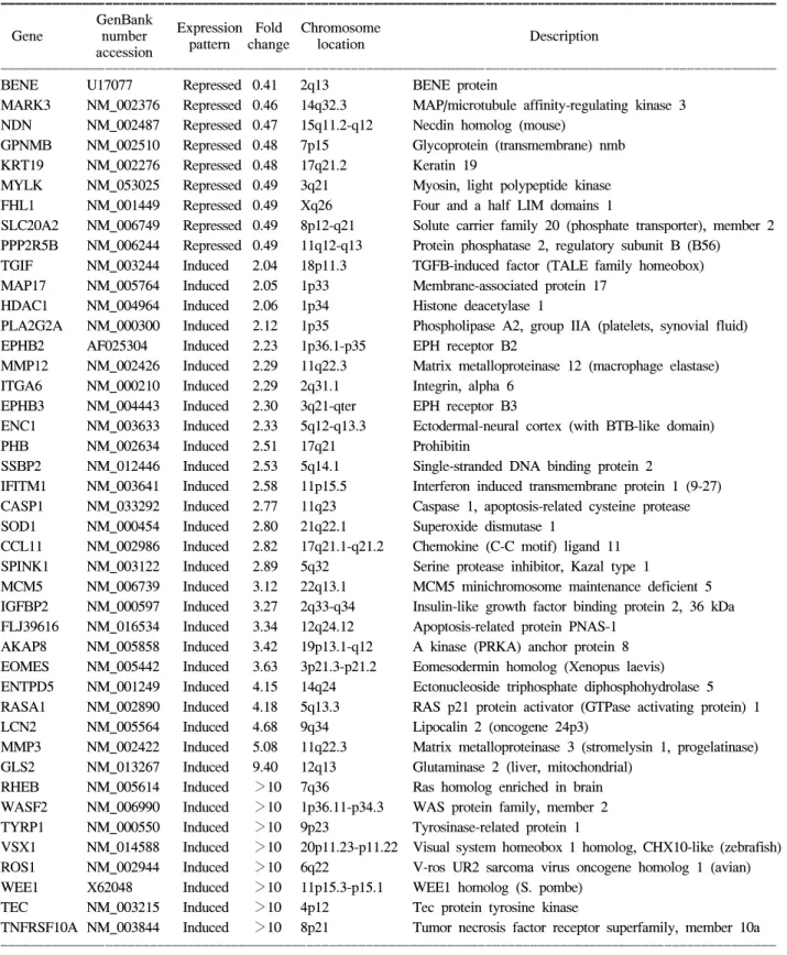
저발현된 양상을 보였다(Table 1). 이 중에서 10배 이상 으로 현저하게 과발현된 유전자는 모두 8개로 RHEB (Ras homolog enriched in brain), WASF2 (WAS protein
family, member 2), TYRP1 (Tyrosinase-related protein 1), VSX1 (Visual system homeobox 1 homolog), ROS1 (V-ros UR2 sarcoma virus oncogene homolog 1), WEE1
Table 1. Continued
ꠚꠚꠚꠚꠚꠚꠚꠚꠚꠚꠚꠚꠚꠚꠚꠚꠚꠚꠚꠚꠚꠚꠚꠚꠚꠚꠚꠚꠚꠚꠚꠚꠚꠚꠚꠚꠚꠚꠚꠚꠚꠚꠚꠚꠚꠚꠚꠚꠚꠚꠚꠚꠚꠚꠚꠚꠚꠚꠚꠚꠚꠚꠚꠚꠚꠚꠚꠚꠚꠚꠚꠚꠚꠚꠚꠚꠚꠚꠚꠚꠚꠚꠚꠚꠚꠚꠚꠚꠚꠚꠚꠚꠚꠚꠚꠚꠚꠚꠚꠚꠚꠚꠚꠚ GenBank
Expression Fold Chromosome
Gene number Description
pattern change location accession
ꠏꠏꠏꠏꠏꠏꠏꠏꠏꠏꠏꠏꠏꠏꠏꠏꠏꠏꠏꠏꠏꠏꠏꠏꠏꠏꠏꠏꠏꠏꠏꠏꠏꠏꠏꠏꠏꠏꠏꠏꠏꠏꠏꠏꠏꠏꠏꠏꠏꠏꠏꠏꠏꠏꠏꠏꠏꠏꠏꠏꠏꠏꠏꠏꠏꠏꠏꠏꠏꠏꠏꠏꠏꠏꠏꠏꠏꠏꠏꠏꠏꠏꠏꠏꠏꠏꠏꠏꠏꠏꠏꠏꠏꠏꠏꠏꠏꠏꠏꠏꠏꠏꠏꠏ
BENE U17077 Repressed 0.41 2q13 BENE protein
MARK3 NM_002376 Repressed 0.46 14q32.3 MAP/microtubule affinity-regulating kinase 3 NDN NM_002487 Repressed 0.47 15q11.2-q12 Necdin homolog (mouse)
GPNMB NM_002510 Repressed 0.48 7p15 Glycoprotein (transmembrane) nmb KRT19 NM_002276 Repressed 0.48 17q21.2 Keratin 19
MYLK NM_053025 Repressed 0.49 3q21 Myosin, light polypeptide kinase FHL1 NM_001449 Repressed 0.49 Xq26 Four and a half LIM domains 1
SLC20A2 NM_006749 Repressed 0.49 8p12-q21 Solute carrier family 20 (phosphate transporter), member 2 PPP2R5B NM_006244 Repressed 0.49 11q12-q13 Protein phosphatase 2, regulatory subunit B (B56) TGIF NM_003244 Induced 2.04 18p11.3 TGFB-induced factor (TALE family homeobox) MAP17 NM_005764 Induced 2.05 1p33 Membrane-associated protein 17
HDAC1 NM_004964 Induced 2.06 1p34 Histone deacetylase 1
PLA2G2A NM_000300 Induced 2.12 1p35 Phospholipase A2, group IIA (platelets, synovial fluid) EPHB2 AF025304 Induced 2.23 1p36.1-p35 EPH receptor B2
MMP12 NM_002426 Induced 2.29 11q22.3 Matrix metalloproteinase 12 (macrophage elastase) ITGA6 NM_000210 Induced 2.29 2q31.1 Integrin, alpha 6
EPHB3 NM_004443 Induced 2.30 3q21-qter EPH receptor B3
ENC1 NM_003633 Induced 2.33 5q12-q13.3 Ectodermal-neural cortex (with BTB-like domain)
PHB NM_002634 Induced 2.51 17q21 Prohibitin
SSBP2 NM_012446 Induced 2.53 5q14.1 Single-stranded DNA binding protein 2
IFITM1 NM_003641 Induced 2.58 11p15.5 Interferon induced transmembrane protein 1 (9-27) CASP1 NM_033292 Induced 2.77 11q23 Caspase 1, apoptosis-related cysteine protease SOD1 NM_000454 Induced 2.80 21q22.1 Superoxide dismutase 1
CCL11 NM_002986 Induced 2.82 17q21.1-q21.2 Chemokine (C-C motif) ligand 11 SPINK1 NM_003122 Induced 2.89 5q32 Serine protease inhibitor, Kazal type 1
MCM5 NM_006739 Induced 3.12 22q13.1 MCM5 minichromosome maintenance deficient 5 IGFBP2 NM_000597 Induced 3.27 2q33-q34 Insulin-like growth factor binding protein 2, 36 kDa FLJ39616 NM_016534 Induced 3.34 12q24.12 Apoptosis-related protein PNAS-1
AKAP8 NM_005858 Induced 3.42 19p13.1-q12 A kinase (PRKA) anchor protein 8 EOMES NM_005442 Induced 3.63 3p21.3-p21.2 Eomesodermin homolog (Xenopus laevis) ENTPD5 NM_001249 Induced 4.15 14q24 Ectonucleoside triphosphate diphosphohydrolase 5 RASA1 NM_002890 Induced 4.18 5q13.3 RAS p21 protein activator (GTPase activating protein) 1 LCN2 NM_005564 Induced 4.68 9q34 Lipocalin 2 (oncogene 24p3)
MMP3 NM_002422 Induced 5.08 11q22.3 Matrix metalloproteinase 3 (stromelysin 1, progelatinase) GLS2 NM_013267 Induced 9.40 12q13 Glutaminase 2 (liver, mitochondrial)
RHEB NM_005614 Induced >10 7q36 Ras homolog enriched in brain WASF2 NM_006990 Induced >10 1p36.11-p34.3 WAS protein family, member 2 TYRP1 NM_000550 Induced >10 9p23 Tyrosinase-related protein 1
VSX1 NM_014588 Induced >10 20p11.23-p11.22 Visual system homeobox 1 homolog, CHX10-like (zebrafish) ROS1 NM_002944 Induced >10 6q22 V-ros UR2 sarcoma virus oncogene homolog 1 (avian) WEE1 X62048 Induced >10 11p15.3-p15.1 WEE1 homolog (S. pombe)
TEC NM_003215 Induced >10 4p12 Tec protein tyrosine kinase
TNFRSF10A NM_003844 Induced >10 8p21 Tumor necrosis factor receptor superfamily, member 10a ꠏꠏꠏꠏꠏꠏꠏꠏꠏꠏꠏꠏꠏꠏꠏꠏꠏꠏꠏꠏꠏꠏꠏꠏꠏꠏꠏꠏꠏꠏꠏꠏꠏꠏꠏꠏꠏꠏꠏꠏꠏꠏꠏꠏꠏꠏꠏꠏꠏꠏꠏꠏꠏꠏꠏꠏꠏꠏꠏꠏꠏꠏꠏꠏꠏꠏꠏꠏꠏꠏꠏꠏꠏꠏꠏꠏꠏꠏꠏꠏꠏꠏꠏꠏꠏꠏꠏꠏꠏꠏꠏꠏꠏꠏꠏꠏꠏꠏꠏꠏꠏꠏꠏꠏ
(WEE1 homolog), TEC (Tec protein tyrosine kinase), TNFRSF10A (Tumor necrosis factor receptor superfamily, member 10a) 등이었다. 10배 이하로 현저하게 발현이 저 하된 유전자는 모두 6개로 SIAT7D (Sialyltransferase 7D), DRD1 (Dopamine receptor D1), SIAT1 (Sialyltransferase 1), ITSN1 (Intersectin 1), TNFSF12 (Tumor necrosis factor superfamily, member 12), CHES1 (Checkpoint suppressor 1) 등이었다(Table 2).
고 찰
기능성 유전체의 분석은 생물 정보학의 발전과 함 께 새로운 학문 분야로 떠올랐다. 빠르게는 반나절 만 에 수 만개의 표본을 분석할 수 있는 수준으로 진보하 였고, 수많은 유전 정보들을 손쉽게 분석하여 목표 질 환에 대한 질병 과정을 예측할 수 있을 뿐 아니라 환자 개개인의 유전적 소인에 따른 진단과 치료가 결정되 는 소위 “맞춤 의학”의 시대를 기대하게 되었다.2 cDNA microarray는 서던 블롯이나 노던 블롯 분석과 같은 배열 기준 부합법(array based hybridization) 원리 를 사용한 것으로서 수백 개에서 수천 개의 DNA를 질 소 셀룰로오스막이 아닌 슬라이드 유리와 같은 기판 위에 고밀도로 정렬하여 고정한다. 이 분석의 장점은 한번의 실험으로 동시에 많은 수의 유전자들에 대한 발현 변이 양상을 탐색할 수 있으며 또한 유전자의 진
단, 돌연변이, 의약품의 효과 확인 및 질병 진단용 등 에 널리 응용될 수 있는 새로운 차원의 HTS (high- throughput screening) 시스템이라고 할 수 있다.6 국내에는 기존의 분자생물학 기술들을 이용하여 밝 혀진 용종에서의 유전자 변이들이 있기는 하지만 조 직병리학적으로 증식성 용종으로 진단된 검체의 유전 자 발현 분포를 알기위한 바이오칩 연구 결과는 아직 까지 자세히 알려져 있지 않다. 임상에서 환자가 나이 가 들어감에 따라 대장내시경을 통해 흔하게 관찰할 수 있는 소형의 용종들이 모두 선종성으로 진행되거 나 암화의 과정을 밟는 것은 아니다. 그렇다면 환자에 따라 종양이 되지 못하고 증식성 용종으로 계속 남아 있는 경우와 선종으로 진행되어 궁극적으로 암화 과 정을 밟게 되는 용종은 유전자 발현이 서로 다를 것이 라고 생각한다. 이 점이 저자들이 본 연구를 착안한 동 기이다.
대장암의 대부분은 이미 존재하고 있던 선종성 용 종에서 발생되고, 선종성 용종을 가지고 있는 환자는 일생 동안 10% 정도에서 암성 용종으로 진행된다고 한다.7 대장에서 1 cm 이하로 미세하게 발견되는 대부 분의 용종은 비신생화성의 단순증식에 의한 돌출로 이루어진 증식성 용종이다.8 과거에 과형성 용종이라 불리어 왔으나 이제는 증식성 용종으로 불리는 이것 은 비신생화성 용종으로 암화의 잠재성이 없어서 임 상적 중요성이 없다고 간주되어 왔다. 즉, 종래의 전통 Table 2. Summarized results of gene expression before and after normalization
ꠚꠚꠚꠚꠚꠚꠚꠚꠚꠚꠚꠚꠚꠚꠚꠚꠚꠚꠚꠚꠚꠚꠚꠚꠚꠚꠚꠚꠚꠚꠚꠚꠚꠚꠚꠚꠚꠚꠚꠚꠚꠚꠚꠚꠚꠚꠚꠚꠚꠚꠚꠚꠚꠚꠚꠚꠚꠚꠚꠚꠚꠚꠚꠚꠚꠚꠚꠚꠚꠚꠚꠚꠚꠚꠚꠚꠚꠚꠚꠚꠚꠚꠚꠚꠚꠚꠚꠚꠚꠚꠚꠚꠚꠚꠚꠚꠚꠚꠚꠚꠚꠚꠚꠚ Normalized Normalized Normalized
Gene Specimen #1 Sepecimen #2 Specimen #3 Fold change
specimen #1 specimen #2 specimen #3
ꠏꠏꠏꠏꠏꠏꠏꠏꠏꠏꠏꠏꠏꠏꠏꠏꠏꠏꠏꠏꠏꠏꠏꠏꠏꠏꠏꠏꠏꠏꠏꠏꠏꠏꠏꠏꠏꠏꠏꠏꠏꠏꠏꠏꠏꠏꠏꠏꠏꠏꠏꠏꠏꠏꠏꠏꠏꠏꠏꠏꠏꠏꠏꠏꠏꠏꠏꠏꠏꠏꠏꠏꠏꠏꠏꠏꠏꠏꠏꠏꠏꠏꠏꠏꠏꠏꠏꠏꠏꠏꠏꠏꠏꠏꠏꠏꠏꠏꠏꠏꠏꠏꠏꠏ
SIAT7D -6.58 -4.38 -4.98 0.01 0.05 0.03 0.03*
DRD1 -5.75 -4.58 -4.34 0.02 0.04 0.05 0.04*
SIAT1 -4.92 -5.49 -3.47 0.03 0.02 0.09 0.05*
ITSN1 -4.79 -5.29 -3.56 0.04 0.03 0.08 0.05*
TNFSF13 -4.94 -5.03 -3.21 0.03 0.03 0.11 0.06*
CHES1 -5.05 -4.41 -2.51 0.03 0.05 0.18 0.09*
RHEB 2.73 2.81 4.53 6.65 6.99 23.15 12.26†
WASF2 2.81 3.32 4.57 6.99 9.98 23.73 13.57†
TYRP1 2.75 5.53 2.61 6.73 46.11 6.07 19.64†
VSX1 4.31 5.17 4.75 19.88 35.91 26.89 27.56†
ROS1 4.45 5.46 5.03 21.99 44.04 32.64 32.89†
WEE1 3.63 5.89 5.09 12.42 59.11 34.19 35.24†
TEC 2.61 4.75 6.29 6.09 26.92 78.14 37.05†
TNFRSF10A 4.73 5.87 5.59 26.51 58.67 48.49 44.56†
ꠏꠏꠏꠏꠏꠏꠏꠏꠏꠏꠏꠏꠏꠏꠏꠏꠏꠏꠏꠏꠏꠏꠏꠏꠏꠏꠏꠏꠏꠏꠏꠏꠏꠏꠏꠏꠏꠏꠏꠏꠏꠏꠏꠏꠏꠏꠏꠏꠏꠏꠏꠏꠏꠏꠏꠏꠏꠏꠏꠏꠏꠏꠏꠏꠏꠏꠏꠏꠏꠏꠏꠏꠏꠏꠏꠏꠏꠏꠏꠏꠏꠏꠏꠏꠏꠏꠏꠏꠏꠏꠏꠏꠏꠏꠏꠏꠏꠏꠏꠏꠏꠏꠏꠏ
*Down-regulated by less than a tenth of the expression as compared with normal colonic mucosa; †Up-regulated by more than 10 times of the expression as compared with normal colonic mucosa.
적인 개념으로는 비종양성이고 악성화로 진행되지 않 는다는 것이 지배적인 의견이었다.3 그러나 최근의 보
고들4,5,9에서 증식성 용종도 대장의 선종과 비교하였을
때, 분자유전학적뿐만 아니라 해부학적 분포의 특성을 공유하고 있어서 선종성 용종의 전구 질환의 가능성 이 대두되었다. 이러한 견해를 뒷받침해주는 근거로서 증식성 용종에서도 암태아성 항원이 증가하고, 종양에 서 주로 나타나는 점액소의 당항원인 Tn이 선종성 용 종뿐만 아니라 증식성 용종에서도 발현한다는 것을 그 예로 들 수 있다.9 Jass10는 증식성 용종 자체가 신생화성 용종은 아니지만, 대장암을 유발하는 인자들에 의해 유 발될 수 있다고 하였다. 그러나 대장 점막의 당단백질 합성과정에서 암과 관련한 신호변경을 초래하는 것이 가능하지만 악성으로의 형질 전환에는 저항성이 있어 증식성 용종이 곧바로 악성 변화를 일으키지는 않는다 고 한다. 따라서 증식성 용종과 선종성 용종 및 대장암 들이 형태학적으로뿐만 아니라 면역 조직화학적인 측 면에서도 상호간에 유전적 연속체일 것이라고 제시하
였다.5,10,11 이것은 종래에 잘 알려진 “선종-대장암 연결”
이론에 추가적으로 “증식상피 - 이형성 - 선종 - 선암종 연결(hyperplastic epithelium - dysplasia - adenoma - carcinoma sequence)이 있다는 것을 의미한다.
2005년에 Snover 등12은 대장의 용종을 증식성 용종, 거치상 선종(serrated adenoma; SA) 및 무경성 거치상 선종(sessile serrated adenoma; SSA) 등으로 분류한 다 음, 이 세가지를 통틀어서 “거치상 용종”이라고 불렀 다. 본 연구에서 사진으로 제시한 증식성 용종의 조직 소견도 거치상 용종에 해당하며, 창자샘이 확장되어 가지 모양으로 갈라지는 거치상 구조를 저명하게 보 여주었다. 거치상 용종은 증식성 용종과 같이 존재하 는 혼합형으로 나타나기도 하며 용종 전체가 거치상 으로 보이기도 한다. 이러한 소견은 세포의 분화적인 측면 보다는 증식의 조절 양상이 다르기 때문에 발생 한다. 특히, SSA는 우측 대장에 많고 대장암과 관련한 생물학적 표지자를 가지고 있다고 한다.12 거치상 용종 의 조직학적 형태는 선종으로부터 종래의 고전적인 증식성 용종과 닮은 형태까지 다양하게 관찰된다. 최 근의 조직학적 연구에서 제안된 세부적인 진단 기준 은 1) 비정상적인 증식과 성숙 장애, 2) 창자샘의 확장 된 증식 구역, 3) 창자샘 기저부의 확장과 톱니 모양의 구조, 4) 세포 성숙의 감소 등이다.13 본 연구에서도 이 점을 거치상 용종을 구분하기 위한 병리 조직학적인 감별 지침으로 사용하였다.
만일 대장암이 한가지만의 원인이 아닌 별개의 서
로 다른 경로를 따라 발생하는 여러 가지 병인론들을 가지고 있다면 지금까지 알려지지 않은 발생 경로가 있을 것이다. 이중에서도 전술한 조직학적 진단 기준 을 충족시키는 용종을 경유하여 암종으로 진행한다는 개념을 “거치상 경로(serrated pathway)”라고 부른다.14 대장 점막에서 이와 같은 경로의 출발점으로 가장 빨 리 나타난 병소를 변종 창자샘 병소(aberrant crypt foci, ACF)라고 하는데, 이것은 차후에 증식성 용종, 선종 및 거치상 선종 등으로 진행한다고 한다.15 이를 알기 위한 근거들은 여러 연구에서 제시되었는데 이 중에 서 대표적인 것으로는, 증식성 용종의 보다 종양적인 형태 즉, SSA가 있을 때 micro satellite instability, high type (MSI-H) 대장암과 관련이 있는 BRAF의 돌연변이 와 DNA 메틸화가 나타난다는 것이다.14,16 BRAF 돌연 변이와 DNA 메틸화는 MSI-H 대장암으로 발전하는 거치상 경로의 초기 변화이다. 거치상 선종과 증식성 용종에서 세포자멸의 억제는 CD95 발현의 감소와 밀 접한 관련이 있었으며 이러한 현상이 특징적인 톱니 모양의 구조를 유도한다고한다.17 거치상 선종으로 진 행할수록 초기에는 SLC5A8유전자가 메틸화 되며, 나 중에는 유전자 내 CpG 섬의 비정상적이고도 광범한 메틸화가 나타난다.18 이와 같은 최근의 연구 결과들은 거치상 선종의 경로를 거쳐 종양으로 발전한다는 소 위 “증식성 용종 - 거치상 선종 - 대장암 연결 현상”이 라는 가설의 가능성을 뒷받침해주는 결과라 할 수 있 다.
본 연구의 분석 결과에서 정상 대장 점막 조직보다 10배 이상으로 과다 발현된 유전자들 중에서 대장암의 형성 과정과 관련이 있다고 타 문헌에서 보고된 유전자 들과 일치하는 것들은 ROS (reactive oxygen species), TNFRSF (tumor necrosis factor receptor superfamily 10A) 및 MMP (matrix metalloproteinase) 일원들이다.
ROS의 과다한 생성은 주요 유전자에 해로운 영향을 미치어 DNA 손상 혹은 불안정을 가져오고 소화관 세 포의 항상성에 지장을 주어 대장암이 발생하는 다단 계 과정 중의 하나에 관여한다고 한다.19 만성염증성 질환이 대장암으로 진행하는 것도 이것과 관련되어 있다.20 대장암 세포주에서 TNFRSF10A의 과다 발현은 p53과 독립적인 세포 자멸사를 유도한다.21 본 연구에 서 과다 발현된 MMP3를 포함한 MMP-1, 7, 13 등의 일원들은 대장암의 성장과 증식에 중요한 역할을 하 는 것으로 알려져 있다.22 본 연구에서 십분의 일 수준 으로 과소 발현된 유전자들 중에서 종양 형성의 과정 과 관련이 있는 것으로 기존에 발표된 유전자들과 일
치하는 것으로는 DRD1 (dopamine receptor D1), SIAT1 (sialyltransferase 1), TNFSF12, CHES1 (checkpoint sup- pressor 1) 및 PDCD4 (programmed cell death protein 4)
등이다.23-26 도파민 수용체는 인간의 대장암 세포에서
대사변화를 유도하고 SIAT는 대장암의 생물학적 공격 성의 좋은 지표이다.23 TNFSF12은 인체 종양 생성에서 조절유전자로 기능하는 것으로 추론 되지만 자세한 기능은 아직까지 잘 알려져 있지 않다.24 CHES1은 종 양 생성에 중요한 유전자를 억제하며 구강암 환자에 서는 이것의 발현이 특징적으로 저하되었음이 증명된 바 있다.25 PDCD4의 발현은 대장암 조직에서 매우 저 하되거나 소실되었다고 한다.26
본 연구에서 과다 또는 과소 발현된 유전자들이 증 식성 용종의 톱니형 형태 즉, 거치상 용종에서 특이성 이 있으며 재현성을 가지고 있는지 여부는 더 많은 증 례를 통해 검증할 필요성이 있다. 저자들이 본 연구에 서는 시행하지 못하였으나 각 유전자 별로 추후에 시 행될 RT-PCR과 면역조직 화학연구를 이용하여 타당 도(validation)를 검증해 보아 할 것으로 생각한다. 또 한, microarray로 얻은 방대한 유전자 발현 패턴 정보를 다른 연구자들의 결과와 비교하는 문제도 남아있다.
저자들의 논제가 초기 선험적 연구인 이유가 여기에 있다. 이러한 문제점들이 보완된다면 본 연구의 결과 는 증식성 용종의 암화 진전에 결정적인 유전자를 발 굴할 수 있으며 이를 이용하여 잠재암 환자의 선별이 나 치료를 위한 분자생물학적인 유도 지표로 응용할 수 있을 것임을 기대한다.
결 론
대장의 증식성 용종에서 나타나는 거치상 용종의 microarray 분석에서 대장암과 관련된 유전자들이 정 상 점막의 것들과 비교하여 유의하게 과다 발현되었 거나 과소 발현되었다. 본 연구의 결과는 증식성 용종 도 악성화 잠재성을 가질 수 있다는 “거치상 경로”의 가능성을 가늠하는 데에 분자생물학적인 특성으로 활 용할 수 있을 것이다.
REFERENCES
1. 황일란, 조성원, 함기백. 소화기 질환에서 cDNA microarray 연구의 응용 및 전망. 대한소화기학회지 2003;41:241-49.
2. Schena M, Shalon D, Davis RW, Brown PO. Quantitative monitoring of gene expression patterns with a comple-
mentary DNA microarray. Science 1995;270:467-70.
3. Achord JL. Hyperplastic colon polyps do not predict adenomas. Gastroenterology 1991;100:1142-3.
4. Ansher AF, Lewis JH, Fleischer DE, Cattau EL Jr, Collen MJ, O'Kieffe DA, et al. Hyperplastic colonic polyps as a marker for adenomatous colonic polyps. Am J Gastroenterol 1989;84:113-7.
5. Rashid A, Houlihan PS, Booker S, Petersen GM, Giardiello FM, Hamilton SR. Phenotypic and molecular charac- teristics of hyperplastic polyposis. Gastroenterology 2000;
119:323-32.
6. Hippo Y, Taniguchi H, Tsutsumi S, Machida N, Chong JM, Fukayama M, et al. Global gene expression analysis of gastric cancer by oligonucleotide microarrays. Cancer Res 2002;62:233-40.
7. Muto T, Bussey HJ, Morson BC. The evolution of cancer of the colon and rectum. Cancer 1975;36:2251-70.
8. 김재선, 송훤택, 지종대, 변관수, 박영태, 김종극 등. 하부 대장 과형성 용종의 의의. 대한소화기학회지 1993;25:
650-58.
9. Itzkowitz SH, Bloom EJ, Lau TS, Kim YS. Mucin associated Tn and sialosyl-Tn antigen expression in colorectal polyps. Gut 1992;33:518-23.
10. Jass JR. Relation between metaplastic polyp and carci- noma of the colorectum. Lancet 1983;1:28-30.
11. Boland CR, Montgomery CK, Kim YS. A cancer-associated mucin alteration in benign colonic polyps. Gastroenterology 1982;82:664-72.
12. Snover DC, Jass JR, Fenoglio-Preiser C, Batts KP. Ser- rated polyps of the large intestine: a morphologic and molecular review of an evolving concept. Am J Clin Pathol 2005;124:380-91.
13. Oberschmid BI, Dietmaier W, Hartmann A, Dahl E, Klopocki E, Beatty BG, et al. Distinct secreted Frizzled receptor protein 1 staining pattern in patients with hyperplastic polyposis coli syndrome. Arch Pathol Lab Med 2004;128:967-73.
14. Kambara T, Simms LA, Whitehall VL, Spring KJ, Wynter CV, Walsh MD, et al. BRAF mutation is associated with DNA methylation in serrated polyps and cancers of the colorectum. Gut 2004;53:1137-44.
15. Jass JR, Whitehall VL, Young J, Leggett BA. Emerging concepts in colorectal neoplasia. Gastroenterology 2002;
123:862-76.
16. Higuchi T, Jass JR. My approach to serrated polyps of the colorectum. J Clin Pathol 2004;57:682-6.
17. Tateyama H, Li W, Takahashi E, Miura Y, Sugiura H, Eimoto T. Apoptosis index and apoptosis-related antigen expression in serrated adenoma of the colorectum: The saw-toothed structure may be related to inhibition of apoptosis. Am J Surg Pathol 2002;26:249-56.
18. Dong SM, Lee EJ, Jeon ES, Park CK, Kim KM. Pro-
gressive methylation during the serrated neoplasia path- way of the colorectum. Mod Pathol 2005;18:170-8.
19. van der Logt EM, Roelofs HM, Wobbes T, Nagengast FM, Peters WH. High oxygen radical production in patients with sporadic colorectal cancer. Free Radic Biol Med 2005;39:182-7.
20. Seidelin JB, Nielsen OH. Continuous cytokine exposure of colonic epithelial cells induces DNA damage. Eur J Gastroenterol Hepatol 2005;17:363-9.
21. Xu SQ, El-Deiry WS. p21(WAF1/CIP1) inhibits initiator caspase cleavage by TRAIL death receptor DR4. Biochem Biophys Res Commun 2000;269:179-90.
22. Roeb E, Arndt M, Jansen B, Schumpelick V, Matern S.
Simultaneous determination of matrix metalloproteinase (MMP)-7, MMP-1, -3, and -13 gene expression by mul- tiplex PCR in colorectal carcinomas. Int J Colorectal Dis 2004;19:518-24.
23. Scemama JL, Ruellan C, Clerc P, Clemente F, Ribet A.
Dopamine receptors in a human colonic cancer cell line (HT29). Some receptor-related biological effects of do- pamine. Int J Cancer 1984;34:675-9.
24. Ho DH, Vu H, Brown SA, Donohue PJ, Hanscom HN, Winkles JA. Soluble tumor necrosis factor-like weak inducer of apoptosis overexpression in HEK293 cells promotes tumor growth and angiogenesis in athymic nude mice. Cancer Res 2004;64:8968-72.
25. Chang JT, Wang HM, Chang KW, Chen WH, Wen MC, Hsu YM, et al. Identification of differentially expressed genes in oral squamous cell carcinoma (OSCC): overex- pression of NPM, CDK1 and NDRG1 and underexpression of CHES1. Int J Cancer 2005;114:942-9.
26. Goke R, Barth P, Schmidt A, Samans B, Lankat-Buttgereit B. Programmed cell death protein 4 suppresses CDK1/cdc2 via induction of p21 (Waf1/Cip1). Am J Physiol Cell Physiol 2004;287:C1541-6.