견관절의 초음파 검사 방법
고려대학교 의과대학 정형외과학교실
정 웅 교
견관절에 동통을 호소하는 환자를 치료하기 위해 서는 정확한 진단이 필수 적이다. 특히 견관절은 골 성 구조보다는 연부 조직의 이상에 의해 통증이 발 생하는 경우가 대부분이므로 이를 검사하기 위해서 는 단순방사선 사진 만으로는 통증의 정확한 원인을 감별하기 어려운 경우가 많다. 따라서 자기 공명 영 상을 비롯한 연부 조직의 상태를 평가할 수 있는 다 양한 영상 진단법 들이 사용되고 있고 최근에는 초 음파에 대한 관심이 높아지고 있다. 초음파는 1979 년 Seltzer 등1)이 처음 소개한 이후 많은 발전을 거 듭해 왔고 특히 7.5 MHz 이상의 고해상도 변환기 (transducer)의 사용 이후 자기 공명 영상에 필적 할 만한 민감도와 특이도를 나타내고 있다2). 초음파 검사의 장점으로는 비침습적이고, 환자의 순응도가 높으며, 검사결과를 바로 알 수 있다. 또한 실시간
검사가 가능하여 동적인 검사(dynamic examina- tion)가 가능하며 반대측을 검사할 수 있다는 점이 다. 하지만 초음파 검사의 가장 큰 문제점은 시술자 의 경험과 숙련도에 따라 영상의 해석이 다양할 수 있다는 점이다3). 본문에서는 견관절 주위의 초음파 검사의 방법에 대하여 기술하고자 한다.
본 문
견관절 주위의 초음파검사는 환자의 뒤 편 혹은 앞 편에서 모두 시행이 가능하나 검사자가 변환기를 자유롭게 견관절의 전, 후, 측 방에 위치할 수 있고 검사자의 요청에 의해 피검자가 팔을 자유롭게 움직 일 수 있도록 등받이나 팔걸이가 없는 회전이 가능 한 의자에 앉게 하는 것이 가장 좋다. 검사는 일반적 으로 상완 이두건 구(bicipital groove) 내의 상완 이두건 장두(long head of biceps tendon)을 관찰 하고 이후 견관절의 전방, 측방, 후방 구조를 차례로 관찰한 다음 견봉쇄골 관절을 관찰하게 된다. 초음 파 검사에서 유의할 점은 관찰하고자 하는 구조의
통신저자: 정 웅 교
서울특별시 성북구 안암동 5가 126-1 고려대학교 안암병원 정형외과 Tel: 02-920-6779, Fax: 02-924-2471 E-mail: [email protected]
Technique of ultrasonographic scanning of the shoulder joint
Woong-Kyo Jeong, M.D.
Department of Orthopedic Surgery, College of Medicine, Korea University
Ultrasonography is a powerful and useful method for the examination of the various shoulder diseases. The use of high-resolution transducer and technical evolution allowed the improvement of the accuracy of detection of various shoulder pathology including the rotator cuff disease. However, its limitation is that there is marked disparity in the interpretation according to the operators’ experience. This article describes the appropriate scan technique of ultra- sonography around shoulder.
Key Words: Shoulder, Ultrasonography, Scan technique
한다. 변환기를 상완골에 수직되게 위치하면 상완 비후 및 관절내 포착 등을 검사할 수도 있다5).
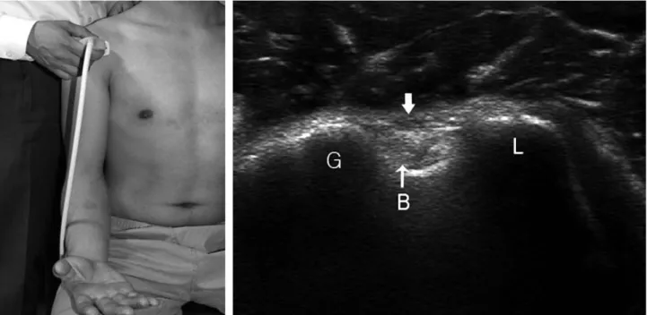
Fig. 1. Short axis scanning of long head of biceps tendon. Scan shows hyperechoic biceps tendon within the bicip- ital groove (B). (G: greater tuberosity, L: lesser tuberosity, arrow: transverse humeral ligament).
Fig. 2. Long axis scanning of long head of biceps tendon. Long axis scan shows biceps tendon(B) demonstrating well-defined fibrillar echotextures.
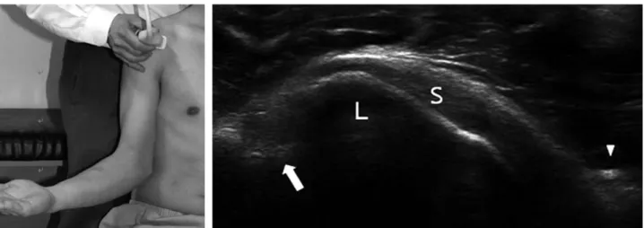
Fig. 3. Long axis scanning of subscapularis tendon. Scan shows subscapularis tendon (S), lesser tuberosity (L) and coracoid process (arrow head). Biceps tendon in the bicipital groove (arrow) appears hypoechoic due to anisotrophy.
Fig. 4. Short axis scan of subscapularis tendon. Transducer is rotated medially to make a perpendicular scan and scan shows multiple hypoechoic cleft (arrow) through the tendon substance (LT: lesser tuberosity).
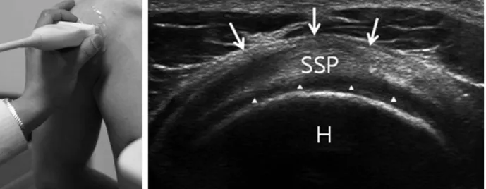
Fig. 5. Appropriate positioning for visualization of the supraspinatus tendon. A. Crass position. B. Modified Crass position.
A B
cle)사이로 근섬유가 위치하여 보이는 현상으로 파 열이 아님을 주의해야 한다. 견갑하건을 관찰할 때
조영 한다. 단축영상에서 극상건은 볼록한 형태를 지닌 균일한 중간 정도의 에코를 나타내게 되며 전
Fig. 6. Short axis scanning of supraspinatus tendon. Scan shows supraspinatus tendon is located between articu- lar cartilage (arrow head) and subdeltoid bulsa (arrow) (SSP: supraspinatus tendon, H: humeral head).
Fig. 7. Long axis scan of supraspinatus tendon. Scan shows convex beak-like shape characterized by fibrillary pattern echogenic lines (SSP: supraspinatus tendon, GT: greater tuberosity, arrow: subdeltoid bulsa).
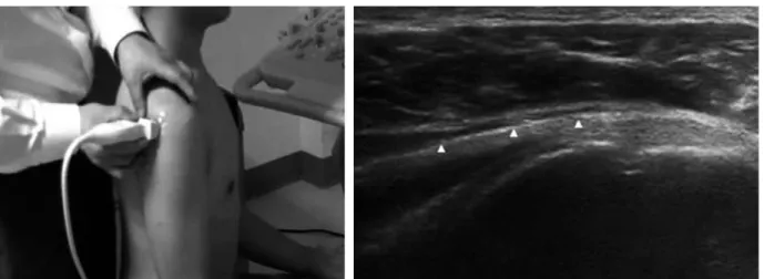
Fig. 8. Long axis scan of infraspinatus tendon. Scan shows the infraspinatus tendon arising within the muscle from thick central aponeurosis (arrow heads).
Fig. 10. Short axis scan of infraspinatus and teres minor tendon. Scan shows lager infraspinatus tendon (ISP) and smaller teres minor tendon (Tm).
Fig. 9. Long axis scan of teres minor tendon. Scan shows the teres minor tendon (arrow heads) arising eccentri- cally relative to the muscle belly.
향이 급격하게 변하게 되므로 비등방성 인공물에 의 한 국소적 저 에코 영역이 관찰될 수 있어 회전근 개
며 중간 부위에 변환기를 견갑골 극을 따라 위치하 고 약간 원위부로 이동 시키면 극하근의 장축 영상
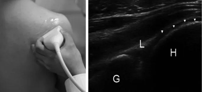
Fig. 11. Scan of posterior labrum and capsule. Scan shows hyperechoic glenoid labrum (L) and hypoechoic articu- lar cartilage (arrow heads) (G: glenoid, H: humeral head).
Fig. 12. Scan of acromioclavicular joint. Scan shows acromion (Ac) and clavicle (Cl) are separated by hypoechoic joint space (arrow: superior acromioclavicular ligament).
을 조 영 할 수 있 으 며 근 육 과 중 심 부 의 건 막 (aponeurosis)을 관찰할 수 있다(Fig. 8). 이 건막 을 따라 외측으로 변환기를 이동하면 대결절 하방에 부착하는 극하건을 관찰할 수 있다. 소원형근의 장 축 조영은 극하근을 조영할 후에 보다 하방으로 변 환기를 위치시키면 관찰할 수 있다. 소원형근의 건 막은 극하근에 비하여 한 쪽으로 치우쳐 있으며 길 이도 짧다. 또한 주행 방향이 극하근 보다 기울어져 있다(Fig. 9). 극하건 및 소원형건의 단축영상을 조 영하기 위해서는 견갑골극에 수직되게 변환기를 위 치한 후 극하근 및 소원형근을 조영하고 근섬유를 따라 외측으로 변환기를 이동시키면서 대결절에 부 착하는 두 건을 관찰할 수 있으며 극하건이 보다 넓 은 부분을 차지한다(Fig. 10)
5. 관절 활액막
견관절의 활액막 중 초음파를 사용한 조영이 용이 한 곳은 후방 관절막이며 전방 이나 하방 관절막은 사이의 연부조직이 많고 깊게 위치하여 효과적인 조 영을 하기 어렵다. 후방 관절막을 검사하기 위해서는 검사하려는 쪽의 손으로 반대측 어깨를 짚는 자세를 취하게 한 상태에서 극하건의 장축 영상을 조영하는 방향으로 변환기를 위치시키며, 극하건 하방의 상완 골두, 견갑와, 후방 관절순을 관찰 할 수 있다(Fig.
11). 삼출액 등이 있는 경우에 극하건과 상완골두 사 이의 저 에코의 부분이 두드러지게 나타난다.
6. 견봉쇄골 관절
견봉쇄골 관절을 관찰하기 위해서는 손가락으로 견봉쇄골 관절을 촉지한 후에 변환기를 원위 쇄골을 따라서 위치시키면 관절의 관상면 영상을 조영할 수 있다(Fig. 12). 상부 견봉쇄골 인대는 비교적 용이 하게 관찰할 수 있으나 하부 견봉쇄골 인대는 관찰 이 되지 않으며 관절내의 디스크 역시 관찰이 되지 않는 경우도 많다.
견봉의 장축을 따라 변환기를 위치 시키면 간혹 견봉 상부 피질골이 분리가 되어 있는 견봉골(os acromiale)을 관찰할 수 있으며 어깨를 움직이면서 원위 분절의 안정성을 평가 할 수 있다8).
맺음말
견관절의 초음파 검사는 견관절 통증 환자에서 병 력 청취, 신체 검사의 연속으로 간편하게 비교적 정 확한 진단을 내릴 수 있는 유용한 도구이다. 하지만 검사자의 주관적 평가에 의해 다양한 결과가 가능하 기에 정확한 해부학적 지식과 병인에 대한 숙지가 있어야 하며 초음파의 특징을 잘 파악하여야 오진을 최소화 할 수 있다.
참고문헌
01. Seltzer SE, Finberg HJ, Weissman BN, Kido DK and Collier BD: Arthrosonography:
gray-scale ultrasound evaluation of the shoulder.
Radiology, 132: 467-468, 1979.
02. Iannotti JP, Ciccone J, Buss DD, et al:
Accuracy of office-based ultrasonography of the shoulder for the diagnosis of rotator cuff tears. J Bone Joint Surg Am, 87: 1305-1311, 2005.
03. O’Connor PJ, Rankine J, Gibbon WW, Richardson A, Winter F and Miller JH:
Interobserver variation in sonography of the painful shoulder. J Clin Ultrasound, 33: 53-56, 2005.
04. Farin PU, Jaroma H, Harju A and Soimakallio S: Medial displacement of the biceps brachii tendon: evaluation with dynamic sonography during maximal external shoulder rotation. Radiology, 195: 845-848, 1995.
05. Pujol N, Hargunani R, Gadikoppula S, Holloway B and Ahrens PM: Dynamic ultra- sound assessment in the diagnosis of intra-articu- lar entrapment of the biceps tendon (hourglass biceps): A preliminary investigation. Int J Shoulder Surg, 3: 80-84, 2009.
06. Crass JR, Craig EV and Feinberg SB: The hyperextended internal rotation view in rotator cuff ultrasonography. J Clin Ultrasound, 15: 416- 420, 1987.
07. Crass JR, Craig EV and Feinberg SB:
Ultrasonography of rotator cuff tears: a review of 500 diagnostic studies. J Clin Ultrasound, 16:
313-327, 1988.
08. Smith J, Dahm DL and Newcomer-Aney KL: Role of sonography in the evaluation of unstable os acromiale. J Ultrasound Med, 27:
1521-1526, 2008.